Introduction
Glycosylphosphatidylinositols (GPIs) are complex
glycolipids that anchor proteins to the outer leaflet of the cell
membrane via their carboxyl terminal (1). The core structure of a GPI consists of a
phosphatidylinositol (PI) moiety, a glucosamine (GlcN) moiety,
three mannoses (Mans) and an ethanolamine-phosphate (EtNP) moiety
(2). Previous studies have shown that
GPIs are able to transduce activation signals into the cell
(3–6).
The molecular mechanisms underlying signal transduction are poorly
understood. However, it is hypothesized that the PI moiety of GPI
may participate in signal transduction via a palmitate group that
is located at the second position of the PI (PI-2nd) (7).
Cluster of differentiation 59 (CD59) is an 18–21 kDa
GPI-anchored glycoprotein (GPIAP), which belongs to the leukocyte
antigen 6 (Ly6) family of proteins and has a high homology to the
mouse protein, Ly6 (8). CD59 was
initially shown to prevent C9 units from binding to the
C5b-8 complex in order to inhibit the formation of
membrane attack complex (9–11). Due to this crucial role, CD59 is
widely expressed in the majority of tissues, including the heart,
liver and kidneys, and circulating cells, such as leukocytes and
red blood cells (12,13). The function of CD59 in complement
regulation has been well documented (9–11);
however, numerous studies have suggested that CD59 also has a role
in T-cell activation and signal transduction (5–7). However,
CD59 is a GPI-anchored protein and does not span the membrane; thus
the mechanisms underlying the CD59-mediated transduction of signals
into the cell remain unclear. It has been suggested that the
function of CD59 in signal transduction is dependent on its
localization to lipid rafts, which act as platforms for the
associations of signaling molecules (14).
Linker for activation of T-cells (LAT) was first
observed in 1990 and, until 1998, it was purified from activated
Jurkat cells and named LAT based on its properties (15). LAT is one of the most important
transmembrane adaptor proteins, and is expressed in mature T-cells,
natural killer cells, mast cells, megakaryocytes and pre-B-cells
(15–18). LAT has no intrinsic enzymatic
activity, but it enables inducible recruitment of effector
molecules to the plasma membrane (19). Human LAT has four extracellular amino
acids, a single transmembrane domain and a long cytoplasmic tail
that contains nine conserved tyrosine motifs. Examination of the
amino acid sequence of LAT showed that the juxtamembrane region of
LAT contains two cysteine (C) residues, C26 and C29 in humans,
which are critical for LAT palmitoylation, raft localization,
phosphorylation and function in T-cell receptor (TCR)-mediated
signaling (20). LAT palmitoylation
is undeniably essential for its function; however, the mechanism
underlying the palmitoylation of LAT is unknown.
Based on the structural characteristics of CD59 and
LAT, the authors of the present study hypothesized that CD59 may
transfer a palmitate group to LAT, causing them to co-localize to
lipid rafts in order to regulate T-cell signal transduction.
Therefore, in the present study, Jurkat cells were transfected with
lentivirus vectors carrying the LAT-enhanced green fluorescent
protein (EGFP) fusion protein, in order to establish a cell line
stably expressing the fusion protein. In addition, the present
study aimed to investigate the biological roles of CD59 in the
proliferation, activation and apoptosis of Jurkat cells via LAT,
and to demonstrate that CD59 may be the candidate protein that
transfers a palmitate group to LAT.
Materials and methods
Materials
Jurkat cells purchased from the cell bank of the
Chinese Academy of Sciences (Beijing, China) were preserved in our
lab. Negative (neg)-EGFP and the LAT-EGFP fusion protein were
constructed by our lab. Lentiviral vectors were constructed by
Shanghai GeneChem Co., Ltd. (Shanghai, China). RPMI-1640 medium was
purchased from HyClone (GE Healthcare Life Sciences, Logan, UT,
USA). The cell counting kit-8 (CCK-8) was purchased from Beijing
Fanbo Biochemicals Co., Ltd. (Beijing, China). Guava Nexin Reagent
was obtained from Merck Millipore (Darmstadt, Germany).
Biotinylated rabbit anti-human CD59 (catalog no., 5516-2; dilution,
1:1,000; Abcam, Cambridge, UK) and phospholipase C-γ1 (PLCG1;
dilution, 1:4,000; catalog no., 2112-1; Abcam) monoclonal
antibodies, and zeta-chain-associated protein kinase 70
(ZAP70)/lymphocyte-specific protein tyrosine kinase (Lck) (C-term)
polyclonal antibodies (dilution, 1,:1,000; catalog no., AB55208;
Sigma-Aldrich; EMD Millipore, Billerica, MA, USA) were used. The
β-actin antibody (dilution, 1:500; catalog no., D110007-0200) was
obtained from Sangon Biotech Co., Ltd. (Shanghai, China). The goat
anti-rabbit IgG (H+L) secondary antibody (catalog no., D111018;
dilution 1:2,000) was purchased from Sangon Biotech Co., Ltd.
TRITC-conjugated goat anti-rabbit IgG and the Mammalian Protein
Extraction reagent were obtained from Beijing ComWin Biotech Co.,
Ltd. (Beijing, China). Enhanced chemiluminescence (ECL) reagent was
purchased from GE Healthcare Life Sciences. The Transcriptor First
Strand cDNA Synthesis kit and the FastStart Essential DNA Green
Master were obtained from Roche Diagnostics (Basel,
Switzerland).
Cell culture
Jurkat cells were maintained in RPMI-1640 medium
supplemented with 10% fetal bovine serum (FBS; Gibco; Thermo Fisher
Scientific, Inc., Waltham, MA, USA) and antibiotics (100 U/ml
penicillin and 100 µg/ml streptomycin). The cells were maintained
at 37°C in a humidified atmosphere containing 5% CO2.
Exponentially growing cells were used in all experiments.
Experimental groups
Jurkat cells were divided into four groups, as
follows: i) Neg group, in which Jurkat cells were transfected with
the neg-EGFP lentivirus; ii) neg + CD59 antibody (CD59Ab) group, in
which the Jurkat cells transfected with the neg-EGFP lentivirus
were treated with the CD59Ab; iii) LAT group, in which Jurkat cells
were transfected with the LAT-EGFP lentivirus; and iv) LAT + CD59Ab
group, in which the Jurkat cells transfected with the LAT-EGFP
lentivirus were treated with the CD59Ab.
Fluorescence microscopy to assess the
transfection efficiencies of neg-EGFP and LAT-EGFP into Jurkat
cells
Jurkat cells were transfected with neg-EGFP or
LAT-EGFP lentiviruses at a multiplicity of infection of 100
particles/cell in RPMI-1640 medium containing 5 µg/ml polybrene and
Eni.S (infection enhancement solution), and then incubated at 37°C
in 5% CO2 in 96-well plates for 12 h, after which the
cells were incubated with fresh medium. After 2 weeks, the
transfected cells expressing EGFP were observed under a
fluorescence microscope.
Analysis of cell viability
Cell viability was measured using CCK-8 assays.
Briefly, exponentially growing neg, neg + CD59Ab, LAT and LAT +
CD59Ab cells (100 µl, 1×10⁵ cells/ml) were seeded into
96-well plates and incubated for 48 h at 37°C in 5% CO2.
Subsequently, 10 µl CCK-8 solution was added to each well followed
by a 4-h incubation. The absorbance was determined at a wavelength
of 490 nm using a microplate reader. All experiments were performed
in quintuplicate.
Analysis of cell apoptosis
Guava Nexin Reagent contains Annexin V-phycoerythrin
(PE) and 7-aminoactinomycin D (7-AAD). Annexin V-PE is used to
detect phosphatidylserine, which is located on the external
membrane of apoptotic cells, and its mean fluorescent intensity
indicates early and late apoptotic cells. 7-AAD is an indicator of
cell membrane structural integrity and binds to late apoptotic and
dead cells. On the histogram, the lower-left quadrant indicates
cells that are not undergoing apoptosis [Annexin V-PE(−),
7-AAD(−)], the lower-right quadrant indicates cells in the early
stages of apoptosis [Annexin V-PE(+), 7-AAD(−)], the upper-right
quadrant indicates cells in the late stages of apoptosis [Annexin
V-PE(+), 7-AAD(+)] and the upper-left quadrant indicates necrotic
cells [Annexin V-PE(−), 7-AAD(+)].
To analyze apoptosis, neg and LAT cells
(106 cells/ml) were cultured in 24-well plates and
treated with CD59Ab at 37°C in a humidified atmosphere containing
5% CO2 for 2 or 6 h. Subsequently, the cells were
re-suspended in 100 µl RPMI-1640 medium containing 10% FBS and
antibiotics, followed by addition of 80 µl Guava Nexin Reagent.
After a 20-min incubation at room temperature in the dark, the
cells were analyzed by flow cytometry. All assays were repeated
three times.
Immunofluorescence staining
Cells (106 cells/ml) were plated onto
glass coverslips, fixed in 4% paraformaldehyde for 20 min at room
temperature, washed with PBS and then blocked with blocking buffer
[10% goat serum (Beyotime Institute of Biotechnology, Shanghai,
China) in PBS] for 30 min at room temperature. Fixed cells were
incubated with rabbit anti-human CD59 (dilution, 1:50) primary
antibody overnight at 4°C, washed with PBS, and incubated for 2 h
with goat anti-rabbit IgG (TRITC) (dilution, 1:100) secondary
antibody at room temperature in the dark, then washed again in PBS.
Coverslips were mounted onto the glass slides using glycerine.
Finally, images of the cells were captured using confocal laser
scanning microscopy.
RNA extraction and reverse
transcription-quantitative polymerase chain reaction (RT-qPCR)
Total RNA was isolated from the cell groups using
Trizol reagent (Beijing ComWin Biotech Co., Ltd.), after which 1 µg
RNA was reverse transcribed into cDNA in a 20 µl reaction volume
using the Transcriptor First Strand cDNA Synthesis kit, according
to the manufacturer's protocol. qPCR was performed using the
FastStart Essential DNA Green Master and the following cycling
conditions: Initial denaturation at 95°C for 5 min, denaturation at
94°C for 15 sec, annealing at 55°C for 30 sec and extension at 70°C
for 30 sec. The primer sequences are shown in Table I. Each sample was analyzed three times
and expression levels were normalized to GAPDH. Data were analyzed
using the 2−ΔΔCq method (21).
 | Table I.Sequences of primers used in this
study. |
Table I.
Sequences of primers used in this
study.
| Gene | Sense primer (5′ to
3′) | Antisense primer
(5′ to 3′) |
|---|
| PLCG1 |
GCAGTGCCTTTGAAGAACAA |
GGGACTGAGGTCACCATTCT |
| PI3K |
TCTACCATGGAGGAGAACCC |
AGCAAATGGAAAGGCAAAGT |
| LCK |
GCATGGCATTCATTGAAGAG |
CCTGGCTGTGTACTCGTTGT |
| LAT |
CTACCCACCTGTCACCTCCT |
CTGTTGGCACCATCAGAATC |
| GAPDH |
GATGACCTTGCCCACAGCCT |
ATCTCTGCCCCCTCTGCTGA |
Western blot analysis
Proteins were extracted from the neg cells, neg +
CD59Ab cells, LAT cells and LAT + CD59Ab cells using Mammalian
Protein Extraction Reagent, according to the manufacturers
protocol. Protein concentrations were measured using the BCA
Protein Assay kit (Beyotime Institute of Biotechnology), after
which protein samples were resolved by 12 and 5% SDS-PAGE and
transferred onto polyvinylidene fluoride membranes. The membranes
were blocked with TBST (containing 5% non-fat milk) or bovine serum
albumin (Solarbio, Beijing, China), then incubated with anti-PLCG1
(dilution, 1:1,000), anti-ZAP70 (dilution, 1:1,000), anti-Lck
(dilution, 1:1,000) and anti-β-actin (dilution, 1:2,000) primary
antibodies at 4°C overnight, followed by incubation with goat
anti-rabbit IgG (H+L) secondary antibody (dilution, 1:5,000) for 2
h at room temperature. Immune complexes were detected using an ECL
solution and visualized on the BioSpectrum 810 Imaging System (UVP,
Inc., Upland, CA, USA).
Statistical analysis
The results are expressed as the mean ± standard
deviation. The software used for statistical analysis was SPSS 17.0
for Windows (SPSS, Inc., Chicago, IL, USA). Multiple samples were
compared with one-way analysis of variance combined with a Tukey's
multiple comparison post-hoc test. P<0.05 was considered to
indicate a significant difference.
Results
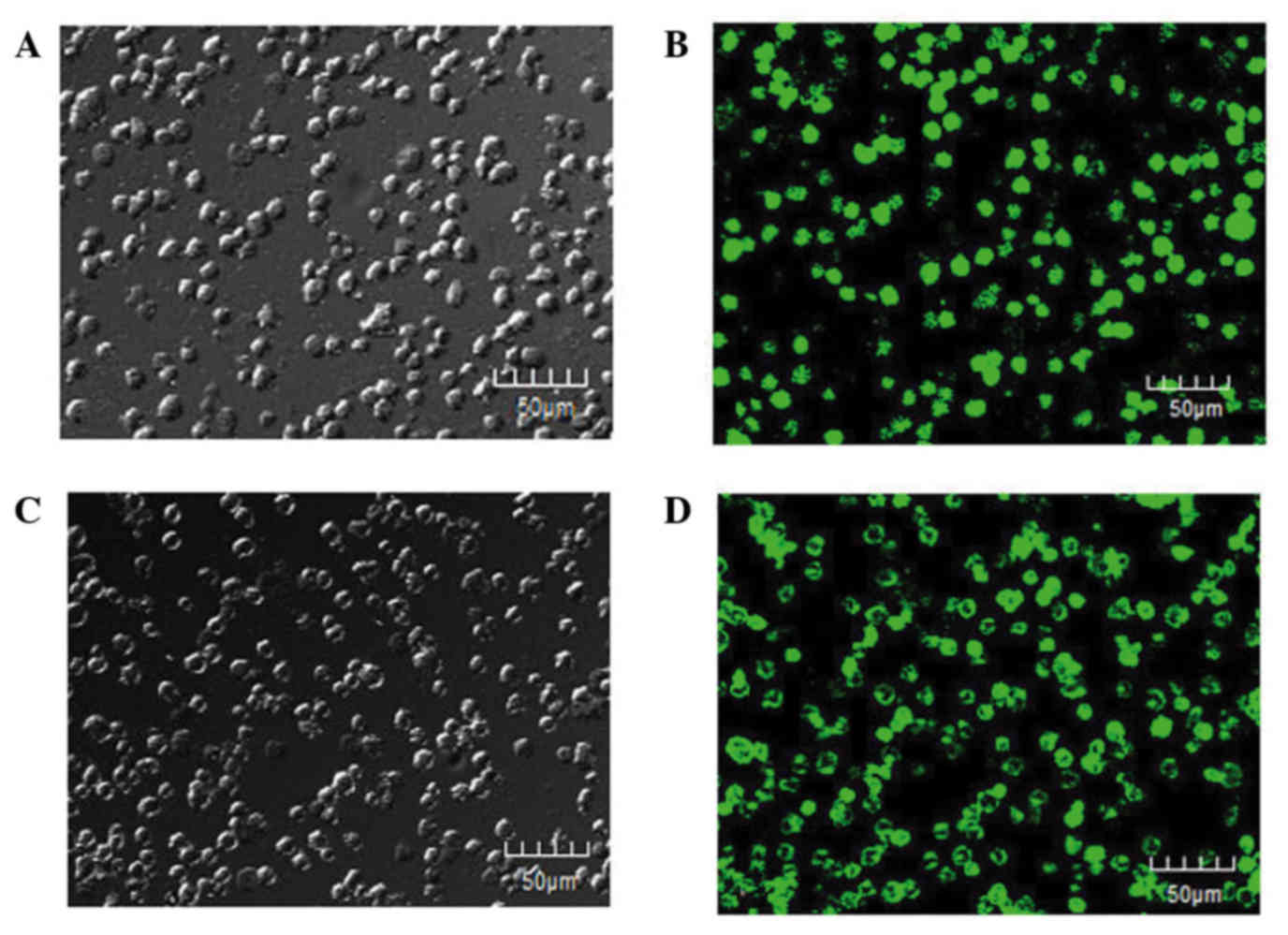
Fluorescence microscopy assessment of
transfection efficiency
Jurkat cells were transfected with neg-EGFP or
LAT-EGFP lentivirus vectors and the detection of green fluorescence
was considered successful transfection. As shown in Fig. 1, green fluorescence was observed
throughout the field of view, with the transfection efficiency
reaching 80%. This suggested the successful establishment of stably
transfected cell lines.
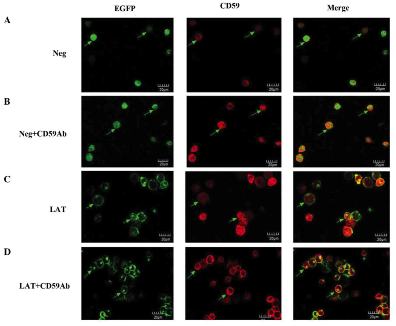
Orientation and distribution of CD59
and LAT in different states of activation
Immunofluorescence staining was used to observe the
orientation and localization of CD59 and LAT. Both were distributed
on the membrane (Fig. 2). The green
fluorescence of the neg group corresponds to the expression of the
EGFP reporter gene, which was diffusely distributed in the
cytoplasm (Fig. 2A). The intensity of
red and green fluorescence indicated the extent of expression of
CD59 and LAT, respectively. As compared with the neg group, neg the
expression of CD59 was enhanced in the neg + CD59Ab group (Fig. 2B). As shown in Fig. 2C and D, LAT was found predominantly in
clusters in the cell membrane. After stimulation with the CD59Ab,
the fluorescence signal of LAT became stronger (Fig. 2D). The red fluorescence of CD59 also
showed clustering and was colocalized with the distribution of LAT
(Fig. 2C and D). These results
suggest that, when Jurkat cells are activated, CD59 is able to
recruit a large number of LAT molecules to lipid rafts. Following
ongoing activation, increasing numbers of CD59 and LAT are
recruited to lipid rafts to regulate downstream signaling.
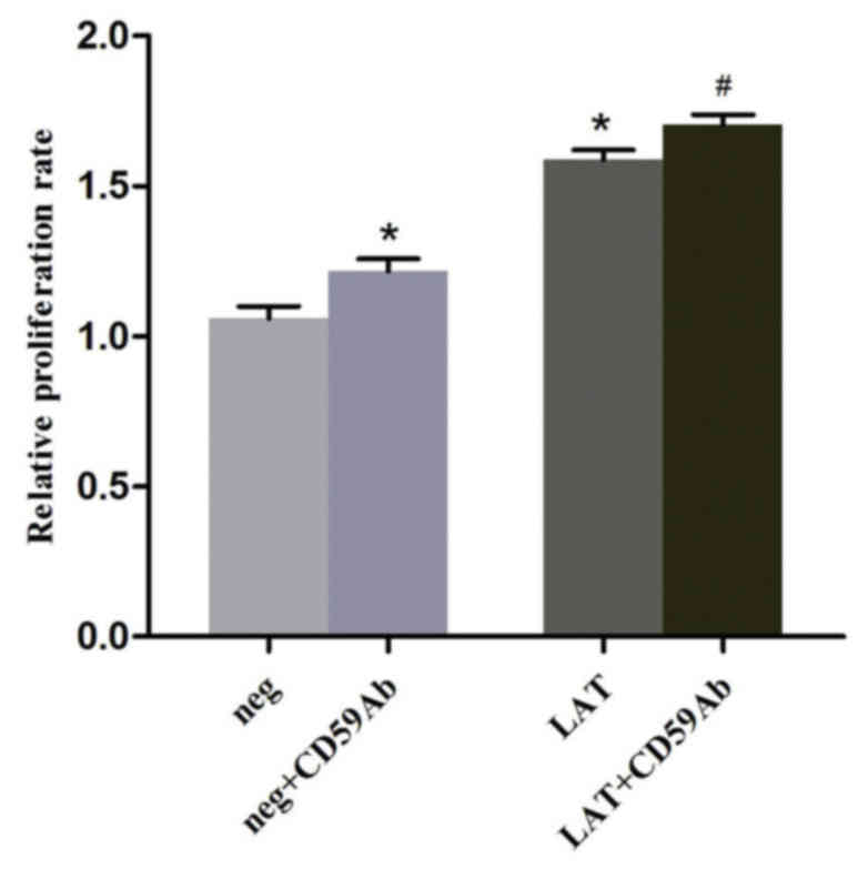
Cross-linking CD59 in lipid rafts
promotes LAT-mediated proliferation of Jurkat cells
CD59 and LAT are crucial molecules that participate
in signal transduction in the membrane (14,22). As
shown in Fig. 2, CD59 and LAT were
colocalized to lipid rafts in Jurkat cells. To further investigate
the relationship between CD59 and LAT, CCK8 assays were performed
to examine the proliferation rate of the various groups. The
results showed that the cell viability of the LAT group was
significantly higher compared with the neg group (P<0.01;
Fig. 3). Cross-linking with CD59
significantly increased the viabilities of both the LAT and neg
group cells (both P<0.01; Fig. 3).
These results suggested that cross-linking CD59 using a monoclonal
antibody or overexpressing LAT were able to significantly increase
the proliferation of Jurkat cells. Notably, the LAT group cells
stimulated with the CD59Ab exhibited the greatest proliferation
ability. These results suggest that GPI-anchored CD59 is able to
recruit LAT into lipid rafts to induce the activation and
proliferation of Jurkat cells.
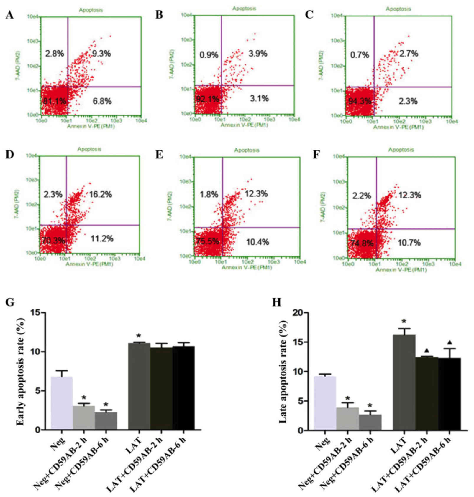
Cross-linking CD59 and overexpressing
LAT reduces apoptosis in Jurkat cells
To observe the apoptotic effects induced by CD59,
the neg and LAT cells treated with CD59Ab were stained with Annexin
V-PE and 7-AAD and analyzed by flow cytometry. As shown in Fig. 4A, the percentages of early apoptotic
(right lower quadrant) and late apoptotic neg cells (right upper
quadrant) were 6.8 and 9.3%, respectively. The apoptosis rates
gradually decreased with a longer incubation with CD59Ab (Fig. 4B and C). The LAT cells showed a higher
percentage of late apoptotic cells compared with the neg group
(16.2 vs. 9.3%; Fig. 4D). However,
the percentage of late apoptotic LAT cells was decreased following
treatment with CD59Ab for 2 or 6 h (12.3 and 12.3%, respectively;
Fig. 4E and F), although the ratio
indicated no significant difference (Fig.
4G). These results, as well as the results of a previous study
(23), suggest that GPI-anchored
proteins, such as CD59, are able to assist SRC family
kinase-mediated signal transduction in lipid rafts, and promote the
proliferation and suppress the apoptosis of Jurkat cells through
cross-linking via its corresponding antibody.
CD59 and LAT induce the upregulation
and activation of crucial protein tyrosine kinases (PTKs)
The present study demonstrated that CD59 and LAT
induced the proliferation of Jurkat cells. To determine the enzymes
or signal transducers involved in the regulatory mechanism, the
mRNA expression levels of PLCG1, phosphoinositide 3-kinase (PI3K),
Lck and LAT, and the protein expression levels of Lck, PI3K, FYN,
ZAP70 and PLCG1, were measured. The mRNA expression levels of
PLCG1, PI3K, Lck and LAT in LAT cells were significantly higher
compared with those in neg cells (Table
II). The expression of PI3K in LAT cells was ~16-fold greater
compared with neg cells. Furthermore, the expression of PI3K was
increased in the neg + CD59Ab group compared with the neg group,
although it was less than the mRNA expression of PI3K in the LAT
group. These results suggest that LAT overexpression exerts
stronger activating effects in Jurkat cells than cross-linking the
GPIAP CD59. Compared with the LAT group, there was no significant
increase in the mRNA expression levels of any of the PTKs in the
LAT + CD59Ab group (Table II).
Therefore, the dual intervention did not enhance the activation of
Jurkat cells compared with overexpresson of LAT alone. Notably, the
mRNA expression levels of LAT in both the neg and LAT cells were
decreased following treatment with CD59Ab (Table II). One possible explanation for this
is that numerous positive regulators set the activation threshold
in T-cells, and SRC kinase-dependent hyperphosphorylation of
inhibitory receptors such as csk binding protein and cytotoxic
T-lymphocyte-associated protein 4, may result in their recruitment
to lipid rafts to displace LAT.
| Table II.Reverse transcription-quantitative
polymerase chain reaction assessment of mRNA expression levels in
the different groups. |
Table II.
Reverse transcription-quantitative
polymerase chain reaction assessment of mRNA expression levels in
the different groups.
|
| ΔCq value |
|---|
|
|
|
|---|
| Gene | Neg | neg+CD59Ab | LAT | LAT+CD59Ab |
|---|
| PLCG1 | 9.74 |
9.09 | 7.95b | 8.63 |
| PI3K | 16.32 | 15.15a | 12.47b | 12.36 |
| LCK | 5.09 |
5.26 | 4.23b | 4.38 |
| LAT | 8.38 |
9.45 | 6.35b | 7.42 |
The expression of various molecules that interact
with LAT in the T-cell signaling pathway were detected. The results
indicated that cross-linking CD59 led to a significant reduction in
the concentrations of upstream PTKs, including ZAP-70, Lck and FYN.
PLCG1 and PI3K are two downstream signaling molecules modulated by
LAT (24). As shown in Fig. 5, there was no change in the
concentration of PLCG1 after cross-linking CD59. However, the band
of PI3K was too light to quantify, which was likely because the
majority of PI3K was phosphorylated to participate in the PI3K/Akt
signaling pathway. These results suggested that the signal
transduction induced by CD59 mainly affected the PI3K/Akt pathway
via LAT, a crucial adaptor (some results are not shown).
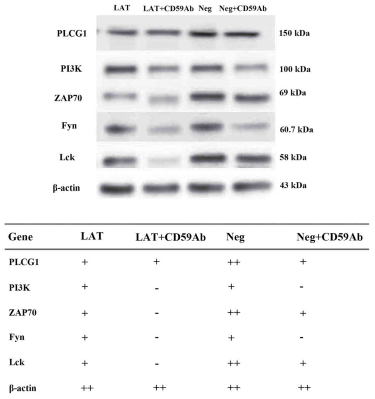 | Figure 5.Western blotting was performed to
detect the expression of PLCG1, PI3K, ZAP70, Fyn and Lck in the
neg, neg + CD59Ab, LAT and LAT + CD59Ab groups. CD59Ab, cluster of
differentiation 59 antibody; LAT, linker for activation of T-cells;
PLCG1, phospholipase C-γ1; PI3K, phosphoinositide 3-kinase; ZAP70,
zeta-chain-associated protein kinase 70; Lck, lymphocyte-specific
protein tyrosine kinase. |
Discussion
CD59 is a GPI-anchored glycoprotein that protects
cells from attack by the complement system (8–11).
Previous studies have shown that cross-linking of CD59 can lead to
a series of intracellular signal transduction events (25–28).
However, the molecular mechanisms underlying CD59-mediated T-cell
signal transduction are unclear. A mechanism has been suggested
based on the structure of the GPI anchor and the positioning of the
CD59 molecule in lipid rafts (14).
Lipid rafts, which mainly contain cholesterol and sphingolipids,
are considered signaling platforms to mediate signal transduction
(29–32). Numerous raft-associated proteins,
including LAT and SRC family kinases, have been shown to have
important roles in signal transduction in lymphocytes (20,33).
GPI-anchored molecules lack transmembrane regions
and are especially enriched in lipid rafts (34). These molecules can induce
intracellular signal transduction events (35). Therefore, there must be a mechanism
connecting the exterior and interior of the plasma membrane.
Subczynski and Kusumi (36)
speculated that this mechanism may occur via a type of non-specific
transmembrane protein. The present study focused on the role of
CD59 in Jurkat cells in order to determine how CD59 may interact
with LAT to transduce signals.
LAT is a 36–38 kDa type III transmembrane adaptor
protein (37), which functions as an
important integration node at the plasma membrane for various
signaling complexes (38).
Microclusters of LAT are composed of signaling complexes that
include adaptors and effectors for T-cell activation (39). Phosphorylation and palmitoylation are
crucial for the localization and function of transmembrane adaptor
proteins (40). LAT can be
phosphorylated by the ZAP70/Syk PTK, which promotes the recruitment
of various signaling molecules to the plasma membrane (41). Protein palmitoylation is an important
form of post-translational covalent modification characterized by
lipid acylation (42). Following
palmitoylation, LAT is recruited to lipid rafts, where it is
phosphorylated to serve as a linker for activated T-cells. In the
GPI structure, a palmitate group is attached to the second carbon
of the inositol ring (43), and the
present study assumed that LAT palmitoylation is associated with
the function of the palmitate group. Lentivirus vectors carrying
neg-EGFP and LAT-EGFP were constructed and transfected into Jurkat
cells. The results of confocal microscopy indicated that CD59 and
LAT colocalized to lipid rafts.
Activation of Jurkat cells through specific
GPI-anchored molecules, such as CD55 and CD59 in humans, results in
the phosphorylation of PTKs, elevation of Ca2+
concentrations and an increase in proliferation (6) and cytokine release (44). Too much calcium can cause damage to
mitochondria and the release of cytochrome c, which
activates caspases to induce apoptosis (45). In order to observe the effect of CD59
on LAT cells and the molecular mechanisms underlying apoptosis, the
effects of CD59 on the proliferation and apoptosis of neg and LAT
cells were examined. The present study demonstrated that CD59 and
LAT molecules were able to promote the growth of Jurkat cells. LAT
cells showed enhanced proliferation and had a relatively high
late-apoptosis rate, which may have been caused by
activation-induced cell death.
TCR engagement triggers signaling cascades that
result in enhanced gene transcription, cell proliferation and
differentiation, and even apoptosis (46,47). The
earliest events in this pathway are the activation of Lck and Fyn,
which are PTKs from the Src family. These enzymes were found to
phosphorylate tyrosine residues within the immunoreceptor
tyrosine-based activation motifs (ITAMs) of the cytosolic domains
of TCR-CD3ξ chains. Phosphorylated ITAMs serve as binding sites for
the Src homology 2 (SH2) domains of ZAP70, which is then
phosphorylated by Lck or Fyn to further activate downstream
substrates (15). LAT is a substrate
of activated ZAP70 and is one of the most important
tyrosine-phosphorylated proteins following TCR activation; LAT is
able to bind to the SH2 domains of growth factor receptor bound
protein 2 and PLCG1 and the p85 subunit of PI3K (48–50). In
the present study, results from immunoblotting and RT-qPCR
indicated that the expression of non-phosphorylated proteins in LAT
and neg cells was decreased following cross-linking of CD59. For
neg cells, the mRNA expression levels of PLCG1 and PI3K were
increased after CD59 cross-linking, while there was a decrease in
the mRNA expression level of LAT following cross-linking. For LAT
cells, there was no significant difference in the mRNA expression
levels of PTKs following cross-linking of CD59, with the exception
of the mRNA expression level of LAT, which decreased markedly
following cross-linking of CD59. These results suggested that CD59
in lipid rafts in combination with palmitoylated LAT were able to
promote T-cell signal transduction, facilitate the phosphorylation
of PTKs and enhance gene transcription. However, the
over-activation of T-cells can trigger and recruit inhibitory
receptors into lipid raft to displace and suppress activators, in
order to maintain T-cell homeostasis (51).
In conclusion, the present study demonstrated the
that GPI-anchored protein CD59 was able to promote TCR signal
transduction, of which the mechanism may involve the CD59-mediated
transfer of a palmitate group to LAT and subsequent regulation of
T-cell signaling. These findings suggest that other GPI-anchored
proteins may be involved in signal transduction in T-cells.
Furthermore, the results of the present study suggested that CD59
and LAT were able to synergistically promote Jurkat cell
proliferation. Future studies should further investigate how
GPI-anchored proteins are able to modulate T-cell signal
transduction.
Acknowledgements
We would like to thank Dr Peng Zhao (Department of
Cancer, Biotherapy Center of the Central Hospital of Qingdao) for
his assistance with recovery of the Jurkat cells. This study was
supported by the National Natural Science Foundation of China
(grant no. 81273206).
References
|
1
|
Lisanti MP, Sargiacomo M, Graeve L,
Saltiel AR and Rodriguez-Boulan E: Polarized apical distribution of
glycosyl-phosphatidylinositol-anchored proteins in a renal
epithelial cell line. Proc Natl Acad Sci USA. 85:pp. 9557–9561.
1988; View Article : Google Scholar : PubMed/NCBI
|
|
2
|
Fujita M and Kinoshita T: GPI-anchor
remodeling: Potential functions of GPI-anchors in intracellular
trafficking and membrane dynamics. Biochim Biophys Acta.
1821:1050–1058. 2012. View Article : Google Scholar : PubMed/NCBI
|
|
3
|
Davis LS, Patel SS, Atkinson JP and Lipsky
PE: Decay-accelerating factor functions as a signal transducing
molecule for human T cells. J Immunol. 141:2246–2252.
1988.PubMed/NCBI
|
|
4
|
Thompson LF, Ruedi JM, Glass A, Low MG and
Lucas AH: Antibodies to 5′-nucleotidase (CD73), a
glycosyl-phosphatidylinositol-anchored protein, cause human
peripheral blood T cells to proliferate. J Immunol. 143:1815–1821.
1989.PubMed/NCBI
|
|
5
|
Presky DH, Low MG and Shevach EM: Role of
phosphatidylinositol-anchored proteins in T cell activation. J
Immunol. 144:860–868. 1990.PubMed/NCBI
|
|
6
|
Korty PE, Brando C and Shevach EM: CD59
functions as a signal-transducing molecule for human T cell
activation. J Immunol. 146:4092–4098. 1991.PubMed/NCBI
|
|
7
|
Corda D, Zizza P, Varone A, Bruzik KS and
Mariggiò S: The glycerophosphoinositols and their cellular
functions. Biochem Soc Trans. 40:101–107. 2012. View Article : Google Scholar : PubMed/NCBI
|
|
8
|
Davies A, Simmons DL, Hale G, Harrison RA,
Tighe H, Lachmann PJ and Waldmann H: CD59, an LY-6-like protein
expressed in human lymphoid cells, regulates the action of the
complement membrane attack complex on homologous cells. J Exp Med.
170:637–654. 1989. View Article : Google Scholar : PubMed/NCBI
|
|
9
|
Farkas I, Baranyi L, Ishikawa Y, Okada N,
Bohata C, Budai D, Fukuda A, Imai M and Okada H: CD59 blocks not
only the insertion of C9 into MAC but inhibits ion channel
formation by homologous C5b-8 as well as C5b-9. J Physiol.
539:537–545. 2002. View Article : Google Scholar : PubMed/NCBI
|
|
10
|
Meri S, Morqan BP, Davies A, Daniels RH,
Olavesen MG, Waldmann H and Lachmann PJ: Human protectin (CD59), an
18,000–20,000 MW complement lysis restricting factor, inhibits
C5b-8 catalysed insertion of C9 into lipid bilayers. Immunology.
71:1–9. 1990.PubMed/NCBI
|
|
11
|
Rollins SA and Sims PJ: The
complement-inhibitory activity of CD59 resides in its capacity to
block incorporation of C9 into membrane C5b-9. J Immunol.
144:3478–3483. 1990.PubMed/NCBI
|
|
12
|
Hideshima T, Okada N and Okada H:
Expression of HEF20, a regulatory molecule of complement
activation, on peripheral blood mononuclear cells. Immunology.
69:396–401. 1990.PubMed/NCBI
|
|
13
|
Meri S, Waldmann H and Lachmann PJ:
Distribution of protectin (CD59), a complement membrane attack
inhibitor, in normal human tissues. Lab Invest. 65:532–537.
1991.PubMed/NCBI
|
|
14
|
Kimberley FC, Sivasankar B and Paul Morgan
B: Alternative roles for CD59. Mol Immunol. 44:73–81. 2007.
View Article : Google Scholar : PubMed/NCBI
|
|
15
|
Zhang W, Sloan-Lancaster J, Kitchen J,
Trible RP and Samelson LE: LAT: The ZAP-70 tyrosine kinase
substrate that links T cell receptor to cellular activation. Cell.
92:83–92. 1998. View Article : Google Scholar : PubMed/NCBI
|
|
16
|
Su YW and Jumaa H: LAT links the pre-BCR
to calcium signaling. Immunity. 19:295–305. 2003. View Article : Google Scholar : PubMed/NCBI
|
|
17
|
Facchetti F, Chan JK, Zhang W, Tironi A,
Chilosi M, Parolini S, Notarangelo LD and Samelson LE: Linker for
activation of T cells (LAT), a novel immunohistochemical marker for
T cells, NK cells, mast, cells and megakaryocytes: Evaluation in
normal and pathological conditions. Am J Pathol. 154:1037–1046.
1999. View Article : Google Scholar : PubMed/NCBI
|
|
18
|
Weber JR, Orstavik S, Torqersen KM,
Danbolt NC, Berg SF, Ryan JC, Taskén K, Imboden JB and Vaage JT:
Molecular cloning of the cDNA encoding pp36, a
tyrosine-phosphorylated adaptor protein selectively expressed by T
cells and natural killer cells. J Exp Med. 187:1157–1161. 1998.
View Article : Google Scholar : PubMed/NCBI
|
|
19
|
Li CY, Peng J, Ren LP, Gan LX, Lu XJ, Liu
Q, Gu W and Guo XJ: Roles of histone hypoacetylation in LAT
expression on T cells and Th2 polarization in allergic asthma. J
Transl Med. 11:262013. View Article : Google Scholar : PubMed/NCBI
|
|
20
|
Zhang W, Trible RP and Samelson LE: LAT
palmitoylation: Its essential role in membrane microdomain
targeting and tyrosine phosphorylation during T cell activation.
Immunity. 9:239–246. 1998. View Article : Google Scholar : PubMed/NCBI
|
|
21
|
Livak KJ and Schmittgen TD: Analysis of
relative gene expression data using real-time quantitative PCR and
the 2(-Delta Delta C(T)) Method. Methods. 25:402–408. 2001.
View Article : Google Scholar : PubMed/NCBI
|
|
22
|
Dustin ML and Depoil D: New insights into
the T cell synapse from single molecule techniques. Nat Rev
Immunol. 11:672–684. 2011. View
Article : Google Scholar : PubMed/NCBI
|
|
23
|
Wange RL: LAT, the linker for activation
of T cells: a bridge between T cell-specific and general signaling
pathways. Sci STKE. 2000:re12000.PubMed/NCBI
|
|
24
|
Chen Y, Thelin WR, Yang B, Milgram SL and
Jacobson K: Transient anchorage of cross-linked
glycosyl-phosphatidylinositol-anchored proteins depends on
cholesterol, Src family kinases, caveolin and phosphoinositides. J
Cell Biol. 175:169–178. 2006. View Article : Google Scholar : PubMed/NCBI
|
|
25
|
Okada H, Nagami Y, Takahashi K, Okada N,
Hideshima T, Takizawa H and Kondo J: 20 KDa homologous restriction
factor of complement resembles T cell activating protein. Biochem
Biophys Res Commun. 162:1553–1559. 1989. View Article : Google Scholar : PubMed/NCBI
|
|
26
|
Lipp AM, Juhasz K, Paar C, Ogris C,
Eckerstorfer P, Thuenauer R, Hesse J, Nimmervoll B, Stockinger H,
Schütz GJ, et al: Lck mediates signal transmission from CD59 to the
TCR/CD3 pathway in Jurkat T cells. PLoS One. 9:e859342014.
View Article : Google Scholar : PubMed/NCBI
|
|
27
|
Suzuki KG: Single-Molecule Imaging of
Signal Transduction via GPI-Anchored Receptors. Methods Mol Biol.
1376:229–238. 2016. View Article : Google Scholar : PubMed/NCBI
|
|
28
|
Stefanová I, Horejsí V, Ansotegui IJ,
Knapp W and Stockinger H: GPI-anchored cell-surface molecules
complexed to protein tyrosine kinases. Science. 254:1016–1019.
1991. View Article : Google Scholar : PubMed/NCBI
|
|
29
|
Simons K and Ikonen E: Functional rafts in
cell membranes. Nature. 387:569–572. 1997. View Article : Google Scholar : PubMed/NCBI
|
|
30
|
Jacobson K, Mouritsen OG and Anderson RG:
Lipid rafts: At a crossroad between cell biology and physics. Nat
Cell Biol. 9:7–14. 2007. View Article : Google Scholar : PubMed/NCBI
|
|
31
|
Goswami D, Gowrishankar K, Bilgrami S,
Ghosh S, Raghupathy R, Chadda R, Vishwakarma R, Rao M and Mayor S:
Nanoclusters of GPI-anchored proteins are formed by cortical
actin-driven activity. Cell. 135:1085–1097. 2008. View Article : Google Scholar : PubMed/NCBI
|
|
32
|
Lee MY, Ryu JM, Lee SH, Park JH and Han
HJ: Lipid rafts play an important role for maintenance of embryonic
stem cell self-renewal. J Lipid Res. 51:2082–2089. 2010. View Article : Google Scholar : PubMed/NCBI
|
|
33
|
Kabouridis PS, Magee AI and Ley SC:
S-acylation of LCK protein tyrosine kinase is essential for its
signaling function in T lymphocytes. EMBO J. 16:4983–4998. 1997.
View Article : Google Scholar : PubMed/NCBI
|
|
34
|
Cinek T and Horejsí V: The nature of large
noncovalent complexes containing
glycosyl-phosphatidylinositol-anchored membrane glycoproteins and
protein tyrosine kinases. J Immunol. 149:2262–2270. 1992.PubMed/NCBI
|
|
35
|
Suzuki KG: Lipid rafts generate
digital-like signal transduction in cell plasma membranes.
Biotechnol J. 7:753–761. 2012. View Article : Google Scholar : PubMed/NCBI
|
|
36
|
Subczynski WK and Kusumi A: Dynamics of
raft molecules in the cell and artificial membranes: Approaches by
pulse EPR spin labeling and single molecule optical microscopy.
Biochim Biophys Acta. 1610:231–243. 2003. View Article : Google Scholar : PubMed/NCBI
|
|
37
|
Fuller DM and Zhang W: Regulation of
lymphocyte development and activation by the LAT family of adapter
proteins. Immunol Rev. 232:72–83. 2009. View Article : Google Scholar : PubMed/NCBI
|
|
38
|
Bartelt RR and Houtman JC: The adaptor
protein LAT serves as an integration node for signaling pathways
that drive T cell activation. Wiley Interdiscip Rev Syst Biol Med.
5:101–110. 2013. View Article : Google Scholar : PubMed/NCBI
|
|
39
|
Balagopalan L, Coussens NP, Sherman E,
Samelson LE and Sommers CL: The LAT story: A tale of cooperativity,
coordination, and choreography. Cold Spring Harb Perspect Biol.
2:a0055122010. View Article : Google Scholar : PubMed/NCBI
|
|
40
|
Lin J, Weiss A and Finco TS: Localization
of LAT in glycolipid-enriched microdomains is required for T cell
activation. J Biol Chem. 274:28861–28864. 1999. View Article : Google Scholar : PubMed/NCBI
|
|
41
|
Jiang Y and Cheng H: Evidence of LAT as a
dual substrate for Lck and Syk in T lymphocytes. Leuk Res.
31:541–545. 2007. View Article : Google Scholar : PubMed/NCBI
|
|
42
|
Tanimura N, Saitoh S, Kawano S, Kosugi A
and Miyake K: Palmitoylation of LAT contributes to its subcellular
localization and stability. Biochem Biophys Res Commun.
341:1177–1183. 2006. View Article : Google Scholar : PubMed/NCBI
|
|
43
|
Ferguson MAJ, Kinoshita T and Hart GW:
Glycosylphosphatidylinositol anchorsEssentials of Glycobiology.
Varki A, Cummings RD, Esko JD, Freeze HH, Stanley P, Bertozzi CR,
Hart GW and Etzler ME: 2nd. Cold Spring Harbor; New York, NY: pp.
3–6. 2009
|
|
44
|
Deckert M, Ticchioni M, Mari B, Mary D and
Bernard A: The glycosylphosphatidylinositol-anchored CD59 protein
stimulates both T cell receptor zeta/ZAP-70-dependent and
-independent signaling pathways in T cells. Eur J Immunol.
25:1815–1822. 1995. View Article : Google Scholar : PubMed/NCBI
|
|
45
|
Li B, Chu X, Gao M and Xu Y: The effects
of CD59 gene as a target gene on breast cancer cells. Cell Immunol.
272:61–70. 2011. View Article : Google Scholar : PubMed/NCBI
|
|
46
|
Weiss A and Littman DR: Signal
transduction by lymphocyte antigen receptors. Cell. 76:263–274.
1994. View Article : Google Scholar : PubMed/NCBI
|
|
47
|
Wange RL and Samelson LE: Complex
complexes: Signaling at the TCR. Immunity. 5:197–205. 1996.
View Article : Google Scholar : PubMed/NCBI
|
|
48
|
Trüb T, Frantz JD, Miyazaki M, Band H and
Shoelson SE: The role of a lymphoid-restricted, Grb2-like
SH3-SH2-SH3 protein in T cell receptor signaling. J Biol Chem.
272:894–902. 1997. View Article : Google Scholar : PubMed/NCBI
|
|
49
|
Fukazawa T, Reedquist KA, Panchamoorthy G,
Soltoff S, Trub T, Druker B, Cantley L, Shoelson SE and Band H: T
cell activation-dependent association between the p85 subunit of
the phosphatidylinositol 3-kinase and Grb2/phospholipase C-gamma
1-binding phosphotyrosyl protein pp36/38. J Biol Chem.
270:20177–20182. 1995. View Article : Google Scholar : PubMed/NCBI
|
|
50
|
Sieh M, Batzer A, Schlessinger J and Weiss
A: GRB2 and phospholipase C-gamma 1 associate with a 36- to
38-kilodalton phosphotyrosine protein after T-cell receptor
stimulation. Mol Cell Biol. 14:4435–4442. 1994. View Article : Google Scholar : PubMed/NCBI
|
|
51
|
Kalland ME, Solheim SA, Skånland SS,
Taskén K and Berge T: Modulation of proximal signaling in normal
and transformed B cells by transmembrane adapter Cbp/PAG. Exp Cell
Res. 318:1611–1619. 2012. View Article : Google Scholar : PubMed/NCBI
|