Introduction
With an incidence rate of 3.19/100,000, glioblastoma
multiforme (GBM) is a highly malignant and notably aggressive
primary tumour of the human brain, and the median survival time for
patients is <12 months in the majority of cases. The invasion of
tumour cells into normal brain tissues means that it is difficult
for surgery to achieve complete resection, and recurrence and
mortality often occur. Different genetic aberrations arising from
GBM lead to genetic heterogeneity of patient tumour cells, which
infers resistance to additional therapies, including post-operative
radiotherapy and chemotherapy (1–3). Epidermal
growth factor receptor variant III (EGFRvIII) is one of the most
common types of mutations of GBM and is responsible for various
biological effects on the generation and development of tumours.
Previous studies have indicated that this variation is
significantly associated with poor prognosis in patients with GBM.
Previously, numerous studies have indicated that EGFRvIII is
associated with multiple characteristics of glioma stem cells,
including self-renewal, abnormal differentiation, tumour initiation
and treatment resistance. Studies have also indicated that high
expression levels of EGFRvIII are positively correlated with the
invasion abilities of GBM cells (1,4). The
amplification of EGFR is often accompanied by upregulated
expression levels of EGFRvIII, which is significantly correlated
with a short survival time for patients with BMG (5,6). EGFRvIII
is also expressed in other types of cancer, including lung cancer,
but has not been identified in normal tissues or benign tumour
tissues (7). Those tumours exhibiting
activated EGFRvIII account for ~50% of patients with BMG (8,9). These
mutations are not detected in the normal brain tissues or in other
normal tissues of adult humans (10,11). Thus,
due to its biological characteristics, EGFRvIII may serve as a good
target for the gene therapy of GBM. If an antibody that
specifically recognizes EGFR and induces apoptosis is designed, it
may have the potential to effectively kill tumour cells through
coupling with cytotoxic drugs. However, only limited studies exist
on the molecular biology of EGFRvIII-associated mechanisms.
Following continuous studies and the development of target drugs
for clinical applications, the combination of surgery and
chemoradiotherapy remains the main measure of current clinical
therapies. Specifically, combined therapy using radiation treatment
and temozolomide is the main regimen for GBM treatment. For GBM
with the EGFRvIII mutation, the response to combined radiation
treatment and temozolomide therapy remains unknown. To the best of
our knowledge, there have been no studies investigating the
mechanisms of the development of resistance. Therefore, the present
study collected clinical materials and used a combination of in
vitro and animal models to examine the effects of combined
radiation treatment and temozolomide therapy on patients with GBM
with the EGFRvIII mutation.
Materials and methods
Cell lines and tissue samples
A total of 57 glioma patients with complete
follow-up results who were treated between January 2013 and
February 2014 in the Affiliated Hospital of Hebei University
(Baoding, China) were randomly selected (Table I). Amongst the patients, 30 were men
and 27 were women, with ages ranging from 22 to 76 years and a
median age of 49 years. The Karnofsky Performance Status (KPS)
scores for these patients ranged from 90 to 20 points, with a mean
of 73 points. A total of 34 patients underwent a complete resection
under surgical microscopy and 23 patients underwent a partial
resection. A total of 26 patients exhibited tumors growing in the
left brain and 31 patients exhibited tumors growing in the right
brain. Post-operative pathological results indicated that 21 tumors
were World Health Organization (WHO) grade I–II, 22 were WHO grade
III and 14 were WHO grade IV (11).
Statistical analyses were conducted on the clinical materials, and
the Affiliated Hospital of Hebei University provided 32 tumor
specimens preserved in liquid nitrogen. Amongst the 32 patients
selected, 13 were men and 19 were women, with ages ranging from 23
to 71 years, with a mean age of 47 years. The post-operative
pathological results indicated that 10 patients were WHO grade II,
5 patients were WHO grade II–III, 8 patients were WHO grade III and
9 patients were WHO grade IV, and 7 exhibited positive EGFRvIII
results. The human GBM U87-EGFRvIII cell lines were obtained from
Dr Webster Cavenee (Ludwig Cancer Institute, San Diego, CA, USA).
The cells were grown in Dulbecco's modified Eagle's medium (Gibco;
Thermo Fisher Scientific, Inc., Waltham, MA, USA) supplemented with
10% fetal bovine serum (Invitrogen; Thermo Fisher Scientific, Inc.)
and 0.4% Geneticin (G-418 sulphate; Gibco; Thermo Fisher
Scientific, Inc.). The cells were incubated at 37°C in a 5%
CO2 and 95% air environment until use. Approval for the
use of the glioma samples was obtained from each patient and the
present study was approved by the Ethics Committee of the Second
Hospital of Tianjin Medical University (Tianjin, China).
 | Table I.Patient characteristics (n=57). |
Table I.
Patient characteristics (n=57).
| Characteristic | Value |
|---|
| Median age at
diagnosis (range), years | 49 (22–76) |
| Age groups by years,
n (%) |
|
|
18–39 | 37 (65) |
|
40–49 | 10 (18) |
|
50–59 | 3 (5) |
|
60–69 | 4 (7) |
|
>70 | 3 (5) |
| Sex |
|
|
Male | 30 (53) |
|
Female | 27 (47) |
| KPS score |
|
|
Average | 73 |
|
Range | 20–90 |
| ≤70, n
(%) | 20 (35) |
| >70,
n (%) | 37 (65) |
| Surgery |
|
| Total
resection | 34 (60) |
| Partial
resection | 23 (40) |
| Side |
|
|
Left | 26 (46) |
|
Right | 31 (54) |
| Survival,
months |
|
|
Average | 11.2 |
|
Range | 2–39 |
| <12,
n (%) | 35 (61) |
| ≥12, n
(%) | 22 (39) |
| WHO grade |
|
|
I–II | 21 (37) |
|
III | 22 (39) |
| IV | 14 (25) |
Cell Counting Kit-8 (CCK-8) assay
The cells were seeded in 96-well plates at a density
of 5×10³ cells/well, with each well containing 100 µl
cell suspension. Subsequent to cell attachment, temozolomide (Tasly
Pharmaceutical Co., Ltd., Tianjin, China) was added at different
concentration gradients (0.1 to 1, at intervals of 0.1 and 1 to 10,
at intervals of 1). After 72 h, 10 µl CCK-8 reagent (Dojindo
Molecular Technologies, Inc., Kumamoto, Japan) was added to each
well, and the plates were incubated at 37°C for 4 h. The optical
absorbance at 450 nm was measured on a microplate reader. The half
maximal inhibitory concentration (IC50) result for 72 h
was calculated using Origin 7.5 software (Microcal Software,
Northampton, MA, USA). The IC50 of the U87-EGFRvIII cell
line following 72 h of exposure to temozolomide, as measured by the
CCK-8 kit was 5 mmol/l.
RNA extraction and polymerase chain
reaction (RT-PCR)
Total RNA from the patient GBM tissues was extracted
using the TRIzol reagent (Invitrogen; Thermo Fisher Scientific,
Inc.) according to the manufacturer's protocol. The concentration
and purity of RNA were determined using a NanoDrop®
ND-1000 spectrophotometer (Thermo Fisher Scientific, Inc.). The
GoScript™ Reverse Transcriptase system (Promega Corporation,
Madison, WI, USA) was used in complementary DNA synthesis according
to the manufacturer's instructions. The expression levels of mature
EGFRvIII were determined by semi-quantitative PCR using a PCR
cycler (Nanjing KeyGenBiotech, Co., Ltd., Beijing, China) with a
forward primer for the mature DNA sequence and a universal adaptor
reverse primer as follows: Forward, 5′-TGACTCCGTCCAGTATTGATCG-3′
and reverse, 5′-GCCCTTCGCACTTCTTACACTT-3′ (Thermo Fisher Scientific
Inc.). All PCR was performed using a PCR kit (PrimeSTAR®
Max DNA Polymerase; cat. no. R045A; Takara Biotechnology Co., Ltd.,
Dalian, China), according to the manufacturer's instructions. The
PCR cycling conditions were as follows: 1 cycle at 95°C for 10 min;
35 cycles at 95°C for 30 sec, 58°C for 30 sec and 72°C for 1 min;
and 1 cycle at 72°C for 10 min. The melting curve was obtained from
72–95°C. The data were derived from three independent
experiments.
Agarose gel electrophoresis
A 0.5% agarose gel was prepared, and poured onto a
taped plate with casting combs in place. A total of 20–30 min was
allowed for the gel to set. The tape and the gel casting combs were
carefully removed, and the gel was placed in a horizontal
electrophoresis apparatus. A 1XTris base, acetic acid and EDTA
electrophoresis buffer was added to the reservoirs until the buffer
covered the agarose gel. Loading dye (ethidium bromide) was added
to each DNA sample, and the samples were mixed and placed into the
wells. The gel was run at 150–200 mA until the required separation
was achieved in 30 min. The DNA fragments were visualized on a
long-wave ultraviolet light box and images were captured with a
camera.
Cell treatment
For the radiation treatment, collected cells were
digested in a sterile 1.5-ml Eppendorf (EP) tube on a super clean
bench and centrifuged for 5 min at ~63 × g (room temperature) with
a tablet centrifuge. Subsequent to standing for 5 min at 37°C to
collect the cells at the tip of the EP tube, T1 and T2 sequence
images were collected via 1.5T nuclear magnetic resonance imaging
(MRI) scans (Siemens AG, Munich, Germany). The data from the MRI
scans were input into Gammaplan 5.3 (Leksell Perfexion Gamma Knife;
Elekta Instrument AB, Stockholm, Sweden) to establish the most
appropriate therapy for each patient. The cell-collecting site was
wiped with 99% isodose, and 6-Gy peripheral doses were applied.
Irradiation was applied with the Leksell Gamma Knife®
Perfexion™, and the cells were collected in 10-cm culture dishes
for continuous culture under sterile conditions subsequent to
irradiation. For temozolomide treatment, the cells were treated
with temozolomide at the IC50 dose (5 mmol/l) for 24
h.
Protein extraction and western-blot
analysis
The cells were harvested 24 h after treatment, and
the total protein from the glioma cell line was extracted using
radioimmunoprecipitation assay lysis buffer with proteinase
inhibitor. The homogenates were clarified by centrifugation at
20,000 × g and 4°C for 15 min, and the protein concentration was
measured using the bicinchoninic acid method. A total of 40 mg
protein mixed with 5X SDS loading buffer was loaded into each lane
and separated by 10 or 12% SDS-PAGE. The separated proteins were
transferred to polyvinylidene fluoride membranes (Roche
Diagnostics, Basel, Switzerland), Membranes were blocked with 5%
non-fat milk in PBST (1 h at 37°C), prior to incubation overnight
at 4°C with primary antibodies against matrix metalloproteinase
(MMP)-9 (1:1,000, cat. no. ab38898), and vascular endothelial
growth factor (VEGF; 1:1,000, cat no. ab32152) (both from Abcam,
Cambridge, UK) and EGFRvIII (rabbit monoclonal antibody, dilution
1:1,000; cat. no. sc-31156; Santa Cruz Biotechnology, Inc., Dallas,
TX, USA) followed by incubation for 1 h at room temperature with a
horseradish peroxidase-conjugated secondary antibody (cat. no.
ZB2010, dilution 1:5,000; Zhongshan Bio Corp., Beijing, China). The
membranes were washed in PBST. Subsequent to washing with stripping
buffer, the membranes were re-probed with antibodies against GAPDH
(dilution 1:5,000; cat. no. SC-47724; Santa Cruz Biotechnology,
Inc.) at room temperature, overnight, and proteins were detected
with an enhanced chemiluminescence detection system (Sigma-Aldrich;
Merck KGaA, Darmstadt, Germany).
Intracranial animal model
In total, 3 groups of 4-week-old (15 g) female nude
mice (Cancer Institute of the Chinese Academy of Medical Science).
Each group contained 8 mice. Mice were kept at 2/cage at a constant
temperature (24°C) with a 12-h light-dark cycle (dark period from
08:00 a.m. to 20:00 p.m.). The mice were fed (specific pathogen
free mice food) once a day (20 g) and were intracranially implanted
with 5×10⁵ U87-EGFRvIII cells to form the following
groups: A group pre-treated with radiosurgery, a group treated with
a combination therapy of 6 Gy radiosurgery and IC50
temozolomide, and a group with a negative control segment, using
the Leksell Perfexion Gamma Knife. Bioluminescence imaging was used
to detect intracranial tumour growth: Mice were anesthetized,
injected intraperitoneally with D-luciferin (Promega Corporation)
at 50 mg/ml. Images were captured with the IVIS Imaging System
(Caliper Life Sciences; PerkinElmer, Inc., Waltham, MA, USA) for
10–120 sec. To quantify the bioluminescence, identical circular
regions of interest were drawn around the entire head of each
animal, and the integrated flux of photons, photons per second, in
each region of interest was determined using the Living Images
software package 3.2 (Caliper Life Sciences; PerkinElmer, Inc.).
Data were normalized to the bioluminescence at the initiation of
treatment for each animal. A tumour volume of 0.6 cm3
and a weight of 20 g were set as the humane end-points. All
protocols involving animals were performed in accordance with an
approved Institutional Animal Care and Use Committee protocol
(Cancer Institute of the Chinese Academy of Medical Science).
Immunohistochemistry analysis
Histological sections of tumour xenografts were
excised and fixed in 10% neutral (4°C) buffered formalin for 3
days. Following paraffin embedding, 5-µm sections were cut. The
paraffin-embedded tissue sections were used to examine EGFRvIII
(cat. no. ab24293), EGFR (cat. no. ab52894), RAC-α
serine/threonine-protein kinase (AKT-1; cat. no. ab81283), MMP-2
(cat. no. ab37150), MMP-9 (cat. no. ab38898) and X-ray repair
cross-complementing protein 6 (Ku70; cat. no. ab92450) expression.
Subsequent to de-waxing, sections were incubated with primary
antibodies (all diluted to 1:100; and all provided by Abcam)
overnight at 4°C, followed by incubation with abiotin-labelled
secondary antibody (dilution 1:100; cat. no. SPN-9001; Zhongshan
Bio Corp., Beijing, China) for 1 h at 37°C, incubation with
diaminobenzidine for 2 h at room temperature, counterstaining with
haematoxylin for 10 min at room temperature, dehydration and
visualization using light microscopy (Olympus Corporation, Tokyo,
Japan).
Statistical analysis
SPSS software (v.16.0; SPSS Inc., Chicago, IL, USA)
was used for all statistical analyses. One-way analysis of variance
with a Tukey's post hoc test or was used to evaluate the
differences between groups. P<0.05 was considered to indicate a
statistically significant difference.
Results
Statistical results from clinical
data
The results (Table
II) indicate that 26 (46%) patients were younger than the
median age of 49 years, and 16 (28%) were older (P=0.426). A total
of 23 patients were men (40%) and 19 were women (33%; P=0.590). The
number of patients with a KPS score lower than the mean score of 70
was 16 (28%), and the number of patients with a KPS score higher
than the mean score was 26 (46%; P=0.426). A total of 29 patients
(51%), underwent complete tumour resection, and 13 (23%) patients
underwent partial tumor resection (P=0.016). There were 19 patients
with tumours growing on the left cerebral hemisphere (33%) and 23
patients with tumours growing on the right cerebral hemisphere
(40%; P=0.924). The number of patients with a survival time of
<12 months, the median survival time was 30 (53%), and the
number of patients with a survival time >12 months was 12 (21%;
P=0.009). The number of patients with WHO grades I–II was 12 (21%)
and the number of patients with WHO grades III–IV was 30 (53%;
P=0.030). Statistical analysis was performed on the data showing
the levels of expression of EGFR and EGFRvIII from the pathology
results of the tumours of 57 patients (Table II). From the 57 patients, 29 (51%)
exhibited high expression of EGFRvIII. Of these patients, 20 and 9
were younger and older than the median age, respectively. In this
group, 16 were men (28%), and 13 were women (23%; P=0.696). The
number of the patients with a KPS score <70, the mean score, was
8 (14%), and the number of patients with a KPS score >70 was 21
(37%; P=0.227). A total of 17 patients (30%) underwent complete
tumour resection and 12 (21%) patients underwent a partial tumour
resection (P=0.872). A total of 16 patients exhibited tumours
growing on the left cerebral hemisphere (28%) and 13 patients
exhibited tumours growing on the right cerebral hemisphere (23%;
P=0.140). The number of patients with a survival time less than the
median survival time was 22 (39%) and the number of patients with a
survival time greater than the median survival time was 7 (12%;
P=0.022). The number of patients with WHO grades I–II was 7 (12%)
and the number of patients with WHO grades III–IV was 22 (39%;
P=0.043).
| Table II.Associations between clinic
pathological features and EGFR and EGFRvIII status (n=57). |
Table II.
Associations between clinic
pathological features and EGFR and EGFRvIII status (n=57).
| Characteristic | EGFR-positive, n
(%) | P-value | EGFRvIII-positive,
n (%) | P-value |
|---|
| Patients | 42 (74) |
| 29 (51) |
|
| Age, years |
|
|
|
|
|
18–49 | 26 (46) | 0.426 | 20 (35) | 0.514 |
|
50–76 | 16 (28) |
| 9 (16) |
|
| Sex |
|
|
|
|
|
Male | 23 (40) | 0.590 | 16 (28) | 0.696 |
|
Female | 19 (33) |
| 13 (23) |
|
| KPS score |
|
|
|
|
|
≤70 | 16 (28) | 0.426 | 8 (14) | 0.227 |
|
>70 | 26 (46) |
| 21 (37) |
|
| Surgery |
|
|
|
|
| Total
resection | 29 (51) | 0.016a | 17 (30) | 0.872 |
| Partial
resection | 13 (23) |
| 12 (21) |
|
| Side |
|
|
|
|
|
Left | 19 (33) | 0.924 | 16 (28) | 0.140 |
|
Right | 23 (40) |
| 13 (23) |
|
| Survival,
months |
|
|
|
|
|
<12 | 30 (53) | 0.009b | 22 (39) | 0.022a |
|
≥12 | 12 (21) |
| 7 (12) |
|
| WHO grade |
|
|
|
|
|
I–II | 12 (21) | 0.030a | 7 (12) | 0.043a |
|
III–IV | 30 (53) |
| 22 (39) |
|
It was revealed that the expression of EGFR and
EGFRvIII demonstrated no significant correlation with the patient
age, gender, KPS score, and growth position. There was a
significant correlation observed between range of surgical
resection and high expression levels of EGFR, EGFRvIII, patient
survival time and tumour grade. It was evident that the high
expression of EGFR and EGFRvIII was negatively correlated with
patient survival time and mostly occurred in high-grade glioma
(P<0.05 and P<0.01, respectively).
Agarose gel electrophoresis of tumour
specimen PCR products
As indicated by the results from agarose gel
electrophoresis (Fig. 1), among the
32 patients with tumours, 9 patients (28%) experienced relatively
marked expression of EGFRvIII, 1 (3%) of whom possessed a WHO grade
II tumour and 8 (25%) of whom possessed a WHO grade III–IV tumour,
which was not completely consistent with the data from the clinical
pathology results. The reason for this observation maybe the
position from which the tumour specimens were obtained. However, it
was observed that the high expression of EGFRvIII was most common
in high-grade glioma, indicating that high expression of EGFRvIII
demonstrates a correlation with glioma malignancy.
Expression of EGFRvIII, EGFR, AKT-1,
MMP-2, MMP-9 and Ku70 in in vitro experiments
In addition to EGFRvIII as the typical marker for
the cell invasion index, MMP may also be detected. Ku70 is a
eukaryotic protein involved in the repair of DNA double-strand
breaks by non-homologous end-joining (12), and maybe a critical biomarker for
cancer cell sensitivity to DNA damage response. The results of
western blot analysis (Fig. 2)
indicated that under radiation treatment at a low dose of 6 Gy, the
expression levels of the EGFRvIII, AKT-1, MMP-2 and MMP-9 proteins
in U87-EGFRvIII cell lines were upregulated to varying extents,
with the exception of EGFR and Ku70. The results from these two
markers requires additional study, which is generally consistent
with the results reported in the literature that radiation
treatment at low doses may lead to an increase in glioma cell
invasiveness (13). However,
subsequent to doubling of the radiation dose, the expression levels
of the proteins demonstrated marked downregulation. Subsequent to
radiosurgery in combination with temozolomide treatment at the
IC50 dose, the expression levels of the EGFRvIII, EGFR,
AKT-1, MMP-2, MMP-9 and Ku70 proteins in the EGFRvIII cell lines
exhibited evident downregulation, and the combined therapy was able
to reverse the condition in which the low dose of 6 Gy promoted the
upregulated expression of the proteins. In the combined therapy
group, the expression of EGFRvIII, EGFR, AKT-1, MMP-2, MMP-9 and
Ku70 in the EGFRvIII cell lines was significantly lower compared
with the simple radiosurgery or simple temozolomide treatment
groups.
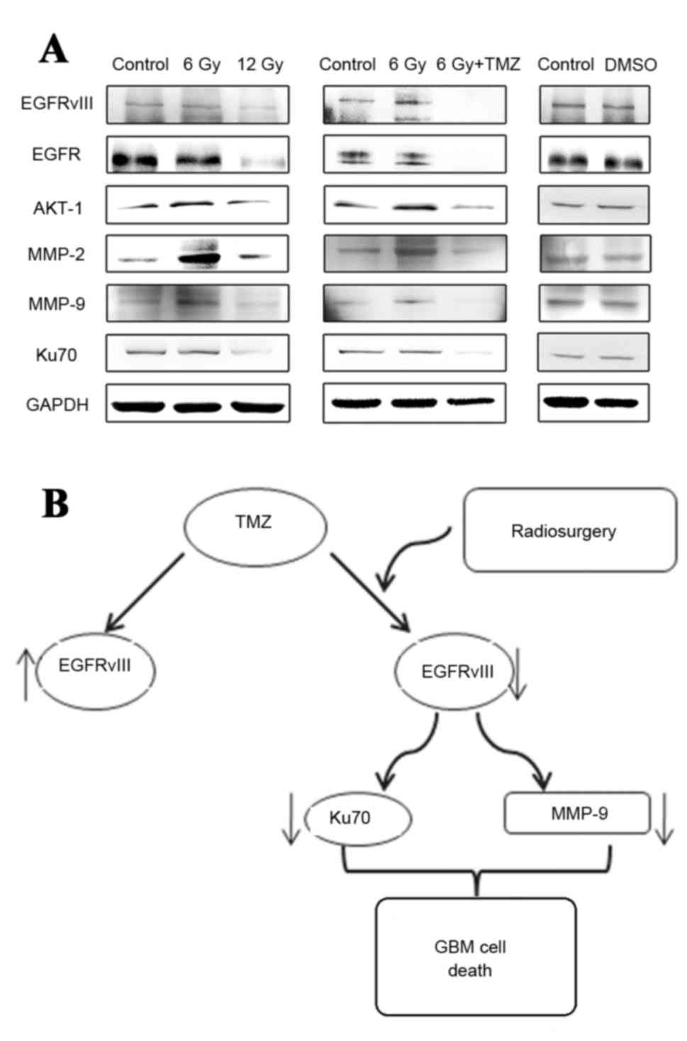 | Figure 2.In vitro expression of
EGFRvIII, EGFR, AKT-1, MMP-2, MMP-9 and Ku70. (A) Levels of
expression of EGFRvIII, EGFR, AKT-1, MMP-2, MMP-9 and Ku70 proteins
in vitro in U87-EGFRvIII cell lines treated with negative
control, 6 Gy radiation, 12 Gy radiation, and combined treatment of
6 Gy radiosurgery and temozolomide. (B) Schematic diagram
illustrating the combined treatment of radiosurgery and
temozolomide. EGFR, epidermal growth factor receptor; EGFRvIII,
EGFR variant III; AKT-1, RAC-α serine/threonine-protein kinase;
MMP-2, matrix metalloproteinase 2; MMP-9; Ku70, X-ray repair
cross-complementing protein 6; GMB, glioblastoma multiforme; DMSO,
dimethyl sulphoxide; TMZ, temozolomide. |
U87-EGFRvIII cells in
heterotransplantation models
In the heterotransplantation animal models, it was
observed that in the U87-EGFRvIII cells treated by radiotherapy at
the low dose of 6 Gy, the tumorigenicity was weaker compared with
the cells treated with the negative control, which differed from
the results of the in vitro experiments. Although radiation
treatment at the low dose may promote the upregulation of
expression of certain invasiveness indices of U87-EGFRvIII cells
such as the AKT-1 and MMP-2, while it exhibits certain inhibitory
effects on tumour proliferation. The mechanism is unclear, but in
animal experiments in the present study, different doses of
radiation therapy have a significant inhibitory effect on the
growth of the tumour (Fig. 3). And,
the size of the tumours formed in the U87-EGFRvIII cells treated
with the combination of radiosurgery and temozolomide were smaller
compared with those in the cells treated with 6 Gy radiation. No
significant change was observed in the weight of the nude mice in
the combined treatment group, and their survival rate was higher
compared with the nude mice with U87-EGFRvIII cells treated with
the 6 Gy radiation treatment. The results from the
immunohistochemical analysis of the tumour cells (Fig. 3) in the heterotransplantation models
indicated that the expression of EGFRvIII and Ki-67 in the tumour
tissues subsequent to tumour formation in the U87-EGFRvIII cells
treated with a combination of radiosurgery and temozolomide
demonstrated a marked downregulation, and subsequent to combined
treatment, the inhibition of MMP-9, VEGF and EGFRvIII was more
evident. These results are different compared with those from the
in vitro experiments, indicating that subsequent to the
treatment of the U87-EGFRvIII cells, the proliferation and tumour
formation abilities decreased significantly, and the invasion
abilities of the formed tumours were also inhibited.
 | Figure 3.U87-EGFRvIII cell line in
heterotransplantation models treated with negative control, 6 Gy
radiation, and combined treatment with radiosurgery and
temozolomide. (A) Bioluminescence images from heterotransplantation
models (magnification, ×40) treated with negative control, 6 Gy
radiation (48 h after treatment with 6 Gy peripheral doses by
radiosurgery with 99% of isodose) and combined treatment with
radiosurgery and temozolomide (cells were treated for 24 h with
temozolomide at the IC50 dose for 24 h after 6 Gy
radiation) at 7, 14 and 21 days subsequent to tumour development.
(B) Histological sections of tumor xenografts were excised and
fixed in 10% neutral buffered formalin. Following paraffin
embedding, 5-µm sections were cut and dried, deparaffinized, and
rehydrated. Next, the nonspecific binding sites were blocked, and
the slides were incubated at 4°C overnight in a 1:100 dilution of
primary antibodies. Finally, the slides were incubated with a
secondary antibody for 30 min. Immunohistochemistry for EGFRvIII
and Ki-67 on xenograft tumour sections (Scale bar, 100 µm). (C)
Evaluation of tumour growth curves. (D) Evaluation of weights of
mice. (E) Improved survival observed in mice treated with the
combined therapy. H&E, hematoxylin and eosin; TMZ, temozolomid;
EGFRvIII, epidermal growth factor receptor variant III. |
In the present study, the EGFRvIII mutation mostly
occurred in GBMs with higher malignancies, and the combined
treatment of radiosurgery and temozolomide may inhibit the
invasiveness of tumour cells compared with other treatment regimens
subsequent to tumour formation in the U87-EGFRvIII cells. The
combined radiosurgery and temozolomide therapy also exerts a
stronger inhibitory effect on the tumorigenicity of the
U87-EGFRvIII cells in the brains of nude mice.
Discussion
GBM is the one of most invasive types of tumours in
the human brain. The invasion capabilities of these tumour cells
results in difficulties in achieving a complete surgical resection
of the tumour, and high rates of recurrence and mortality.
Therefore, patients with GMB exhibit poor prognoses and notably
short median survival times. The EGFR-targeting tyrosine kinase
inhibitor possesses a high degree of immune activity, which is
usually associated with drug resistance and may lead to
chemotherapy failure. EGFR is the tyrosine kinase receptor on the
cell surface and exhibits significantly high expression levels and
mutations in GBM. The most common type of EGFR mutation is
EGFRvIII: The deletion of exon 2 to 7, which leads to the fusion of
exon 1 and 8 and produces tumour-specific surface antigens on the
cell surface (14,15). EGFRvIII has also been identified in
other types of cancer, including lung, breast and prostate cancer,
and may also be present in head and neck squamous carcinoma, but it
is not exhibited in normal tissues or benign tumour tissues
(14–16). The amplification of EGFR is often
accompanied by the upregulated expression of EGFRvIII, which is
significantly correlated with short survival times for patients
with BMG. Previous studies have indicated that a high expression
level of EGFRvIII is positively correlated with the invasion
abilities of GBM cells. Previous studies have also indicated that
30–60% of GBM patients exhibit high expression levels of EGFRvIII,
and these mutations are not detected in normal brain tissues of
adult humans or other normal tissues (17–19).
Therefore, EGFRvIII is a good target for GBM gene therapy. However,
limited studies are available on the molecular biology of
EGFRvIII-associated mechanisms (20–22).
Numerous previous studies have examined the mechanisms and
development of EGFRvIII-associated target drugs, such as the use of
the protein Jagged 1-Notch signalling pathway and antibodies that
specifically identify EGFR DNA oligonucleotide aptamers, among
others, for the development of a more effective therapeutic regimen
due to the poor effectiveness of current therapeutic tools
(21,22). At present, surgical resection combined
with chemotherapy remains the best treatment, although it is
difficult to implement the enhanced radiation treatment combined
with temozolomide to improve the therapeutic effect (23,24). In
the present study, data from the clinical cases in the combined
treatment group indicated that in the low-grade, WHO I–II, gliomas,
33% of the tumours possessed the EGFRvIII mutation. In the
high-grade, WHO III–IV, gliomas, 61% of the tumours exhibited the
EGFRvIII mutation. It was revealed that the probability of
occurrence of high-grade glioma with the EGFRvIII mutation was
greater, indicating that the EGFRvIII mutation was associated with
tumour malignancy. PCR analysis of the tumour cells indicated that
9 patients (28%) exhibited EGFRvIII expression in tumour tissues, 1
(11%) of which possessed a WHO grade I–II tumour and 8 (89%) of
which possessed WHO grade III–IV tumours. This proportion was lower
compared with the results from pathological tests, which may be due
to uncertainty in the sampling positions, but this method also
illustrated the correlation between EGFRvIII mutation and tumour
malignancy.
In the glioma radiation treatment group, the
observation that therapeutic efficacy is positively correlated with
radiation dose was verified. In the present study, the analysis of
high-dose radiotherapy was not performed as the results are well
established. In addition, under the conditions that normal brain
tissues are protected and the radiation reactions are reduced,
little improvement remains to be achieved in current radiotherapy
measures (25,26). Certain previous studies have indicated
that radiation treatment at low doses between 5–8 Gy exhibits a
promotional effect on the invasion abilities of tumour cells.
Radiation treatment may promote the invasion abilities of glioma
cells (13,27,28).
Therefore, 6 Gy was selected as the dose for the present study to
increase the invasion abilities of glioma. The results of the
western blot analysis indicated that the 6-Gy dose may increase the
expression levels of the EGFRvIII, EGFR, AKT-1, MMP-2, MMP-9 and
Ku70 proteins in the U87-EGFRvIII cell lines, but subsequent to
combined therapy with temozolomide, the expression levels of these
proteins in the U87-EGFRvIII cell lines was significantly inhibited
and was evidently lower compared with the normal negative control
group. The animal experiments illustrated the marked difference.
The 6-Gy radiation dose of the in vitro experiment increased
the invasion capabilities of the U87-EGFRvIII cell lines, but the
tumorigenicity and the invasive indices in the tumour tissues
subsequent to tumour formation were not significantly improved and
were inhibited, indicating that low-dose radiation, although
increasing the invasiveness of the U87-EGFRvIII cells, did not
significantly increase the rates of proliferation and the growth of
the U87-EGFRvIII cells, but demonstrated an inhibitory effect.
Subsequent to combined radiotherapy and temozolomide treatment, the
tumour growth in the brains of the nude mice was significantly
lower and the survival time was greatly extended compared with
those in the negative control treatment group and the
radiation-only treatment group. These results suggested that
combined treatment with radiotherapy and temozolomide exhibits a
clear inhibitory effect on the tumourigenicity of U87-EGFRvIII
cells.
Overall, gene-targeted therapy based on molecular
biology and epigenetics is a potential prospect for GBM treatment,
but the application of this approach in clinical treatment is not
available at present. Therefore, to improve the existing treatment
options and to enhance the efficacy of current therapeutics, the
development of more accurate radiotherapy treatments or the use of
additional chemotherapeutical drugs, including bevacizumab and
valproic acid (VPA) (29–31), should be investigated alongside the
development of novel therapeutic techniques and treatments.
Acknowledgements
The temozolomide was donated by Tasly Pharmaceutical
Co., Ltd. (Tianjin, China).
Funding
The present study was supported by the National
High-tech Research and Development Program of China (863 Program;
grant no. 2014AA021102), the General Item of the Tianjin Natural
Science Fund (grant no. 13JCYBJC24000), the Science and Technology
Research and Development Program of Hebei Province of China (grant
no. 13277785D), and the Science and Technology Fund of the Tianjin
Health Bureau (grant no. 2011KZ95).
Availability of data and materials
All data generated or analyzed during this study are
included in this published article.
Authors' contributions
All authors read and approved the final manuscript.
Cell lines and tissue samples:JLX, YLT, GKW and CF. Cell treatment:
YGL, GKW, ZYZ, YLG, YQZ, DSX and XML. Immunohistochemistry
analysis: YGL, KZ, XML and CSK. Intracranial animal model: YGL,
JHZ, KZ and CSK. Protein extraction and western-blot analysis: YGL
and KZ. RT-PCR: YGL, JHZ and KZ. Statistical analysis: YGL, JLX,
JHZ and KZ. Writing the manuscript: YGL, JHZ, JLX and XML.
Ethics approval and consent to
participate
The present study was approved by the Ethics
Committee of the Second Hospital of Tianjin Medical University
(Tianjin, China). Approval for the use of glioma samples was
obtained from each patient.
Consent for publication
Not applicable.
Competing interests
The authors declare that they have no competing
interests.
References
|
1
|
Furnari FB, Fenton T, Bachoo RM, Mukasa A,
Stommel JM, Stegh A, Hahn WC, Ligon KL, Louis DN, Brennan C, et al:
Malignant astrocytic glioma: Genetics biology, and paths to
treatment. Genes Dev. 21:2683–2710. 2007. View Article : Google Scholar : PubMed/NCBI
|
|
2
|
Maher EA, Furnari FB, Bachoo RM, Rowitch
DH, Louis DN, Cavenee WK and DePinho RA: Malignant glioma: Genetics
and biology of a grave matter. Genes Dev. 15:1311–1333. 2001.
View Article : Google Scholar : PubMed/NCBI
|
|
3
|
Wen PY and Kesari S: Malignant gliomas in
adults. N Engl J Med. 359:492–507. 2008. View Article : Google Scholar : PubMed/NCBI
|
|
4
|
Jin X, Yin J, Kim SH, Sohn YW, Beck S, Lim
YC, Nam DH, Choi YJ and Kim H: EGFR-AKT Smad signaling promotes
formation of glioma stem-like cells and tumor angiogenesis by
ID3-driven cytokine induction. Cancer Res. 71:7125–7134. 2011.
View Article : Google Scholar : PubMed/NCBI
|
|
5
|
Jeon HM, Kim SH, Jin X, Park JB, Kim SH,
Joshi K, Nakano I and Kim H: Cross talk between glioma-initiating
cells and endothelial cells drives tumor progression. Cancer Res.
74:4482–4492. 2014. View Article : Google Scholar : PubMed/NCBI
|
|
6
|
Moscatello DK, Holgado-Madruga M, Godwin
AK, Ramirez G, Gunn G, Zoltick PW, Biegel JA, Hayes RL and Wong AJ:
Frequent expression of a mutant epidermal growth factor receptor in
multiple human tumors. Cancer Res. 55:5536–5539. 1995.PubMed/NCBI
|
|
7
|
Libermann TA, Nusbaum HR, Razon N, Kris R,
Lax I, Soreq H, Whittle N, Waterfield MD, Ullrich A and
Schlessinger J: Amplification, enhanced expression and possible
rearrangement of EGF receptor gene in primary human brain tumours
of glial origin. Nature. 313:144–147. 1985. View Article : Google Scholar : PubMed/NCBI
|
|
8
|
Hatanpaa KJ, Burma S, Zhao D and Habib AA:
Epidermal growth factor receptor in glioma: Signal transduction,
neuropathology, imaging, and radioresistance. Neoplasia.
12:675–684. 2010. View Article : Google Scholar : PubMed/NCBI
|
|
9
|
Pelloski CE, Ballman KV, Furth AF, Zhang
L, Lin E, Sulman EP, Bhat K, McDonald JM, Yung WK, Colman H, et al:
Epidermal growth factor receptor variant III status defines
clinically distinct subtypes of glioblastoma. J Clin Oncol.
25:2288–2294. 2007. View Article : Google Scholar : PubMed/NCBI
|
|
10
|
Yoshimoto K, Dang J, Zhu S, Nathanson D,
Huang T, Dumont R, Seligson DB, Yong WH, Xiong Z, Rao N, et al:
Development of a real-time RT-PCR assay for detecting EGFRvIII in
glioblastoma samples. Clin Cancer Res. 14:488–493. 2008. View Article : Google Scholar : PubMed/NCBI
|
|
11
|
Louis DN, Perry A, Reifenberger G, von
Deimling A, Figarella-Branger D, Cavenee WK, Ohgaki H, Wiestler OD,
Kleihues P and Ellison DW: The 2016 World Health Organization
classification of tumors of the central nervous system: A summary.
Acta Neuropathol. 131:803–820. 2016. View Article : Google Scholar : PubMed/NCBI
|
|
12
|
Jia Q, Li Y, Xu D, Li Z, Zhang Z, Zhang Y,
Liu D, Liu X, Pu P and Kang C: Radiosensitivity of glioma to Gamma
Knife treatment enhanced in vitro and in vivo by RNA interfering
Ku70 that is mediated by a recombinant adenovirus. J Neurosurg. 113
Suppl:S228–S235. 2010.
|
|
13
|
Madani I, De Neve W and Mareel M: Does
ionizing radiation stimulate cancer invasion and metastasis? Bull
Cancer. 95:292–300. 2008.PubMed/NCBI
|
|
14
|
Ge H, Gong X and Tang CK: Evidence of high
incidence of EGFRvIII expression and coexpression with EGFR in
human invasive breast cancer by laser capture microdissection and
immunohistochemical analysis. Int J Cancer. 98:357–361. 2002.
View Article : Google Scholar : PubMed/NCBI
|
|
15
|
Olapade-Olaopa EO, Moscatello DK, MacKay
EH, Horsburgh T, Sandhu DP, Terry TR, Wong AJ and Habib FK:
Evidence for the differential expression of a variant EGF receptor
protein in human prostate cancer. Br J Cancer. 82:186–194. 2000.
View Article : Google Scholar : PubMed/NCBI
|
|
16
|
Khattri A, Zuo Z, Brägelmann J, Keck MK,
El Dinali M, Brown CD, Stricker T, Munagala A, Cohen EE, Lingen MW,
et al: Rare occurrence of EGFRvIII deletion in head and neck
squamous cell carcinoma. Oral Oncol. 51:53–58. 2015. View Article : Google Scholar : PubMed/NCBI
|
|
17
|
Tan Y, Shi YS, Wu XD, Liang HY, Gao YB, Li
SJ, Zhang XM, Wang F and Gao TM: DNA aptamers that target human
glioblastomamultiforme cells overexpressing epidermal growth factor
receptor variant III in vitro. Acta Pharmacol Sin. 34:1491–1498.
2013. View Article : Google Scholar : PubMed/NCBI
|
|
18
|
Kim J, Lee JS, Jung J, Lim I, Lee JY and
Park MJ: Emodin suppresses maintenance of stemness by augmenting
proteosomal degradation of epidermal growth factor
receptorepidermal growth factor receptor variant III in glioma stem
cells. Stem Cells Dev. 24:284–295. 2015. View Article : Google Scholar : PubMed/NCBI
|
|
19
|
Congdon KL, Gedeon PC, Suryadevara CM,
Caruso HG, Cooper LJ, Heimberger AB and Sampson JH: Epidermal
growth factor receptor and variant III targeted immunotherapy.
Neuro Oncol. 16 Suppl 8:viii20–viii25. 2014. View Article : Google Scholar : PubMed/NCBI
|
|
20
|
Zhang X, Liang H, Tan Y, Wu X, Li S and
Shi Y: U87-EGFRvIII cell-specific aptamer mediates small
interfering RNA delivery. Biomed Rep. 2:495–499. 2014. View Article : Google Scholar : PubMed/NCBI
|
|
21
|
Kim EJ, Kim SO, Jin X, Ham SW, Kim J, Park
JB, Lee JY, Kim SC and Kim H: Epidermal growth factor receptor
variant III renders glioma cancer cells less differentiated by
JAGGED1. Tumour Biol. 36:2921–2928. 2015. View Article : Google Scholar : PubMed/NCBI
|
|
22
|
Katanasaka Y, Kodera Y, Kitamura Y,
Morimoto T, Tamura T and Koizumi F: Epidermal growth factor
receptor variant type III markedly accelerates angiogenesis and
tumor growth via inducing c-myc mediated angiopoietin-like 4
expression in malignant glioma. Mol Cancer. 12:312013. View Article : Google Scholar : PubMed/NCBI
|
|
23
|
Senft C, Bink A, Franz K, Vatter H, Gasser
T and Seifert V: Intraoperative MRI guidance and extent of
resection in glioma surgery: A randomised, controlled trial. Lancet
Oncol. 12:997–1003. 2011. View Article : Google Scholar : PubMed/NCBI
|
|
24
|
Gilbert MR, Dignam JJ, Armstrong TS, Wefel
JS, Blumenthal DT, Vogelbaum MA, Colman H, Chakravarti A, Pugh S,
Won M, et al: A randomized trial of bevacizumab for newly diagnosed
glioblastoma. N Engl J Med. 370:699–708. 2014. View Article : Google Scholar : PubMed/NCBI
|
|
25
|
Binello E, Green S and Germano IM:
Radiosurgery for high-grade glioma. Surg Neurol Int. 3 Suppl
2:S118–S126. 2012. View Article : Google Scholar : PubMed/NCBI
|
|
26
|
Waters JD, Rose B, Gonda DD, Scanderbeg
DJ, Russell M, Alksne JF, Murphy K, Carter BS, Lawson J and Chen
CC: Immediate post-operative brachytherapy prior to irradiation and
temozolomide for newly diagnosed glioblastoma. J Neurooncol.
113:467–477. 2013. View Article : Google Scholar : PubMed/NCBI
|
|
27
|
Kwak SY, Kim BY, Ahn HJ, Yoo JO, Kim J,
Bae IH and Han YH: Ionizing radiation-inducible miR-30e promotes
glioma cell invasion through EGFR stabilization by directly
targeting CBL-B. FEBS J. 282:1512–1525. 2015. View Article : Google Scholar : PubMed/NCBI
|
|
28
|
Zhang X, Li X, Zhang N, Yang Q and Moran
MS: Low doses ionizing radiation enhances the invasiveness of
breast cancer cells by inducing epithelial-mesenchymal transition.
Biochem Biophys Res Commun. 412:188–192. 2011. View Article : Google Scholar : PubMed/NCBI
|
|
29
|
Iuchi T, Hatano K, Uchino Y, Itami M,
Hasegawa Y, Kawasaki K, Sakaida T and Hara R: Methionine Uptake and
required radiation dose to control glioblastoma. Int J Radiat Oncol
Biol Phys. 93:133–140. 2015. View Article : Google Scholar : PubMed/NCBI
|
|
30
|
Hawkins-Daarud A, Rockne R, Corwin D,
Anderson AR, Kinahan P and Swanson KR: In silico analysis suggests
differential response to bevacizumab and radiation combination
therapy in newly diagnosed glioblastoma. J R Soc Interface.
12:201503882015. View Article : Google Scholar : PubMed/NCBI
|
|
31
|
Krauze AV, Myrehaug SD, Chang MG, Holdford
DJ, Smith S, Shih J, Tofilon PJ, Fine HA and Camphausen K: A phase
2 study of concurrent radiation therapy, temozolomide, and the
histone deacetylase inhibitor valproic acid for patients with
glioblastoma. Int J Radiat Oncol Biol Phys. 92:986–992. 2015.
View Article : Google Scholar : PubMed/NCBI
|


