Introduction
Osteosarcoma is the most frequent primary malignant
bone tumor, which mainly occurs in children and adolescents
(1–3).
Previous study has shown that osteosarcoma accounts for
approximately 19 and 5% of all malignant bone tumors and childhood
neoplasm, respectively (4). Despite
the recently advances in multi-modal therapeutics, the prognosis
and 5 years survival rate of osteosarcoma remains unsatisfactory
(5). Therefore, it is urgent for us
to explore the molecular mechanisms and find new therapeutic
strategies to target this disease.
MicroRNAs (miRNAs/miRs) are short, endogenous
noncoding and highly conserved RNAs, which can restrain the
expression of target genes through binding to the 3′-untranslated
region (3′-UTR) mRNA (6–8). According to previous studies, miRNAs
have taken part in a great deal of cellular processes, including
cell proliferation, invasion, apoptosis, chemo-resistance (9–11).
Furthermore, many studies have already proved that abnormal
expression of miRNAs play significantly parts in the occurrence and
progression of cancers, including hepatocellular carcinoma, breast
cancer, gastric carcinoma, prostate cancer, and also osteosarcoma
(12–14). Numerous researches have suggested that
miR-192-5p was dramatically downregulated in malignant tumors,
while overexpression of miR-192-5p could inhibit tumorigenesis
through different mechanisms (15,16). In
human osteosarcoma, miR-192-5p functioned as a major role in
inhibiting the tumorigenesis in osteosarcoma (17). Nevertheless, the potential mechanisms
of miR-192-5p in regulating the development and progression of
osteosarcoma remains largely unknown.
Ubiquitination is a critical posttranslational
modification, which modulates cellular processes, including cell
cycle regulation, chromatin remodeling, DNA damage response and so
forth (18). Ubiquitination
modification is a dynamic reversible process, which is catalyzed by
deubiquitinases (DUBs) (19).
Ubiquitin-specific protease 1 (USP1), one of the best characterized
member of the DUBs family, is famous for its regulation of cellular
response to DNA damage (20).
Recently, more and more studies reported that USP1 played an
important role in oncogenesis and tumorigenesis in human malignant
cancers, including osteosarcoma (21–24).
Given the crucial parts of miR-192-5p and USP1 in
regulating the initiation and progression of osteosarcoma, we
performed this study to identify whether miR-192-5p could
negatively regulates osteosarcoma by directly targeting USP1.
Materials and methods
Tissue collection
A total of 25 samples of osteosarcoma and matched
the adjacent non-tumor tissues were collected from surgical
resection between June 2009 and June 2012 in Renmin Hospital of
Wuhan University. The parameters of patients were showed in
Table I. Tissues were obtained and
then frozen in liquid nitrogen immediately and stored at −80°C
until being used. All patients were further followed up every 3–5
months until 5 years. The patients participated in this study did
not undergo any chemotherapy, radiotherapy or immunotherapy before
surgery. This study was approved by the Medical Ethics Committee of
Renmin Hospital of Wuhan University. All patients and their
families provided written informed consent to take part in this
research. The study did not contain any identifying information
about any participants. All the data was kept by the administrator
of the study team in a confidential manner and was not used by any
other purposes. We confirm that all experiments were performed in
accordance with relevant guidelines and regulations.
 | Table I.Associations between microRNA-192-5p
levels and clinicopathological variables of osteosarcoma
patients. |
Table I.
Associations between microRNA-192-5p
levels and clinicopathological variables of osteosarcoma
patients.
|
|
| Relative miR-192-5p
expression |
|
|---|
|
|
|
|
|
|---|
| Variable | Total no.
(n=25) | Low (n=13) | High (n=12) | P-value |
|---|
| Sex |
|
|
| 0.870 |
|
Male | 15 | 8 | 7 |
|
|
Female | 10 | 5 | 5 |
|
| Age (years) |
|
|
| 0.568 |
|
<20 | 18 | 10 | 8 |
|
|
≥20 | 7 | 3 | 4 |
|
| Histologic
subtype |
|
|
| 0.238 |
|
Osteoblastic | 13 | 6 | 7 |
|
|
Chondroblastic | 9 | 5 | 4 |
|
|
Fibroblastic | 3 | 2 | 1 |
|
| Anatomical
site |
|
|
| 0.920 |
|
Femure | 11 | 6 | 5 |
|
|
Tibia | 9 | 5 | 4 |
|
|
Humerus | 3 | 1 | 2 |
|
|
Other | 2 | 1 | 1 |
|
| Tumor grade |
|
|
| 0.007b |
|
Low | 8 | 1 | 7 |
|
|
High | 17 | 12 | 5 |
|
| Enneking stage |
|
|
| 0.009b |
| I | 6 | 1 | 5 |
|
| II | 12 | 7 | 5 |
|
|
III | 7 | 5 | 2 |
|
| Tumor size
(cm) |
|
|
| 0.027a |
|
<8 | 13 | 4 | 9 |
|
| ≥8 | 12 | 9 | 3 |
|
Cell culture and transfection
143B and U2OS (human osteosarcoma cell lines) and
hFOB (normal human osteoblast cell line) were used in the present
study, which were gained from Cell Bank of Type Culture Collection
(Shanghai, China). We used DMEM (HyClone, Logan, UT, USA)
supplemented with 10% fetal bovine serum (FBS; Gibco, Grand Island,
NY, USA), 100 U/ml penicillin and 100 µg/ml streptomycin to culture
the above cells in an atmosphere of 5% CO2 at 37°C.
miR-192-5p mimic was purchased from GeneCreate Biological
Engineering Co., Ltd. (Wuhan, China). Then we transfected
miR-192-5p mimic and miR-NC (negative control) into cells at 60%
confluence by Lipofectamine 2000 (Invitrogen, Carlsbad, CA, USA).
Overexpression USP1 plasmid (pCDNA3.1-USP1) or empty plasmid
(GeneCreate Biological Engineering Co., Ltd.) were transfected into
cells at 80% confluence using Lipofectamine 2000 in accordance with
the manufacturer's instructions.
Cell proliferation assay
CCK-8 (Dojindo Molecular Technologies, Inc.,
Kumamoto, Japan) assay was performed to evaluate cell proliferation
capacity. Briefly, transfected cells were seeded into individual
well plates at 1×10⁵ cells per/well, then incubated for
0, 24, 48 and 72 h. Subsequently, microplate reader (Bio-Rad
Laboratories, Inc., Hercules, CA, USA) was used to detect the OD
value at 450 nm.
Cell cycle assay
For cell cycle assay, transfected cells were
inoculated in 6-well plates for 24 h. Then 143B and U2OS cells were
fixed with 75% cold ethanol at 4°C for 24 h. Subsequently, both
cell lines were stained with a propidium iodide (PI; BD
Biosciences, San Jose, CA, USA) for 30 min in the dark. Then,
stained cells were analyzed by using flow cytometer (FACS Calibur;
BD Biosciences). At last, ModFitLT V2.0 software (BD Biosciences)
was applied to analyze the above data.
Cell apoptosis assay
Apoptosis was assessed by flow cytometric. Cells
were harvested and washed with ice-cold PBS twice. Then we
resuspended cells with 300 µl of binding buffer. After being
stained with Annexin V-FITC and PI (BD Biosciences), FACS Calibur
was performed to analyze cell apoptosis.
Cell invasion assay
In order to investigate the cell invasion ability,
Matrigel (BD Biosciences) were precoated into 8 micron Transwells
(Corning Incorporated, Corning, NY, USA), and then 143B and U2OS
cells were added into the upper chambers with serum free medium.
DMEM (500 µl) with 10% FBS was placed into the lower chamber for
chemical induction. After incubating for 24 h, we carefully wiped
out remaining cells which did not invade. Matrigel membranes were
fixed with paraformaldehyde and then stained with crystal violet
solution. The invaded cells were counted under a phrase contrast
microscope (Olympus, Tokyo, Japan).
Cell migration assay
Cell migration was evaluated by wound healing assay.
The transfected cells were cultured in 6-well plates for 24 h.
Then, artificial wound was scratched in confluent cell monolayers
by sterile 10 ul pipette tip. Photographs were taken at 0 and 24 h
by using inverse microscope.
Luciferase reporter assay
The mutant (mut) or wild-type (wt) 3′UTR of USP1 was
inserted into pGL4 luciferase promoter vector (Promega Corporation,
Madison, WI, USA). Subsequently, miR-192-5p mimics or miR-NC and
the vectors carrying USP1 mut or wt 3′UTR were co-transfected into
143B and U2OS cells by lipofectamine 2000 (Invitrogen). After 24 h,
the dual luciferase reporter assay system (Promega Corporation) was
performed to determine luciferase values.
Reverse transcription-quantitative
polymerase chain reaction (RT-qPCR)
Total RNAs were extract from tissues and cell lines
using TRIzol reagent (Invitrogen). Reverse transcription reactions
were conducted via Takara RNA PCR kit (Takara, Kyoto, Japan). qPCR
was performed using a SYBR-Green detection system (Takara).
Relative gene expression was calculated using 2−ΔΔCq
method. The expression of U6 and β-actin were acted as the internal
control for the expression of miR-192-5p and USP1, respectively.
The primers for miR-192-5p were forward,
5′-GCGGCGGCTGACCTATGAATTG-3′ and reverse,
5′-ATCCAGTGCAGGGTCCGAGG-3′; U6 forward, 5′-TCCGATCGTGAAGCGTTC-3′
and reverse, 5′-GTGCAGGGTCCGAGGT-3′; USP1 mRNA forward,
5′-AGGTTGCTAGTACAGCGTTTGC-3′ and reverse,
5′-CACTGGATTCCTTGTTTCTATCAGA-3′; and β-actin forward,
5′-GGCACTCTTCCAGCCTTCC-3′ and reverse, 5′-GAGCCGCCGATCCACAC-3′.
Western blot analysis
We extracted total proteins from transfected cells
by using RIPA lysis buffer (Beyotime Institute of Biotechnology,
Shanghai, China). All proteins were separated by 10% SDS-PAGE gel
and transferred them onto the PVDF membrane (EMD Millipore Corp.,
Bedford, MA, USA). The membrane were blocked in 10% non-fat dried
milk for 2 h and then incubated with primary anti-USP1 and GAPDH
(both from Abcam, Cambridge, UK) at 4°C overnight. After being
washed with TBST five times, the membrane was incubated with
secondary antibody for 1 h at room temperature. Then the membrane
was washed three times with TBST again. Finally, the proteins were
detected by using enhanced chemiluminescent. GAPDH was performed as
an endogenous control.
Statistical analysis
All statistical analyses were conducted by using
SPSS 20.0 (IBM Corp., Armonk, NY, USA) and Graph Pad Prism 5
software (GraphPad Software, Inc., San Diego, CA, USA). Data which
were followed the Gaussian distribution, calculated as mean ± SD.
Differences between two groups were measured by Student's t-test
(for the migration, invasion, apoptosis and cell cycle assays),
while one-way ANOVA (for CCK-8 assay) followed by Tukey's post hoc
test was performed for comparisons between more than two groups.
Correlation between miR-192-5p levels and clinicopathological
variables of osteosarcoma patients were assessed by Chi-square
test. The Pearson' procedure method was used in the patients. And
the Kaplan-Meier survival analysis was applied in the survival
experiments. P<0.05 was considered as statistically significant.
Each experiment was performed three times.
Results
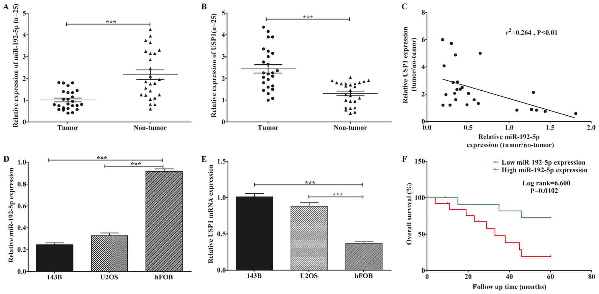
Reduced expression of miR-192-5p and
elevated expression of USP1 in osteosarcoma tissues and cell
lines
The expression levels of miR-192-5p and USP1 mRNA
were detected in tissues and cell lines of osteosarcoma. MiR-192-5p
was distinctly downregulated in osteosarcoma tissues (Fig. 1A). Whereas, USP1 mRNA showed an
opposite trend (Fig. 1B). Moreover,
Pearson's correlation assay indicated that the expression levels of
USP1 and miR-192-5p were inversely correlated in osteosarcoma
tissues (Fig. 1C). Next, two classic
osteosarcoma cell lines: 143B and U2OS were performed to further
confirm the findings above. Here we found that lower expressions of
miR-192-5p were observed in both cell lines compared to hFOB
(Fig. 1D). In contrast, the
expression levels of USP1 mRNA was markedly higher in both
osteosarcoma cell lines compared to hFOB (Fig. 1E). Furthermore, to evaluate the
association between the expression level of miR-192-5p and
clinicopathological variables, we divided all patients into two
groups (low expression group and high expression group) based on
the median expression level of tumor tissues. As showed in Table I, the low expression of miR-192-5p was
statistically correlated with tumor grade, Enneking stage (25) and tumor size, while not associated
with sex, age, histologic subtype and anatomical site in patients
with osteosarcoma. Besides, the Kaplan-Meier survival analysis
suggested that osteosarcoma patients with low expression of
miR-192-5p presented to have a shorter overall survival (Fig. 1F). These finding indicated that the
low expression of miR-192-5p was closely associated with the high
expression of USP1.
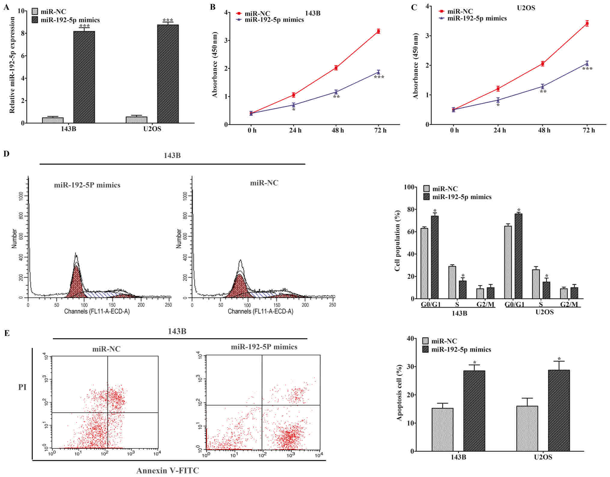
Upregulation of miR-192-5p inhibited
osteosarcoma cell proliferation
We transfected 143B and U2OS cells with miR-192-5p
mimics or miR-NC. After being cultured, we found that the
expression profile of miR-192-5p was dramatically increased
compared to miR-NC group (Fig. 2A).
Then, CCK-8 assay was conducted to evaluate the ability of cell
proliferation in 143B and U2OS cells. The results indicated that
cell proliferation was statistically suppressed when miR-192-5p was
upregulated (Fig. 2B and C). Flow
cytometric assay was used to further identify whether cell
proliferation was inhibited by miR-192-5p through altering
cell-cycle progression or inducing cell apoptosis. Cell cycle
analysis showed that miR-192-5p mimics brought about a higher
G0⁄G1-phase and a lower S-phase arrest in 143B and U2OS cells
compared to miR-NC group (Fig. 2D).
Subsequently, cell apoptosis analysis was performed to explore the
influence of miR-192-5p mimics on cell apoptosis, which revealed
that cell apoptosis was remarkably induced in 143B and U2OS cells
compared to miR-NC group (Fig. 2E).
These findings demonstrated that overexpression of miR-192-5p
repressed osteosarcoma cell proliferation through regulating cell
cycle during G1 to S phase and inducing cell apoptosis.
Overexpression of miR-192-5p
suppressed osteosarcoma cell migration and invasion and enhanced
the sensitivity of osteosarcoma cells to cisplatin
Wound-healing and Transwell chamber assays were
performed to elucidated cell migration and invasion in 143B and
U2OS cells. Wound-healing assay showed that the migration capacity
of cells in the miR-192-5p mimics group was obviously inhibited
after wounding (Fig. 3A). Transwell
chamber assay showed that ectopic expression of miR-192-5p
inhibited cell invasion (Fig. 3B).
These findings suggested that upregulation of miR-192-5p weaken the
migration and invasion capability of 143B and U2OS cells. CCK-8
assay was performed to evaluate the effect of miR-192-5p mimics on
the chemo-sensitivity of osteosarcoma cells. The response of 143B
and U2OS cells to cisplatin enhanced after treated with the
miR-192-5p mimic compared to miR-NC group (Fig. 3C and D). The data demonstrated that
miR-192-5p mimics reduced chemo-resistance of osteosarcoma cells to
cisplatin. And it had been reported that the cisplatin drug was
presented the chemio-sensitivity in osteosarcoma cells (26).
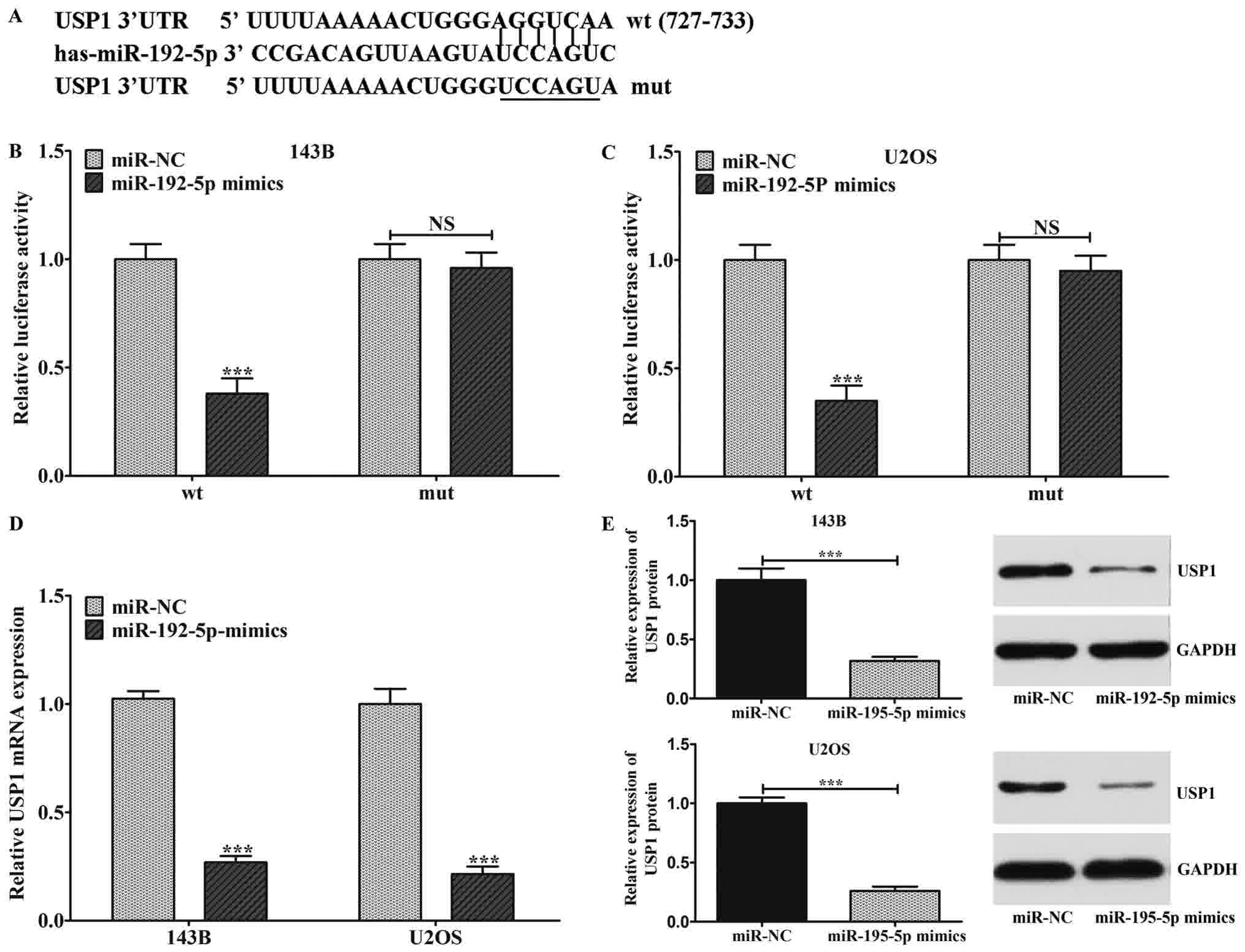
USP1 was a direct target of miR-192-5p
in osteosarcoma cell
In order to verify the relationships between
miR-192-5p and USP1, TargetScan tools was performed to predict the
target sites for miR-192-5p. It was demonstrated that there was a
combination of target sequences between the USP1 3′-UTR and
miR-192-5p (Fig. 4A). Then,
dual-luciferase reporter system assay was carried out to affirm the
above prediction. In detail, the 3′-UTR of USP1 containing
wild-type (wt) or mutant-type (mut) miR-192-5p target sequences was
inserted into the plasmid. Then, we co-transfected 143B and U2OS
cells with these reporters plasmid and miR-192-5p. Luciferase
activity was detected 48 h after transfection. As showed in
Fig. 4B and C, miR-192-5p mimics
significantly restrained wild-type 3′UTR-USP1 reporter activity
while there was no repression on the mutant 3′UTR-USP1 reporter
activity, which revealed that miR-192-5p most likely suppressed
gene expression via miR-192-5p binding sequences at the 3′-UTR of
USP1. Besides, we observed that Ectopic expression of miR-192-5p
statistically inhibited the expression of USP1 on mRNA and protein
level (Fig. 4D and E).
miR-192-5P regulated osteosarcoma cell
proliferation, apoptosis, migration, invasion and chemo-sensitivity
through USP1
To further ascertain whether upregulation of
miR-192-5p inhibited OS cells biological functions through USP1. We
transfected USP1 vector into 143B and U2OS cells. After cultivated
in vitro, the USP1 mRNA and protein expressions were
restored in 143B and U2OS cells (Fig. 5A
and B). Ectopic expression of USP1 statistically enhanced cell
proliferation, migration and invasion abilities and decreased cell
apoptosis and chemo-sensitivity in 143B and U2OS cells. However,
when co-transfected with miR-192-5p mimics and USP1, overexpression
of miR-192-5p partially abolished the effects of USP1 on cells
proliferation, chemo-sensitivity, migration, invasion and apoptosis
(Fig. 5C-I). Taken together,
miR-192-5p inhibited osteosarcoma cell biological functions
primarily through downregulating USP1.
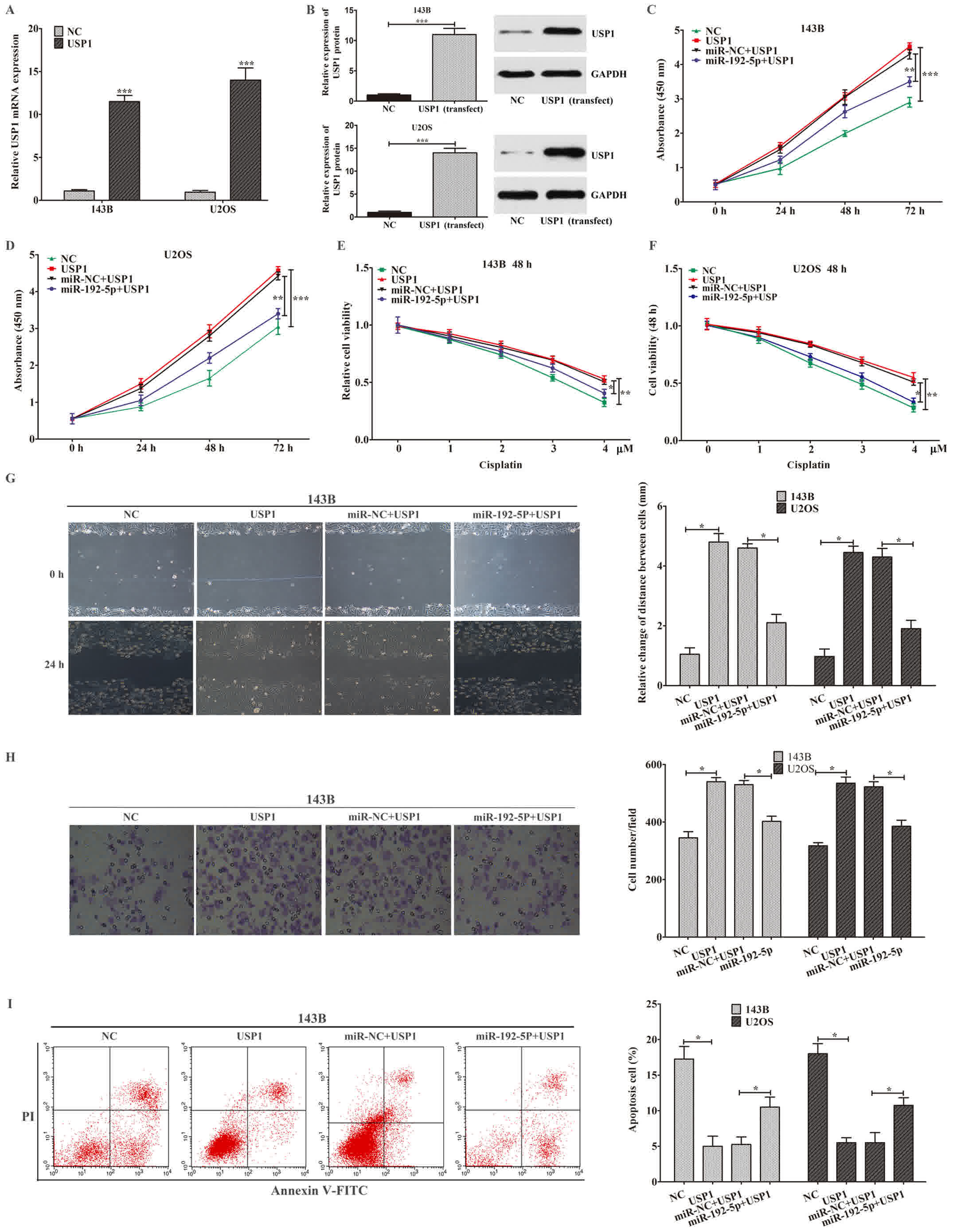 | Figure 5.miR-192-5p regulated osteosarcoma
cell proliferation, apoptosis, migration, invasion and
chemo-sensitivity through USP1. (A) The USP1 mRNA expression was
detected by reverse transcription-quantitative polymerase chain
reaction. (B) The USP1 protein expression was detected by western
blot analysis. (C-I) Ectopic expression of USP1 statistically
enhanced cell (C and D) proliferation, decreased cell (E and F)
chemo-sensitivity, promoted cell (G) migration and (H) invasion
(magnification, ×100), and reduced cell (I) apoptosis in 143B and
U2OS cells, which could be partially abolished by miR-192-5p
following co-transfection with miR-192-5p mimics and USP1. Data are
presented as the mean ± standard deviation of 3 independent assays.
*P<0.05, **P<0.01 and ***P<0.001 vs. NC, or as indicated.
miR-NC. miR, microRNA; NC, negative control; USP1,
ubiquitin-specific protease 1; PI, propidium iodide. |
Discussion
Increasing evidence have suggested that miRNAs could
regulate cell proliferation, apoptosis, migration, invasion and
chemo-sensitivity in malignant tumors, including osteosarcoma
(27–30). For instance, Chen et al
(31) revealed that miR-211-5p was
downregulated in triple-negative breast cancer (TNBC), which
inhibit TNBC cell biological functions via targeting SETBP1. Our
previous study also showed that miR-335 was statistically
downregulated in osteosarcoma stem cells. Moreover, overexpression
of miR-335 suppressed stem cell-like characteristics by targeting
POU5F1 (32). Our study indicated
that miR-192-5p was significantly downregulated and USP1 was
remarkably upregulated in 25 osteosarcoma samples and tow cell
lines. Pearson's correlation assay indicated that the expression
levels of miR-192-5p was inversely associated with USP1. Moreover,
low expression of miR-192-5p in patients was statistically
correlated with tumor grade, Enneking stage and tumor size. And
osteosarcoma patients with low expression of miR-192-5p presented
to have a shorter overall survival. Then, biological functions of
miR-192-5p were explored in 143B and U2OS. The results shown that
upregulation of miR-192-5p inhibited cell proliferation through
cell cycle arrest and inducing cell apoptosis, suppressed cell
migration and invasion and enhanced cell chemo-sensitivity in
osteosarcoma cells. To gain insight into the detail relationship
between miR-192-5p and USP1, we used TargetScan software (33) to predict that there was a highly
conservative binding site between USP1 and miR-192-5p. Then the
prediction was further proved by luciferase activity assay.
Furthermore, we observed that USP1 acted as an opposite role in
regulating cell biological function compared that with miR-192-5p.
Moreover, when co-transfected with miR-192-5p mimics and USP1
simultaneously, overexpression of miR-192-5p partially abolished
the effects of USP1 on cells proliferation, apoptosis, migration,
invasion and chemo-sensitivity in osteosarcoma cells.
Aberrant expression of miR-192 played a crucial role
in the development and progression of multiple malignant tumors
(34–36). Previous studies have proved that
miR-192 was significantly downregulated in many cancers, and
miR-192 also has been reported to regulate cell biological
functions in tumors, including proliferation, migration, invasion
and apoptosis (37,38). For instance, Feng et al
(15) suggested that miR-192-5p was
significantly low in lung cancer. They further demonstrated that
miR-192-5p suppressed cell proliferation and induced cell apoptosis
through RB1. Lian et al (16)
also proved that miR-192-5p reduced tumor metastasis by targeting
the SLC39A6/SNAIL pathway in HCC cells. Although previous study had
researched the effect of miR-192 on human osteosarcoma, which
indicated that miR-192 was downregulated in osteosarcoma and
miR-192 could suppressed the progression of osteosarcoma (17,39), the
exact molecular mechanism remained largely unclear. Consistent with
the above studies, we identified that upregulation of miR-192-5p
inhibited cell proliferation by preventing cell cycle from G1 to S
phase and inducing cell apoptosis in osteosarcoma cells. We also
found that miR-192-5p repressed cell migration and invasion, and
increased cells more sensitivity to cisplatin in osteosarcoma
cells. Taken together, we came to the conclusion that miR-192-5p
played an important role in suppressing osteosarcoma.
To clarify the potential molecular mechanism about
miR-192-5p regulates cell biological function in osteosarcoma.
Based on open-target prediction programs (TargetScan software), we
found that USP1 may be a target gene of miR-192-5p. Recently,
several studies indicated that USP1 had been found to be
upregulated in many kinds of tumors, and they also found that
deregulated USP1 could suppress cell proliferation, migration,
invasion, chemo-resistance (40,41).
Increasing studies suggested that USP1 played a role part in
osteosarcoma. For example, Williams et al (24) indicated that USP1 was upregulated in
osteosarcoma cells, which promoted cell proliferation, suppressed
osteoblastic differentiation and stabilized ID proteins. Liu et
al (23) confirmed that USP1 was
upregulated in osteosarcoma. Silencing of USP1 inhibited cell
proliferation and invasion through reducing expression of some
downstream proteins, including Notch signaling pathway. Previous
studies of our team suggested that Notch signaling pathway played a
key role in the development and progression in osteosarcoma.
Moreover Notch pathway could negatively regulated osteosarcoma stem
cell-like properties, like cell proliferation, apoptosis,
chemo-resistance (42,43). In the present research, we initially
used the TargetScan software to suggest that USP1 was a putative
binding site of miR-192-5p. Subsequently, overexpression of
miR-192-5p restrained wild-type 3′UTR-USP1 reporter activity, while
not in mutant 3′UTR-USP1 reporter activity in U2OS and 143B cells.
Overexpression of miR-192-5p significantly repressed the USP1
expression on mRNA and protein level. Moreover, we also found that
ectopic expression of USP1 promoted cell proliferation and
migration, decreased cell chemo-sensitivity, which could be
partially reversed by overexpression of miR-192-5p. All above data
support that ectopic expression of miR-192-5p repressed OS cell
proliferation, migration and invasion and increased the sensitivity
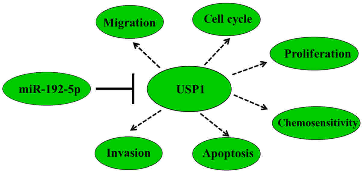
of osteosarcoma cells to cisplatin via targeting USP1. The
mechanism of how the miR-192-5p/USP1 axis regulates the initiation
and progression of osteosarcoma was presented in Fig. 6. Considering that Notch signal pathway
act as important downstream pathway of USP1, further research
should be performed to explore whether miR-192-5p can suppress
osteosarcoma oncogenicity by targeting USP1 through inactivation of
Notch signal pathway.
However, there are still some limitations in our
study. First of all, our sample size is too small, and we need a
larger sample size experiment to verify the conclusions in our
study. Secondly, transient transfection of miR-192-5p instead of
stable expression is used in our research. Thirdly, in order to
make the composing of figure more concise, we only put the pictures
of 143B cell line instead of two cell lines in figures. Forthly, in
our study, we only explored the expression of USP1 at the level of
mRNA. And we will further investigate its expression at the level
of protein, which will further demonstrate our point of view.
Finally, the downstream signal pathway of USP1 in our study need to
be further illustrated.
In conclusion, in addition to the above limitations,
our study elaborates the relationship between miR-192-5p and USP1
in osteosarcoma for the first time. Moreover, we provide evidence
to prove that miR-192-5p inhibited the progression of osteosarcoma
by targeting USP1. Therefore, miR-192-5p may serve as a valuable
biomarker and miR-192-5p/USP1 axis may function as a novel
therapeutic target for osteosarcoma.
Acknowledgements
Not applicable.
Funding
The present study was supported in part by a grant
from the Natural Science Foundation of China (grant no.
81502575).
Availability of data and materials
All data generated or analyzed during the present
study are included in this published article.
Authors' contributions
SZ, WG, MX, JC and GD performed the experiments. SZ,
LY, ZZ and GD analyzed the data. SZ, ZZ and LY contributed
reagents, materials and analytical tools. SZ, JC and WG reviewed
all data and wrote the paper. All authors read and approved the
final manuscript.
Ethics approval and consent to
participate
The present study was approved by the Medical Ethics
Committee of Renmin Hospital of Wuhan University (Hubei, China).
All patients and their families provided written informed consent
to take part in this research. All of the data was kept by the
administrator of the study team in a confidential manner and was
not used for any other purposes. We confirm that all experiments
were performed in accordance with relevant guidelines and
regulations.
Consent for publication
All participants provided written informed consent
for the publication of their data.
Competing interests
The authors declare that they have no competing
interests.
References
|
1
|
Geng S, Gu L, Ju F, Zhang H, Wang Y, Tang
H, Bi Z and Yang C: MicroRNA-224 promotes the sensitivity of
osteosarcoma cells to cisplatin by targeting Rac1. J Cell Mol Med.
20:1611–1619. 2016. View Article : Google Scholar : PubMed/NCBI
|
|
2
|
Zhang M, Wang D, Zhu T and Yin R:
miR-214-5p targets ROCK1 and suppresses proliferation and invasion
of human osteosarcoma cells. Oncol Res. 25:75–81. 2017. View Article : Google Scholar : PubMed/NCBI
|
|
3
|
Zhang Z, Zhang M, Chen Q and Zhang Q:
Downregulation of microRNA-145 promotes epithelial-mesenchymal
transition via regulating Snail in osteosarcoma. Cancer Gene Ther.
24:83–88. 2017. View Article : Google Scholar : PubMed/NCBI
|
|
4
|
Mirabello L, Troisi RJ and Savage SA:
Osteosarcoma incidence and survival rates from 1973 to 2004: Data
from the surveillance, epidemiology, and end results program.
Cancer. 115:1531–1543. 2009. View Article : Google Scholar : PubMed/NCBI
|
|
5
|
Chen B, Huang Z, Zhang Y, Chen Y and Li Z:
MicroRNA-145 suppresses osteosarcoma metastasis via targeting
MMP16. Cell Physiol Biochem. 37:2183–2193. 2015. View Article : Google Scholar : PubMed/NCBI
|
|
6
|
Bartel DP: MicroRNAs: Genomics,
biogenesis, mechanism, and function. Cell. 116:281–297. 2004.
View Article : Google Scholar : PubMed/NCBI
|
|
7
|
Wu W, Dang S, Feng Q, Liang J, Wang Y and
Fan N: MicroRNA-542-3p inhibits the growth of hepatocellular
carcinoma cells by targeting FZD7/Wnt signaling pathway. Biochem
Biophys Res Commun. 482:100–105. 2017. View Article : Google Scholar : PubMed/NCBI
|
|
8
|
Tormo E, Adam-Artigues A, Ballester S,
Pineda B, Zazo S, González-Alonso P, Albanell J, Rovira A, Rojo F,
Lluch A and Eroles P: The role of miR-26a and miR-30b in
HER2+ breast cancer trastuzumab resistance and
regulation of the CCNE2 gene. Sci Rep. 7:413092017. View Article : Google Scholar : PubMed/NCBI
|
|
9
|
Yang T, Zhao P, Rong Z, Li B, Xue H, You
J, He C, Li W, He X, Lee RJ, et al: Anti-tumor efficiency of
lipid-coated cisplatin nanoparticles co-loaded with MicroRNA-375.
Theranostics. 6:142–154. 2016. View Article : Google Scholar : PubMed/NCBI
|
|
10
|
Zeng JF, Ma XQ, Wang LP and Wang W:
MicroRNA-145 exerts tumor-suppressive and chemo-resistance lowering
effects by targeting CD44 in gastric cancer. World J Gastroenterol.
23:2337–2345. 2017. View Article : Google Scholar : PubMed/NCBI
|
|
11
|
Wei R, Yang Q, Han B, Li Y, Yao K, Yang X,
Chen Z, Yang S, Zhou J, Li M, et al: microRNA-375 inhibits
colorectal cancer cells proliferation by downregulating JAK2/STAT3
and MAP3K8/ERK signaling pathways. Oncotarget. 8:16633–16641.
2017.PubMed/NCBI
|
|
12
|
Miao Y, Zheng W, Li N, Su Z, Zhao L, Zhou
H and Jia L: MicroRNA-130b targets PTEN to mediate drug resistance
and proliferation of breast cancer cells via the PI3K/Akt signaling
pathway. Sci Rep. 7:419422017. View Article : Google Scholar : PubMed/NCBI
|
|
13
|
Yan X, Zhu Z, Xu S, Yang LN, Liao XH,
Zheng M, Yang D, Wang J, Chen D, Wang L, et al: MicroRNA-140-5p
inhibits hepatocellular carcinoma by directly targeting the unique
isomerase Pin1 to block multiple cancer-driving pathways. Sci Rep.
7:459152017. View Article : Google Scholar : PubMed/NCBI
|
|
14
|
Li B, Zhang S, Shen H and Li C:
MicroRNA-144-3p suppresses gastric cancer progression by inhibiting
epithelial-to-mesenchymal transition through targeting PBX3.
Biochem Biophys Res Commun. 484:241–247. 2017. View Article : Google Scholar : PubMed/NCBI
|
|
15
|
Feng S, Cong S, Zhang X, Bao X, Wang W, Li
H, Wang Z, Wang G, Xu J, Du B, et al: MicroRNA-192 targeting
retinoblastoma 1 inhibits cell proliferation and induces cell
apoptosis in lung cancer cells. Nucleic Acids Res. 39:6669–6678.
2011. View Article : Google Scholar : PubMed/NCBI
|
|
16
|
Lian J, Jing Y, Dong Q, Huan L, Chen D,
Bao C, Wang Q, Zhao F, Li J, Yao M, et al: miR-192, a prognostic
indicator, targets the SLC39A6/SNAIL pathway to reduce tumor
metastasis in human hepatocellular carcinoma. Oncotarget.
7:2672–2683. 2016. View Article : Google Scholar : PubMed/NCBI
|
|
17
|
Wang Y, Zhang S, Xu Y, Zhang Y, Guan H, Li
X, Li Y and Wang Y: Upregulation of miR-192 inhibits cell growth
and invasion and induces cell apoptosis by targeting TCF7 in human
osteosarcoma. Tumour Biol. 37:15211–15220. 2016. View Article : Google Scholar : PubMed/NCBI
|
|
18
|
Grabbe C, Husnjak K and Dikic I: The
spatial and temporal organization of ubiquitin networks. Nat Rev
Mol Cell Biol. 12:295–307. 2011. View
Article : Google Scholar : PubMed/NCBI
|
|
19
|
Liang Q, Dexheimer TS, Zhang P, Rosenthal
AS, Villamil MA, You C, Zhang Q, Chen J, Ott CA, Sun H, et al: A
selective USP1-UAF1 inhibitor links deubiquitination to DNA damage
responses. Nat Chem Biol. 10:298–304. 2014. View Article : Google Scholar : PubMed/NCBI
|
|
20
|
Sourisseau T, Helissey C, Lefebvre C,
Ponsonnailles F, Malka-Mahieu H, Olaussen KA, André F, Vagner S and
Soria JC: Translational regulation of the mRNA encoding the
ubiquitin peptidase USP1 involved in the DNA damage response as a
determinant of Cisplatin resistance. Cell Cycle. 15:295–302. 2016.
View Article : Google Scholar : PubMed/NCBI
|
|
21
|
Garcia-Santisteban I, Peters GJ,
Giovannetti E and Rodríguez JA: USP1 deubiquitinase: Cellular
functions, regulatory mechanisms and emerging potential as target
in cancer therapy. Mol Cancer. 12:912013. View Article : Google Scholar : PubMed/NCBI
|
|
22
|
Lee JK, Chang N, Yoon Y, Yang H, Cho H,
Kim E, Shin Y, Kang W, Oh YT, Mun GI, et al: USP1 targeting impedes
GBM growth by inhibiting stem cell maintenance and radioresistance.
Neuro-Oncol. 18:37–47. 2016. View Article : Google Scholar : PubMed/NCBI
|
|
23
|
Liu J, Zhu H, Zhong N, Jiang Z, Xu L, Deng
Y, Jiang Z, Wang H and Wang J: Gene silencing of USP1 by lentivirus
effectively inhibits proliferation and invasion of human
osteosarcoma cells. Int J Oncol. 49:2549–2557. 2016. View Article : Google Scholar : PubMed/NCBI
|
|
24
|
Williams SA, Maecker HL, French DM, Liu J,
Gregg A, Silverstein LB, Cao TC, Carano RA and Dixit VM: USP1
deubiquitinates ID proteins to preserve a mesenchymal stem cell
program in osteosarcoma. Cell. 146:918–930. 2011. View Article : Google Scholar : PubMed/NCBI
|
|
25
|
Enneking WF, Spanier SS and Goodman MA: A
system for the surgical staging of musculoskeletal sarcoma. Clin
Orthop Relat Res. 106–120. 1980.PubMed/NCBI
|
|
26
|
Zhang Z, Yu L, Dai G, Xia K, Liu G, Song
Q, Tao C, Gao T and Guo W: Telomerase reverse transcriptase
promotes chemoresistance by suppressing cisplatin-dependent
apoptosis in osteosarcoma cells. Sci Rep. 7:70702017. View Article : Google Scholar : PubMed/NCBI
|
|
27
|
Wei CH, Wu G, Cai Q, Gao XC, Tong F, Zhou
R, Zhang RG, Dong JH, Hu Y and Dong XR: MicroRNA-330-3p promotes
cell invasion and metastasis in non-small cell lung cancer through
GRIA3 by activating MAPK/ERK signaling pathway. J Hematol Oncol.
10:1252017. View Article : Google Scholar : PubMed/NCBI
|
|
28
|
Mahati S, Xiao L, Yang Y, Mao R and Bao Y:
miR-29a suppresses growth and migration of hepatocellular carcinoma
by regulating CLDN1. Biochem Biophys Res Commun. 486:732–737. 2017.
View Article : Google Scholar : PubMed/NCBI
|
|
29
|
Xiao Q, Yang Y, An Q and Qi Y:
MicroRNA-100 suppresses human osteosarcoma cell proliferation and
chemo-resistance via ZNRF2. Oncotarget. 8:34678–34686. 2017.
View Article : Google Scholar : PubMed/NCBI
|
|
30
|
Zhang P, Tang WM, Zhang H, Li YQ, Peng Y,
Wang J, Liu GN, Huang XT, Zhao JJ, Li G, et al: MiR-646 inhibited
cell proliferation and EMT-induced metastasis by targeting FOXK1 in
gastric cancer. Br J Cancer. 117:525–534. 2017. View Article : Google Scholar : PubMed/NCBI
|
|
31
|
Chen LL, Zhang ZJ, Yi ZB and Li JJ:
MicroRNA-211-5p suppresses tumour cell proliferation, invasion,
migration and metastasis in triple-negative breast cancer by
directly targeting SETBP1. Br J Cancer. 117:78–88. 2017. View Article : Google Scholar : PubMed/NCBI
|
|
32
|
Guo X, Yu L, Zhang Z, Dai G, Gao T and Guo
W: miR-335 negatively regulates osteosarcoma stem cell-like
properties by targeting POU5F1. Cancer Cell Int. 17:292017.
View Article : Google Scholar : PubMed/NCBI
|
|
33
|
Fromm B, Billipp T, Peck LE, Johansen M,
Tarver JE, King BL, Newcomb JM, Sempere LF, Flatmark K, Hovig E and
Peterson KJ: A uniform system for the annotation of vertebrate
microRNA genes and the evolution of the human microRNAome. Annu Rev
Genet. 49:213–242. 2015. View Article : Google Scholar : PubMed/NCBI
|
|
34
|
Botla SK, Savant S, Jandaghi P, Bauer AS,
Mücke O, Moskalev EA, Neoptolemos JP, Costello E, Greenhalf W,
Scarpa A, et al: Early epigenetic downregulation of microRNA-192
expression promotes pancreatic cancer progression. Cancer Res.
76:4149–4159. 2016. View Article : Google Scholar : PubMed/NCBI
|
|
35
|
Geng L, Chaudhuri A, Talmon G, Wisecarver
JL, Are C, Brattain M and Wang J: MicroRNA-192 suppresses liver
metastasis of colon cancer. Oncogene. 33:5332–5340. 2014.
View Article : Google Scholar : PubMed/NCBI
|
|
36
|
Pichiorri F, Suh SS, Rocci A, De Luca L,
Taccioli C, Santhanam R, Zhou W, Benson DM Jr, Hofmainster C, Alder
H, et al: Downregulation of p53-inducible microRNAs 192, 194, and
215 impairs the p53/MDM2 autoregulatory loop in multiple myeloma
development. Cancer Cell. 30:349–351. 2016. View Article : Google Scholar : PubMed/NCBI
|
|
37
|
Yang SY, Choi SA, Lee JY, Park AK, Wang
KC, Phi JH, Koh EJ, Park WY, Park SH, Hwang DW, et al: miR-192
suppresses leptomeningeal dissemination of medulloblastoma by
modulating cell proliferation and anchoring through the regulation
of DHFR, integrins, and CD47. Oncotarget. 6:43712–43730. 2015.
View Article : Google Scholar : PubMed/NCBI
|
|
38
|
Sun J, Fan Z, Lu S, Yang J, Hao T and Huo
Q: miR-192 suppresses the tumorigenicity of prostate cancer cells
by targeting and inhibiting nin one binding protein. Int J Mol Med.
37:485–492. 2016. View Article : Google Scholar : PubMed/NCBI
|
|
39
|
Wang Y, Jia LS, Yuan W, Wu Z, Wang HB, Xu
T, Sun JC, Cheng KF and Shi JG: Low miR-34a and miR-192 are
associated with unfavorable prognosis in patients suffering from
osteosarcoma. Am J Transl Res. 7:111–119. 2015.PubMed/NCBI
|
|
40
|
Das DS, Das A, Ray A, Song Y, Samur MK,
Munshi NC, Chauhan D and Anderson KC: Blockade of deubiquitylating
enzyme USP1 inhibits DNA repair and triggers apoptosis in multiple
myeloma cells. Clin Cancer Res. 23:4280–4289. 2017. View Article : Google Scholar : PubMed/NCBI
|
|
41
|
Zhiqiang Z, Qinghui Y, Yongqiang Z, Jian
Z, Xin Z, Haiying M and Yuepeng G: USP1 regulates AKT
phosphorylation by modulating the stability of PHLPP1 in lung
cancer cells. J Cancer Res Clin Oncol. 138:1231–1238. 2012.
View Article : Google Scholar : PubMed/NCBI
|
|
42
|
Cao Y, Yu L, Dai G, Zhang S, Zhang Z, Gao
T and Guo W: Cinobufagin induces apoptosis of osteosarcoma cells
through inactivation of Notch signaling. Eur J Pharmacol.
794:77–84. 2017. View Article : Google Scholar : PubMed/NCBI
|
|
43
|
Yu L, Fan Z, Fang S, Yang J, Gao T, Simões
BM, Eyre R, Guo W and Clarke RB: Cisplatin selects for stem-like
cells in osteosarcoma by activating Notch signaling. Oncotarget.
7:33055–33068. 2016.PubMed/NCBI
|