Introduction
Colorectal cancer (CRC) is the third leading cause
of cancer-related death in Japan (1).
The number of patients with CRC, and the number CRC-related
mortalities have increased in Japan, probably as a result of the
increase in popularity of a western lifestyle (2,3). Some
dietary ingredients such as fatty acids have been proposed to
increase the risk of CRC (4).
Some long-chain fatty acids (LCFAs) possess
pro-tumoral activity. Linoleic acid enhances colon carcinogenesis
and metastasis by activating the receptor for advanced glycation
end-products (RAGE) and high mobility group box-1 in the
azoxymethan-induced rat colon cancer model (5,6). Linoleic
acid also inhibits proliferation of cancer cells and induces
quiescence (7,8). Elaidic acid (EA), a trans-fatty acid,
enhances metastatic potential in colon cancer cells by induction of
stemness and promoting epithelial mesenchymal transition (EMT)
(9,10).
The LCFAs bind to, and mediate functional effects
through, specific membrane-bound receptors: the G-protein coupled
receptor 40 (GPR40) and GPR120 receptors (11). The GPR40 is expressed in the beta
cells of the pancreatic islets to affect insulin secretion
(12), and binds to various LCFAs.
This receptor exhibits ligand-biased signaling, and binding to
oleic acid causes it to couple to Gq type of G-protein, stimulating
calcium ion influx and inositol phosphate synthesis (13). In contrast, EA-induced GPR40-signaling
transactivates epidermal growth factor receptor (EGFR) and c-SRC
signaling (10). Activation of GPR40
is essential for multifunctional effects of LCFAs.
In the body, LCFAs are supplied as part of
triglycerides (TGs) and reach various tissues as well as tumor
cells from circulating blood (14).
Thus, evaluation of GPR40 expression status and plasma TG levels in
CRC patients would be useful in elucidating the role of LCFAs in
CRC. In the present study, we examined GPR40 expression and plasma
TG levels in 36 CRC patients and sought to understand if fatty
acids affect CRC progression and survival outcomes.
Patients and methods
Patients
We randomly selected 36 patients with pT3-CRC
diagnosed pathologically in the Department of Molecular Pathology,
Nara Medical University (Kashihara, Japan) from 2012 to 2015. All
cases were treated with curative resection, without a history of
diabetes mellitus. Written informed consent was not required as any
identifying information was removed from the samples prior to
analysis, to ensure strict privacy protection (unlinkable
anonymization). All procedures were performed in accordance with
the Ethical Guidelines for Human Genome/Gene Research enacted by
the Japanese Government, which was approved by the Ethics Committee
of the Nara Medical University (approval no. 937).
Immunohistochemistry
Consecutive 4-µm sections of resected tissue were
immunohistochemically stained using the immunoperoxidase technique
described previously (15).
Anti-GPR40 antibody (Abnova, Walnut, CA, USA) was used at a
concentration of 0.2 µg/ml. Secondary antibodies (Medical and
Biological Laboratories, Nagoya, Japan) were used at a
concentration of 0.2 µg/ml. Tissue sections were color-developed
with diamine benzidine hydrochloride (DAKO, Glastrup, Denmark), and
counterstained with Meyer's hematoxylin (Sigma Chemical Co., St.
Louis, MO, USA). A GPR40 expression score was calculated by
multiplying the staining strength score (0–2) with the staining
area (0–5), which yielded scores ranging from 0 to 10.
Statistical analysis
Statistical analyses of experimental data were
carried out using the Spearman r test, analysis of variance
(ANOVA), and the two-tailed chi-squared test (InStat; Graphpad
Software Inc., La Jolla, CA, USA). A Bonferoni test was performed
after ANOVA as a post hoc test. Survival analysis was performed
using the Kaplan-Meier method along with the log-rank test.
Univariate and multivariate analyses were performed using the
log-rank trend test and the Cox's hazard model, respectively (SPSS
Statistics, IBM Japan, Tokyo, Japan). Statistical significance was
defined as a two-sided P-value <0.05.
Results
Expression of GPR40
In 36 CRCs with sub-serosal or sub-adventitial layer
invasion (pT3), (patients without a history of diabetes mellitus),
GPR40 expression was examined immunohistochemically (Fig. 1). Eighteen cases displayed relatively
high GPR40 expression, and were attributed a ‘GPR40-high’ status,
while 18 other cases with relatively low GPR40 expression were
given a ‘GPR40-low’ status (Table I).
In GPR40-high samples, GPR40 immunoreactivity was identified in the
plasma membrane (Fig. 1A). In
contrast, in GPR40-low samples, a signal of weak intensity was
observed in relatively few cells.
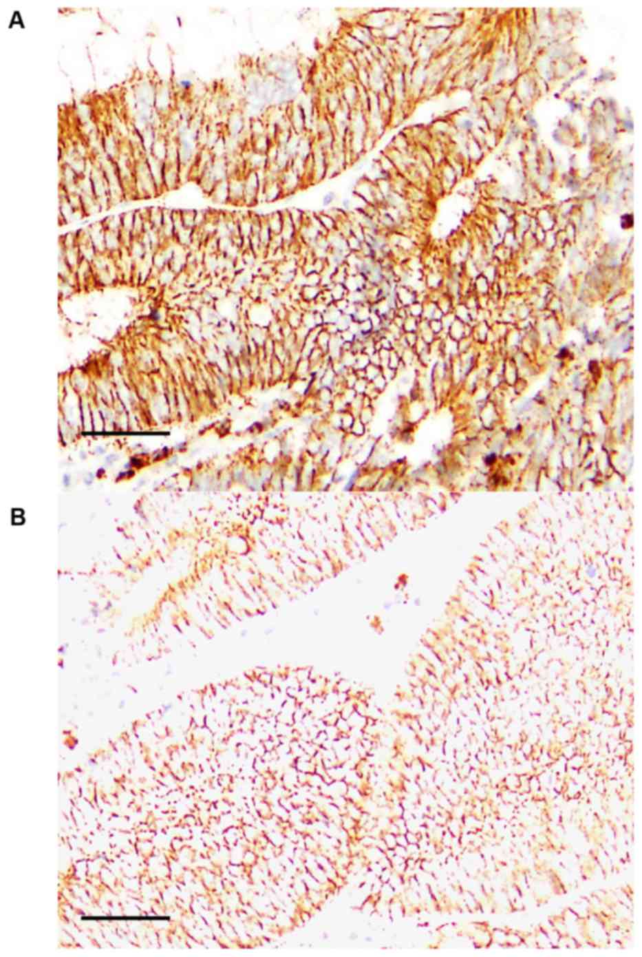 | Figure 1.Expression of GPR40 in colorectal
cancer. GPR40 expression was examined by immunohistochemistry. (A)
A case of tub2, stage IV, pT3, pN2, pM1 (liver). GPR40 expression
was 10. (B) A case of tub1, stage II, pT3, pN0, pM0. GPR40
expression was 0.5. Scale bar, 100 µm. Pathological parameters were
described according to the tumor, node, metastasis pathological
classification system (31). GPR40,
G-protein coupled receptor 40. |
 | Table I.Expression of GPR40 and
clinicopathological parameters in 36 pT3 colorectal cancer
cases. |
Table I.
Expression of GPR40 and
clinicopathological parameters in 36 pT3 colorectal cancer
cases.
|
| GPR40
expressiona |
|
|---|
|
|
|
|
|---|
| Characteristic | Low | High | P-value |
|---|
| n | 18 | 18 |
|
| GPR40 levels | 2.7 | 6.7 | <0.0001 |
| Age (mean ± SD) | 68.6±11.9 | 69.1±10.8 | NS |
| Sex (M:F) | 8:10 | 10:8 | NS |
| Histological
gradeb |
|
|
|
| G1 | 6 | 6 |
|
| G2 | 12 | 12 | NS |
|
pT3 | 18 | 18 |
|
| pN
0 | 15 | 3 |
|
| pN
1–2 | 3 | 15 | 0.0002 |
| pM
0 | 18 | 12 |
|
| pM
1 | 0 | 6 | 0.0191 |
| Stageb |
|
|
|
| II | 15 | 3 |
|
|
III | 3 | 9 |
|
| IV | 0 | 6 | 0.0002 |
| TG (mean ± SD) | 121.9±23.9 | 190.8±65.7 | 0.0002 |
| LDL (mean ±
SD) | 124.2±15.7 | 126.8±25.6 | NS |
Interestingly, a ‘GPR40-high’ status was associated
significantly with lymph node metastasis, liver metastasis, and
stage, but not with histological differentiation.
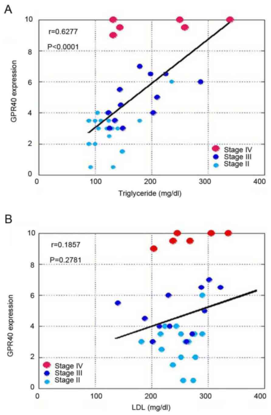
GPR40 expression and blood TG
levels
Next, we compared GPR40 expression with TG or
low-density lipoprotein (LDL) levels (Fig. 2). A ‘GPR40-high’ status was associated
with higher blood TG levels, but not with blood low density
lipoprotein (LDL) levels. Moreover, TG levels were associated with
stage, whereas LDL levels were not associated with stage (Table II).
| Table II.Association between stage and TG or
LDL in patients with colorectal cancer. |
Table II.
Association between stage and TG or
LDL in patients with colorectal cancer.
| Parameter | n | TG | LDL |
|---|
| Stagea |
|
|
|
| II | 18 | 124.8±36.0 | 124.8±15.4 |
|
III | 12 | 177.2±49.2 | 122.8±27.0 |
| IV | 6 | 209.3±86.0 | 133.2±24.0 |
| P-value |
| 0.0018 | NS |
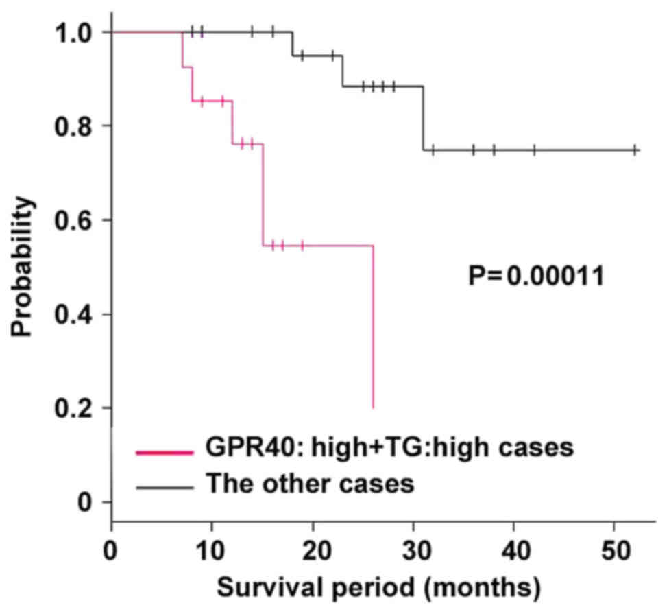
The samples were divided into the two groups by
GPR40 expression status and blood TG levels. The first group
consisted of samples with both high GPR40 status as well as high TG
levels (GPR40:high+TG:high) and the rest of the samples constituted
the second group. The two groups were compared with regard to
stage, TG levels, and survival outcomes (Table III). The GPR40:high+TG:high group
showed more advanced stage and shorter survival periods. Survival
analysis showed that the GPR40:high+TG:high group showed worse
prognosis compared to that of the other group (Fig. 3).
| Table III.Comparison of disease stage and
survival between patient groups classified by TG and GPR40
expression status. |
Table III.
Comparison of disease stage and
survival between patient groups classified by TG and GPR40
expression status.
| Group | GPR40:high
+TG:high | Other cases | P-value |
|---|
| n | 13 | 23 |
|
| Stagea |
|
|
|
| II | 1 | 17 | 0.0010 |
|
III | 8 | 4 |
|
| IV | 4 | 2 |
|
| TG | 216.9±58.4 | 122.1±21.2 | <0.0001 |
| Survival period
(months) | 15.6±5.4 | 25.2±10.4 | 0.0039 |
Table IV shows the
results of univariate analysis of clinicopathological parameters.
The GPR40 expression status and GPR40+TG levels were the two
parameters with the highest statistical significance, followed by
liver metastasis (pM). Table V shows
results of multivariate analysis; the GPR40 expression status was
highly statistically significant, followed by the GPR40+TG
parameter, among the clinicopathological parameters analyzed.
| Table IV.Univariate analyses of
clinicopathologic parameters. |
Table IV.
Univariate analyses of
clinicopathologic parameters.
| Parameter | Chi-squared | P-value |
|---|
| Stagea |
|
|
| II,
III, IV | 11.3000 | 0.000760 |
| pN
(0,1–2) | 14.0000 | 0.000130 |
| pM
(0,1) | 20.2000 | 0.000007 |
| TG (high, low) | 7.8700 | 0.005000 |
| GPR40 | 18.7000 | 0.000015 |
| GPR40+TG | 13.3000 | 0.000270 |
|
(GPR40:high+TG:high, others) |
|
|
| Table V.Multivariate analyses of
clinicopathologic parameters. |
Table V.
Multivariate analyses of
clinicopathologic parameters.
| Parameter | Hazard ratio | Lower 95% | Upper 95% | P-value |
|---|
| GPR40 | 4.787 | 1.453 | 15.77 | 0.01004 |
| GPR40+TG | 2.649 | 1.123 | 6.249 | 0.02613 |
| (GPR40:high+ TG:
high, others) |
|
|
|
|
| Stage (II, III,
IV)a | 0.0627 | 0.004189 | 0.9387 | 0.04488 |
Discussion
The incidence of CRC is on the rise in Japan
(1). Life style, especially a western
style diet, is implicated as an important causative factor in CRC
(2,3).
Recent studies revealed a strong relationship between CRC and the
metabolic syndrome (16,17) and showed that diabetes mellitus is a
risk factor for CRC (18,19). Several factors linking CRC risk with
diabetes have been proposed, such as increased expression of
insulin-like growth factors; oxidative stress (18); advanced glycation end-products (AGE;
which may enhance malignant potential of colorectal cells)
(5,20); and activation of the renin-angiotensin
system (21). Hypertriglyceridemia or
hyper low-density lipoproteinemia is associated with colonic
adenomas, though the risk contribution of these factors is
controversial (22,23).
The membrane-bound receptors of LCFA (which are
components of TGs) are G-protein coupled receptors (GPCRs), of
which GPR40 and GPR120 are well known (11,24).
However, the differential biological activities of fatty acids
cannot be fully explained by the activity of these receptors alone.
To fully explore the spectrum of fatty acid functionality, the
activities of cytoplasmic receptors, bioactive metabolites of fatty
acids, and those of fatty acids integrated into the plasma membrane
need to be studied (7,8,25). In a
previous study on trans fatty acids, we showed that EA
transactivates EGFR from GPR via c-SRC to increase ‘stemness’ and
induce the epidermal-to-mesenchymal transition in cancer cells
(10). Oleic acid also transactivates
EGFR via c-SRC signaling (26). The
diverse activities of fatty acids might thus be a result of
activation of multiple GPCR-dependent and GPCR-independent
pathways.
In the present study, expression of GPR40 was
associated with blood TG levels but not with blood LDL levels. The
GPR40 expression was also associated with nodal metastasis, distant
metastasis (especially to the liver), stage, and poor prognosis.
High GPR40 expression and high TG levels prognosticated worse
survival outcomes in our study. These results suggest that GPR40
expression might be linked to CRC progression. Univariate analysis
revealed that GPR40 expression status alone showed a stronger
association with patient survival than did GPR40:high+TG:high
status. This result suggests that heterogeneity in LCFA content of
TGs might be a confounding factor and may be masking the
significant influence of GPR40 expression in the case of patients
with a GPR40:high+TG:high status.
Our recent reports show different effect of some
long chain fatty acids. Linoleic acid, EA and oleic acid are
examined on their effects to cancer cells. Linoleic acid provides
increased stemness with dormancy (7).
EA enhanced metastability by increased proliferative stemness
(10). In. contrast, oleic acid does
not increase metastability (10).
Univariate and multivariate analyses showed GPR40 is more
significant than GPR40+TG. As described above, different fatty acid
provides different effect. TGs is a mixture of many fatty acids,
which might cancel the effects each other. Then it should be needed
clarifying content of each fatty acid. It does not seem that GPR40
and TG levels in stage IV cases. In stage IV cases, other
pro-metastatic factors, such as growth factors might cause more
relevant contributions to metastasis.
Our previous studies show that linoleic acid (an n-6
LCFA) and EA (a trans fatty acid), enhance colon carcinogenesis or
metastability of CRC (5,6,9,10), whereas docosahexaenoic acid (DHA) and
eicosapentaenoic acid (EPA) (n-3 LCFAs) suppress colon
carcinogenesis and enhance efficacy of chemotherapy regimens
(27). The n-3 LCFAs also activate
the hippo system via GPR40 and GPR120 to inhibit proliferation of
CRC (28). Although blood TG level is
widely utilized as a predictive clinical parameter, the identity of
the fatty acids that constitute the TGs might be an important
factor that determines the relationship between TGs and cancer
risk.
Metabolic syndrome is a collection of risk factors,
such as hypertension, higherglycemia, hyperlipidemia, and visceral
fat based on insulin resistance (29). Diets of patients with the metabolic
syndrome are reported to contain fat with a high n-6/n-3 fatty acid
ratio (30) as well as a high content
of trans fatty acids (31). From
these findings, metabolic syndrome-associated hypertriglyceridemia
might be suggested to be pro-tumoral for CRC. In the cases examined
here, the blood TG level was associated with survival outcomes of
the patients, which suggests that hypertriglyceridemia needs to be
closely examined with reference to CRC progression.
The association between GPR40 with blood TG levels
may imply that TGs induce GPR40 expression in CRC. In contrast,
LCFAs suppress GPR40 expression in pancreatic islet beta cells to
adversely affect diabetes (12). The
mechanism of regulation of GPR40 expression has not been fully
elucidated, though LCFAs have been implicated. The signaling
pathways leading to GPR40 overexpression and the effect of
dyslipidemia on such signaling need to be examined in future
studies.
Acknowledgements
The authors would like to thank Ms. Tomomi Masutani
for assistance with the preparation of this manuscript.
Funding
The present study was supported by MEXT KAKENHI
(grant nos. 16H05164, 17K15648, 17K19923 and 16K19087).
Availability of data and materials
All data generated or analyzed during the present
study are included in this published article.
Authors' contributions
HK designed the study. CN, KS, RFT and YL collected
and analyzed the data. IK, KG, TS, HO and KF analyzed the data. CN
and HO prepared and had final approval of the manuscript.
Ethics approval and consent to
participate
The present study was approved by the Ethics
Committee of the Nara Medical University (approval no. 937).
Consent for publication
Not applicable.
Competing interests
The authors confirm that they have no competing
interests.
Glossary
Abbreviations
Abbreviations:
|
CRC
|
colorectal cancer
|
|
LCFA
|
long-chain fatty acid
|
|
GPCR or GPR
|
G-protein coupled receptor
|
|
TG
|
triglyceride
|
|
LDL
|
low-density lipoprotein
|
|
EA
|
elaidic acid
|
|
RAGE
|
receptor for advanced glycation
end-products
|
|
EMT
|
epithelial-mesenchymal transition
|
|
EGFR
|
epidermal growth factor receptor
|
|
AGE
|
advanced glycation end-products
|
References
|
1
|
Wakao F, Nishimoto H, Kataonoda K, Tsukuma
H and Mikami H: Cancer statistics in Japan, 2013National Cancer
Research Institute. Tokyo: 2013
|
|
2
|
Kimura Y, Kono S, Toyomura K, Nagano J,
Mizoue T, Moore MA, Mibu R, Tanaka M, Kakeji Y, Maehara Y, et al:
Meat, fish and fat intake in relation to subsite-specific risk of
colorectal cancer: The Fukuoka colorectal cancer study. Cancer Sci.
98:590–597. 2007. View Article : Google Scholar : PubMed/NCBI
|
|
3
|
Mizoue T, Tanaka K, Tsuji I, Wakai K,
Nagata C, Otani T, Inoue M and Tsugane S: Research Group for the
Development and Evaluation of Cancer Prevention Strategies in
Japan: Alcohol drinking and colorectal cancer risk: An evaluation
based on a systematic review of epidemiologic evidence among the
Japanese population. Jpn J Clin Oncol. 36:582–597. 2006. View Article : Google Scholar : PubMed/NCBI
|
|
4
|
Bultman SJ: Interplay between diet, gut
microbiota, epigenetic events, and colorectal cancer. Mol Nutr Food
Res. 61:2017. View Article : Google Scholar : PubMed/NCBI
|
|
5
|
Shimomoto T, Luo Y, Ohmori H, Chihara Y,
Fujii K, Sasahira T, Denda A and Kuniyasu H: Advanced glycation end
products (AGE) induce the receptor for AGE in the colonic mucosa of
azoxymethane-injected Fischer 344 rats fed with a high-linoleic
acid and high-glucose diet. J Gastroenterol. 47:1073–1083. 2012.
View Article : Google Scholar : PubMed/NCBI
|
|
6
|
Ohmori H, Luo Y, Fujii K, Sasahira T,
Shimomoto T, Denda A and Kuniyasu H: Dietary linoleic acid and
glucose enhances azoxymethane-induced colon cancer and metastases
via the expression of high-mobility group box 1. Pathobiology.
77:210–217. 2010. View Article : Google Scholar : PubMed/NCBI
|
|
7
|
Ohmori H, Sasahira T, Fujii K, Luo Y,
Shimomoto T and Kuniyasu H: Linoleic acid-induced growth
suppression induces quiescent cancer cell nests in nude mice.
Pathobiology. 75:226–232. 2008. View Article : Google Scholar : PubMed/NCBI
|
|
8
|
Sasaki T, Fujii K, Yoshida K, Shimura H,
Sasahira T, Ohmori H and Kuniyasu H: Peritoneal metastasis
inhibition by linoleic acid with activation of PPARgamma in human
gastrointestinal cancer cells. Virchows Arch. 448:422–427. 2006.
View Article : Google Scholar : PubMed/NCBI
|
|
9
|
Ohmori H, Fujii K, Kadochi Y, Mori S,
Nishiguchi Y, Fujiwara R, Kishi S, Sasaki T and Kuniyasu H: Elaidic
acid, a trans-fatty acid, enhances the metastasis of colorectal
cancer cells. Pathobiology. 84:144–151. 2017. View Article : Google Scholar : PubMed/NCBI
|
|
10
|
Fujii K, Luo Y, Fujiwara-Tani R, Kishi S,
He S, Yang S, Sasaki T, Ohmori H and Kuniyasu H: Pro-metastatic
intracellular signaling of the elaidic trans fatty acid. Int J
Oncol. 50:85–92. 2017. View Article : Google Scholar : PubMed/NCBI
|
|
11
|
Hara T, Kashihara D, Ichimura A, Kimura I,
Tsujimoto G and Hirasawa A: Role of free fatty acid receptors in
the regulation of energy metabolism. Biochim Biophys Acta.
1841:1292–1300. 2014. View Article : Google Scholar : PubMed/NCBI
|
|
12
|
Tomita T, Masuzaki H, Iwakura H, Fujikura
J, Noguchi M, Tanaka T, Ebihara K, Kawamura J, Komoto I, Kawaguchi
Y, et al: Expression of the gene for a membrane-bound fatty acid
receptor in the pancreas and islet cell tumours in humans: Evidence
for GPR40 expression in pancreatic beta cells and implications for
insulin secretion. Diabetologia. 49:962–968. 2006. View Article : Google Scholar : PubMed/NCBI
|
|
13
|
Mizuta K, Zhang Y, Mizuta F, Hoshijima H,
Shiga T, Masaki E and Emala CW Sr: Novel identification of the free
fatty acid receptor FFAR1 that promotes contraction in airway
smooth muscle. Am J Physiol Lung Cell Mol Physiol. 309:L970–L982.
2015. View Article : Google Scholar : PubMed/NCBI
|
|
14
|
Newsholme EA: The glucose/fatty acid cycle
and physical exhaustion. Ciba Found Symp. 82:89–101.
1981.PubMed/NCBI
|
|
15
|
Kuniyasu H, Yasui W, Shinohara H, Yano S,
Ellis LM, Wilson MR, Bucana CD, Rikita T, Tahara E and Fidler IJ:
Induction of angiogenesis by hyperplastic colonic mucosa adjacent
to colon cancer. Am J Pathol. 157:1523–1535. 2000. View Article : Google Scholar : PubMed/NCBI
|
|
16
|
O'Flanagan CH, Bowers LW and Hursting SD:
A weighty problem: Metabolic perturbations and the obesity-cancer
link. Horm Mol Biol Clin Investig. 23:47–57. 2015.PubMed/NCBI
|
|
17
|
Schoenberg MH: Physical activity and
nutrition in primary and tertiary prevention of colorectal cancer.
Visc Med. 32:199–204. 2016. View Article : Google Scholar : PubMed/NCBI
|
|
18
|
Kasuga M, Ueki K, Tajima N, Noda M, Ohashi
K, Noto H, Goto A, Ogawa W, Sakai R, Tsugane S, et al: Report of
the Japan diabetes society/Japanese cancer association joint
committee on diabetes and cancer. Cancer Sci. 104:965–976. 2013.
View Article : Google Scholar : PubMed/NCBI
|
|
19
|
Shi J, Xiong L, Li J, Cao H, Jiang W, Liu
B, Chen X, Liu C, Liu K, Wang G and Cai K: A linear dose-response
relationship between fasting plasma glucose and colorectal cancer
risk: Systematic review and meta-analysis. Sci Rep. 5:175912015.
View Article : Google Scholar : PubMed/NCBI
|
|
20
|
Kuniyasu H, Chihara Y and Kondo H:
Differential effects between amphoterin and advanced glycation end
products on colon cancer cells. Int J Cancer. 104:722–727. 2003.
View Article : Google Scholar : PubMed/NCBI
|
|
21
|
Shimomoto T, Ohmori H, Luo Y, Chihara Y,
Denda A, Sasahira T, Tatsumoto N, Fujii K and Kuniyasu H:
Diabetes-associated angiotensin activation enhances liver
metastasis of colon cancer. Clin Exp Metastasis. 29:915–925. 2012.
View Article : Google Scholar : PubMed/NCBI
|
|
22
|
Yang MH, Rampal S, Sung J, Choi YH, Son
HJ, Lee JH, Kim YH, Chang DK, Rhee PL, Kim JJ, et al: The
association of serum lipids with colorectal adenomas. Am J
Gastroenterol. 108:833–841. 2013. View Article : Google Scholar : PubMed/NCBI
|
|
23
|
Tian Y, Wang K, Li J, Wang J, Wang Z, Fan
Y, Ye Y, Ji G and Li Y: The association between serum lipids and
colorectal neoplasm: A systemic review and meta-analysis. Public
Health Nutr. 18:3355–3370. 2015. View Article : Google Scholar : PubMed/NCBI
|
|
24
|
Yonezawa T, Kurata R, Yoshida K, Murayama
MA, Cui X and Hasegawa A: Free fatty acids-sensing G
protein-coupled receptors in drug targeting and therapeutics. Curr
Med Chem. 20:3855–3871. 2013. View Article : Google Scholar : PubMed/NCBI
|
|
25
|
Kuniyasu H: Linoleic acidEncyclopedia of
Cancer. 2nd edition. Springer-Verlag; Berlin: pp. 1691–1693.
2008
|
|
26
|
Soto-Guzman A, Robledo T, Lopez-Perez M
and Salazar EP: Oleic acid induces ERK1/2 activation and AP-1 DNA
binding activity through a mechanism involving Src kinase and EGFR
transactivation in breast cancer cells. Mol Cell Endocrinol.
294:81–91. 2008. View Article : Google Scholar : PubMed/NCBI
|
|
27
|
Lee JY, Sim TB, Lee JE and Na HK:
Chemopreventive and chemotherapeutic effects of fish oil derived
Omega-3 polyunsaturated fatty acids on colon carcinogenesis. Clin
Nutr Res. 6:147–160. 2017. View Article : Google Scholar : PubMed/NCBI
|
|
28
|
Zhang K, Hu Z, Qi H, Shi Z, Chang Y, Yao
Q, Cui H, Zheng L, Han Y, Han X, et al: G-protein-coupled receptors
mediate ω-3 PUFAs-inhibited colorectal cancer by activating the
Hippo pathway. Oncotarget. 7:58315–58330. 2016.PubMed/NCBI
|
|
29
|
World Health Organization: Definition,
diagnosis and classification of diabetes mellitus and its
complications: Report of a WHO Consultation. World Health
Organization; Geneva: 1999
|
|
30
|
Simopoulos AP: Essential fatty acids in
health and chronic disease. Am J Clin Nutr. 70 3 Suppl:560S–569S.
1999. View Article : Google Scholar : PubMed/NCBI
|
|
31
|
Mozaffarian D, Aro A and Willett WC:
Health effects of trans-fatty acids: Experimental and observational
evidence. Eur J Clin Nutr. 63 Suppl 2:S5–S21. 2009. View Article : Google Scholar : PubMed/NCBI
|
|
32
|
Brierley JD, Gospodarowicz MK and
Wittekind C: TNM Classification of Malignant Tumours. Wiley
Blackwell; Oxford, UK: 2017
|