Introduction
Malignant glioma is the most common type of cancer
in the brain, accounting for 80% of all malignant brain tumors
worldwide (1,2). In previous years, the deregulation of
numerous oncogenes and tumor suppressors including microRNAs (miRs)
has been observed in glioma, and investigating the underlying
molecular mechanism may be beneficial for developing effective
therapeutic strategies for this disease (2–4). miRs are
a class of non-coding RNAs that are 18–25 nucleotides in length,
and have been demonstrated to act as key regulators of gene
expression by directly binding to the complementary regions of
their target mRNA, leading to mRNA degradation or translational
inhibition (5). Through the
inhibition of the expression of their target mRNA, a multitude of
miRs participate in various physiological and pathological
biological processes, including differentiation, development,
angiogenesis and tumorigenesis (5–7). Glioma,
like other types of cancer, possesses a distinct miR expression
signature, and previous studies have identified that a number of
miRs are involved in the regulation of glioma cell proliferation,
survival, cell cycle progression, migration and invasion (8–10).
Furthermore, a number of miRs have been suggested as potential
therapeutic targets in the treatment of glioma (8,10,11).
miR-423 is involved in a number of physiological and
pathological progresses, including skeletal muscle development
(12), childhood obesity (13), heart failure (14), idiopathic pulmonary fibrosis (15) and acute graft-versus-host disease
(16). Furthermore, the deregulation
of miR-423 has been identified to be involved in several types of
human cancer (17,18). For instance, miR-423 was upregulated
in head and neck squamous cell carcinoma tissues compared with
normal tissues (17). It may also
promote the proliferation of hepatocellular carcinoma cells through
regulating the G1/S transition by targeting
cyclin-dependent kinase inhibitor 1 (p21Cip1/Waf1)
(18). Additionally, miR-423 promotes
cell proliferation in breast cancer cell lines through its
miR-423-3p strand as opposed to its miR-423-5p strand (19). Previously, miR-423-5p was demonstrated
to be significantly upregulated in glioma, and promoted the
malignant phenotypes of glioma cells as well as their temozolomide
resistance (20), suggesting that
miR-423-5p serves an oncogenic function in glioma. However, the
regulatory function of miR-423-3p in glioma remains unclear.
In the present study, the molecular mechanisms
underlying the effect of miR-423-3p on glioma growth were
investigated.
Materials and methods
Tissue collection
The present study was approved by the Ethics
Committee of Xiangya Hospital, Central South University (Changsha,
China). A total of 58 cases of glioma specimens and 10 cases of
normal brain tissues were obtained from Xiangya Hospital, Central
South University (Changsha, China) between January 2010 and March
2012. Written informed consent was obtained from all patients prior
to the study. Tissues were snap-frozen in liquid nitrogen following
surgical resection, and stored in liquid nitrogen until use. The
clinicopathological information of the patients with glioma
included in the present study is summarized in Table I.
 | Table I.Associations between miR-423-3p
expression levels and the clinicopathological characteristics of
patients with glioma. |
Table I.
Associations between miR-423-3p
expression levels and the clinicopathological characteristics of
patients with glioma.
| Variable | Cases (n=58) | Low miR-423-3p level
(n=31) | High miR-423-3p level
(n=27) | P-value |
|---|
| Age, years |
|
|
| 0.596 |
|
<55 | 25 | 12 | 13 |
|
| ≥55 | 33 | 19 | 14 |
|
| Sex |
|
|
| 0.270 |
| Male | 39 | 23 | 16 |
|
|
Female | 19 | 8 | 11 |
|
| WHO grade |
|
|
| 0.026 |
|
I–II | 20 | 15 | 5 |
|
|
III–IV | 38 | 16 | 22 |
|
| KPS |
|
|
| 0.013 |
|
>90 | 21 | 16 | 5 |
|
|
≤90 | 37 | 15 | 22 |
|
Reverse transcription-quantitative
polymerase chain reaction (RT-qPCR)
Total RNA was extracted from the tissues and cells
using TRIzol® reagent (Thermo Fisher Scientific, Inc.,
Waltham, MA, USA), according to the manufacturer's protocol. The
MirVana™ RT-PCR microRNA detection kit (Thermo Fisher Scientific,
Inc.) was used to examine miR expression, according to the
manufacturer's protocol. U6 was used as an internal reference. The
standard SYBR Green RT-PCR Kit (Takara Bio, Inc., Otsu, Japan) was
used to examine mRNA expression, according to the manufacturer's
protocol. GAPDH was used as an internal reference. The primers used
were as follows: miR-423 forward,
5′-ATGGTTCGTGGGTGAGGGGCAGAGAGCGAGAGCAGGGTCCGAGGTATTCG-3′ and
reverse, 5′-GTGCAGGGTCCGAGGT-3′; U6 forward,
5′-CTCGCTTCGGCAGCACA-3′ and reverse, 5′-AACGCTTCACGAATTTGCGT-3′;
PANX2 forward, 5′-CCAAGAACTTCGCAGAGGAAC-3′ and reverse,
5′-GGGCAGGAACTTGTGCTCA-3′; GAPDH forward,
5′-GGAGCGAGATCCCTCCAAAAT-3′; and reverse,
5′-GGCTGTTGTCATACTTCTCATGG-3′. The reaction mixture included cDNA
solution (1 µl), PCR master mix (10 µl), primers (2 µl) and water
(7 µl). The reaction conditions were 9°C for 3 min, followed by 40
cycles at 95°C for 15 sec and 60°C for 30 sec. The relative
expression level was quantified using the 2−ΔΔCq method
(21).
Cell culture
Human glioma U251 and U87MG Uppsala cell lines were
obtained from the Cell Bank of Type Culture Collection of the
Chinese Academy of Sciences (Shanghai, China). Cells were cultured
in Dulbecco's modified Eagle's medium (Thermo Fisher Scientific,
Inc.) with 10% fetal bovine serum (Thermo Fisher Scientific, Inc.)
and were maintained at 37°C in a humidified incubator (Thermo
Fisher Scientific, Inc.) containing 5% CO2.
Cell transfection
Lipofectamine 2000 transfection reagent (Thermo
Fisher Scientific, Inc.) was used to perform transfection,
according to the manufacturer's protocol. Briefly, U251 and U87MG
Uppsala cells were transfected with a negative control (NC)
inhibitor, miR-423-3p inhibitor, non-specific small interfering RNA
(siRNA) or PANX2-specific siRNA, respectively were purchased from
Guangzhou FulenGen Co. Ltd. (Guangzhou, China). Following
transfection at 37°C for 48 h, the expression assay was
performed.
Western blotting
U251 and U87MG Uppsala cells were lysed in RIPA
Lysis Buffer (Beyotime Institute of Biotechnology, Haimen, China).
The protein concentration was quantified using a bicinchinonic acid
protein assay kit (Thermo Fisher Scientific, Inc.), according to
the manufacturer's protocol. Protein (50 µg) was separated by
SDS-PAGE (12% gel), transferred onto a polylvinylidene fluoride
membrane (Thermo Fisher Scientific, Inc.), and then blocked using
5% non-fat dried milk (Yili Group, Beijing, China) in Tris-buffered
saline with Tween-20 (TBST; Beyotime Institute of Biotechnology) at
room temperature for 3 h. The membrane was incubated with rabbit
polyclonal anti-PANX2 primary antibody (1:100; cat no. ab55917;
Abcam, Cambridge, MA, USA) or rabbit polyclonal anti-GAPDH primary
antibody (1:100; cat no. ab9485; Abcam) at room temperature for 3
h, and then washed three times using TBST. Subsequently, the
membrane was incubated with goat monoclonal anti-rabbit secondary
antibody (1:5,000; cat no. ab190492; Abcam) for 1 h at room
temperature, and washed three times using TBST. The immune
complexes were detected using an enhanced chemiluminescence western
blotting kit (Thermo Fisher Scientific, Inc.), according to the
manufacturer's protocol. Image J software (version 1.0, National
Institutes of Health, Bethesda, MD, USA) was used to analyze the
relative protein expression, represented as the density ratio
relative to GAPDH.
Cell proliferation analysis
U251 and U87MG Uppsala cells (2×10³ cells
per well) were seeded in 96-well plates, and 100 µl fresh
serum-free DMEM with 0.5 g/l MTT solution (Sigma-Aldrich; Merck
KGaA, Darmstadt, Germany) was added. Following incubation at 37°C
for 0, 24, 48 and 72 h, the medium containing MTT solution was
removed, and 50 µl dimethylsulfoxide (Sigma-Aldrich; Merck KGaA)
was added. Following incubation at 37°C for 10 min, the absorbance
at 570 nm of each sample was determined using a plate reader
(Bio-Rad Laboratories, Inc., Hercules, CA, USA).
Cell apoptosis analysis
A flow cytometer was used to determine the cell
apoptosis with an Annexin V-Fluorescein Isothiocyanate (FITC)
Apoptosis Detection kit (Sigma-Aldrich; Merck KGaA). Cells were
harvested and washed with ice-cold PBS twice, and 106
cells were resuspended in 200 µl binding buffer with 10 µl
annexin-V-FITC and 5 µl propidium iodide-phycoerythrin, prior to
incubation in the dark at 4°C for 30 min. Subsequently, 300 µl
binding buffer was added, followed by analysis using flow cytometry
(BD Accuri C6 software 1.0, C6; BD Biosciences, Franklin Lakes, NJ,
USA).
Bioinformatics analysis and luciferase
reporter assay
TargetScan Human 5.1 software (22) (www.targetscan.org) was used to determine the putative
target of miR-423-3p. The wild-type (WT) PANX2 3′-untranslated
region (UTR) was constructed by PCR, which was performed by
Yearthbio (Changsha, China), and inserted into the pMIR-REPORT
miRNA Expression Reporter vector (Thermo Fisher Scientific, Inc.),
according to the manufacturer's protocol. The mutant type (MT) of
PANX2 3′-UTR was constructed using the Easy Mutagenesis System kit
(Promega Corporation, Madison, WI, USA), according to the
manufacturer's protocol, and then inserted into the pMIR-REPORT
miRNA Expression Reporter vector (Thermo Fisher Scientific, Inc.).
U251 and U87MG Uppsala cells were co-transfected with WT
PANX2-3′UTR plasmid or MT PANX2-3′UTR plasmid, and miR-NC or
miR-423-3p mimics, using Lipofectamine 2000 transfection reagent
(Thermo Fisher Scientific, Inc.). Following transfection at 37°C
for 48 h, the activity of Renilla luciferase and firefly
luciferase were determined using a dual-luciferase reporter assay
system (Promega Corporation), 48 h after transfection. The activity
of Renilla luciferase was normalized to that of the firefly
luciferase.
Statistical analysis
Data are expressed as the mean ± standard deviation.
The differences between two groups were analyzed using two-tailed
Student's t-test. Statistical analysis was performed using SPSS
software (version 17.0; SPSS, Inc., Chicago, IL, USA). Pearson's
correlation analysis was performed to examine the correlation
between the miR-423-3p and PANX2 expression in glioma tissues.
Kaplan Meier analysis with log rank tests were used for the
survival analysis. P<0.05 was considered to indicate a
statistically significant difference.
Results
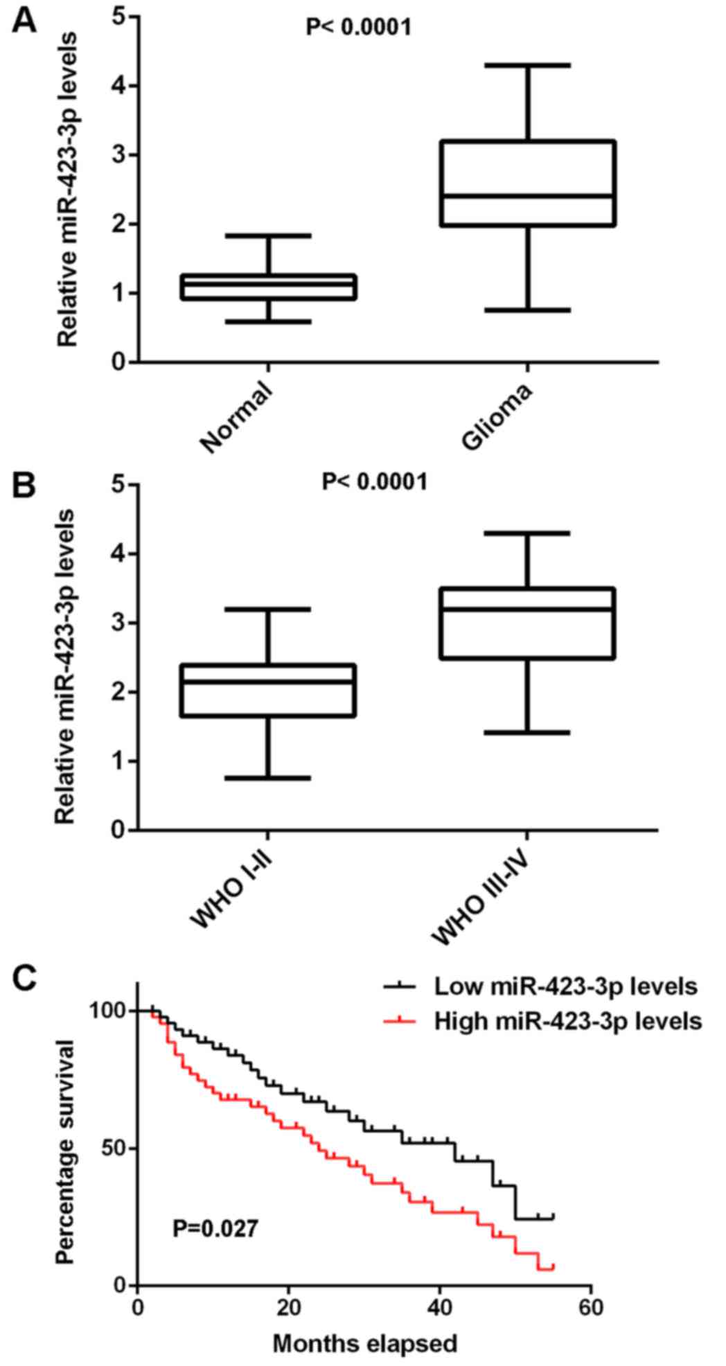
Upregulation of miR-423-3p is
associated with glioma progression
In the present study, qPCR was used to determine the
expression of miR-423-3p in glioma tissues. Normal brain tissues
were used as controls. As indicated in Fig. 1A, miR-423-3p expression levels were
significantly increased in glioma tissues compared with normal
brain tissues. Furthermore, the expression of miR-423-3p was
increased in World Health Organization (WHO) III–IV grade glioma
compared with WHO I–II grade glioma (Fig.
1B). These glioma tissues were further divided into two groups,
a high miR-423-3p group and a low miR-423-3p group, on the basis of
mean expression value. It was observed that high expression of
miR-423-3p was significantly associated with an advanced grade of
glioma as well as a low Karnofsky performance score (KPS), but not
with age and sex (Table I).
Furthermore, as presented in Fig. 1C,
the patients with glioma with high miR-423-3p expression levels had
a shorter survival time, compared with those with low miR-423-3p
expression levels. Therefore, miR-423-3p upregulation may
contribute to the malignant progression of glioma as well as poor
prognosis in patients with glioma.
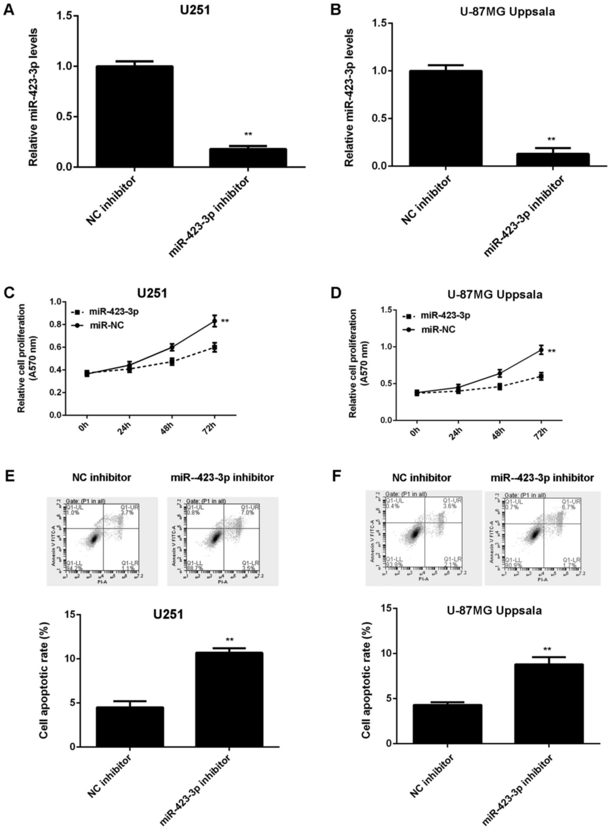
Knockdown of miR-423-3p decreases U251
and U87MG Uppsala cell proliferation and induces cell
apoptosis
As miR-423-3p was upregulated in glioma, glioma U251
and U87MG Uppsala cells were transfected with an miR-423-3p
inhibitor in order to knock down miR-423-3p expression.
Transfection with an NC inhibitor was used as the control group.
Following transfection with miR-423-3p inhibitor, miR-423-3p
expression levels were significantly decreased compared with the NC
inhibitor group (Fig. 2A and B). An
MTT assay further indicated that the inhibition of miR-423-3p led
to the decreased proliferation of U251 and U87MG Uppsala cells
(Fig. 2C and D). Cell apoptosis was
further examined and it was revealed that the knockdown of
miR-423-3p resulted in a significant increase in U251 and U87MG
Uppsala cell apoptosis (Fig. 2E and
F). Therefore, the results of the present study demonstrated
that the knockdown of miR-423-3p decreases U251 and U87MG Uppsala
cell proliferation and induces cell apoptosis.
PANX2 is a novel target of miR-423-3p
in U251 and U87MG Uppsala cells
Bioinformatics analysis indicated that PANX2 was a
putative target gene of miR-423-3p (Fig.
3A). To the best of our knowledge, this targeting association
has never previously been reported. In the present study,
luciferase reporter plasmids were constructed containing WT or MT
PANX2 3′-UTR (Fig. 3B and C). The
luciferase reporter gene assay was then performed in U251 and U87MG
Uppsala cells. The results of the present study indicated that
luciferase activity was significantly decreased in U251 and U87MG
Uppsala cells co-transfected with the WT PANX2 3′-UTR plasmid and
miR-423-3p mimic compared with the control group, which was
eliminated during transfection with the MT PANX2 3′-UTR plasmid
(Fig. 3D and E). Subsequently, it was
revealed that the inhibition of miR-423-3p significantly increased
the expression of PANX2 protein in U251 and U87MG Uppsala cells
(Fig. 3F and G). Therefore, PANX2 is
a potential novel target gene of miR-423-3p in U251 and U87MG
Uppsala cells.
 | Figure 3.PANX2 was identified as a target of
miR-423-3p, and luciferase activity was compared between cells with
miR-423-3p, miR-NC and control with PANX2 protein levels compared
between cells treated with a miR-423-3p inhibitor and with a NC
inhibitor. (A) TargetScan software demonstrated that PANX2 was a
putative target of miR-423-3p. (B) WT or MT PANX2 3′UTR was (C)
cloned into a luciferase reporter vector. The luciferase activity
was significantly decreased in (D) U251 and (E) U87MG Uppsala cells
co-transfected with WT-PANX2-3′UTR plasmid and miR-423-3p mimics
compared with the control group, which was eliminated during
transfection with the MT-PANX2-3′UTR plasmid. Western blotting was
used to examine the protein levels of PANX2 in (F) U251 and (G)
U87MG Uppsala cells transfected with miR-423-3p inhibitor or NC
inhibitor. **P<0.01 vs. control, ##P<0.01 vs. NC
inhibitor. miR, microRNA; PANX2, pannexin 2; NC, negative control;
WT, wild-type; MT, mutant type; UTR, untranslated region. |
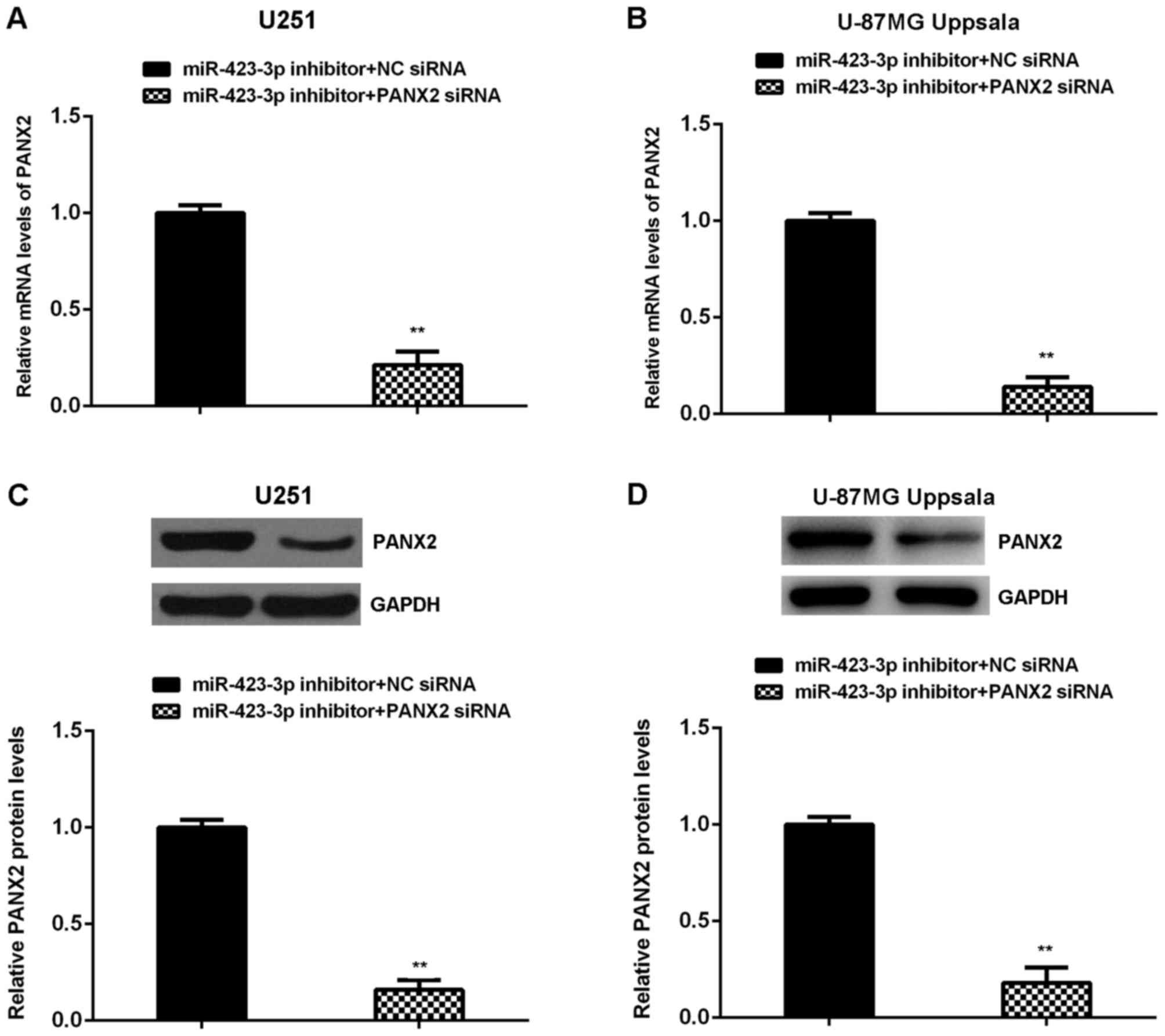
Knockdown of PANX2 attenuates the
effects of miR-423-3p inhibition on U251 and U87MG Uppsala
cells
On the basis of the aforementioned results, it was
hypothesized that PANX2 may be involved in miR-423-3p-mediated
glioma growth. To investigate this hypothesis, U251 and U87MG
Uppsala cells were co-transfected with miR-423-3p inhibitor and
PANX2 siRNA, or miR-423-3p inhibitor and NC siRNA. Following
transfection, the mRNA and protein levels of PANX2 were
significantly decreased in the miR-423-3p inhibitor + PANX2 siRNA
group, compared with the miR-423-3p inhibitor + NC siRNA group
(Fig. 4A-D). An MTT assay further
demonstrated that the proliferation of U251 and U87MG Uppsala cells
was significantly increased in the miR-423-3p inhibitor + PANX2
siRNA group, compared with the miR-423-3p inhibitor + NC siRNA
group (Fig. 5A and B), indicating
that knockdown of PANX2 attenuates the suppressive effects of
miR-423-3p inhibition on glioma cell proliferation. Cell apoptosis
was then assessed. It was revealed that the apoptosis of U251 and
U87MG Uppsala cells was significantly decreased in the miR-423-3p
inhibitor + PANX2 siRNA group, compared with the miR-423-3p
inhibitor + NC siRNA group (Fig. 5C and
D), indicating that the knockdown of PANX2 attenuates the
effect of miR-423-3p inhibition on glioma cell apoptosis. As a
result, it may be suggested that the knockdown of miR-423-3p at
least partially inhibits proliferation and induces the apoptosis of
glioma cells, via the direct targeting of PANX2.
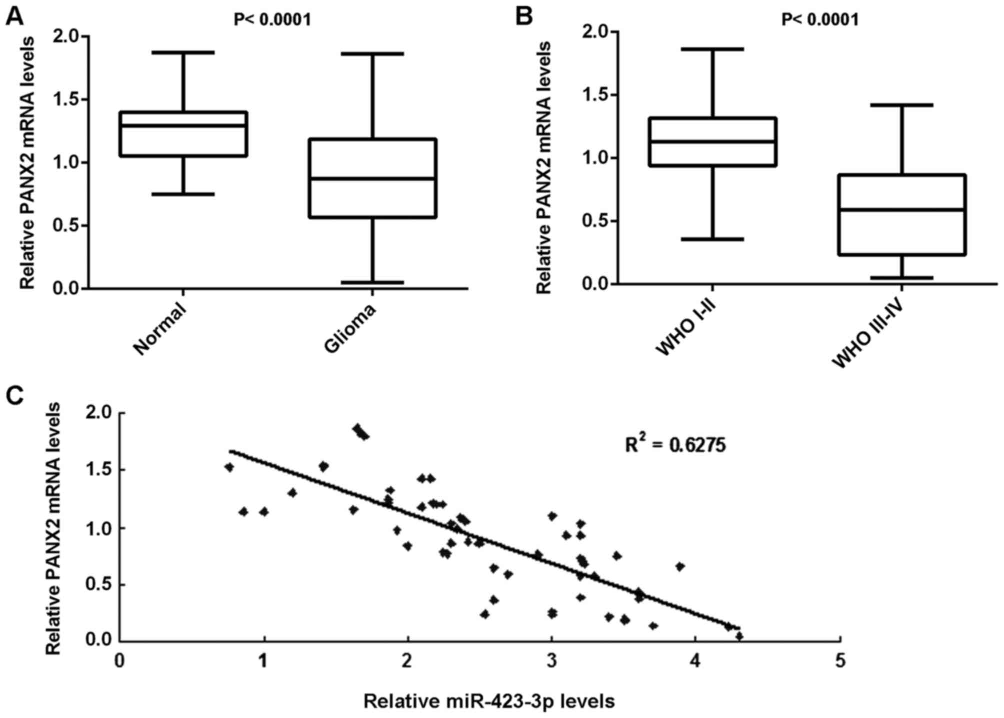
PANX2, which is downregulated in
glioma, is inversely correlated with the miR-423-3p expression
Finally, RT-qPCR was performed to examine the mRNA
expression levels of PANX2 in glioma. The expression of PANX2 mRNA
was demonstrated to be significantly decreased in glioma tissues
compared with normal brain tissues (Fig.
6A). Furthermore, PANX2 mRNA expression levels in WHO III–IV
grade glioma were decreased compared with in WHO I–II grade glioma
(Fig. 6B). Notably, the PANX2 mRNA
expression levels were inversely correlated with the miR-423-3p
expression levels in glioma tissues (Fig.
6C). The decreased expression of PANX2 may be due to the
upregulation of miR-423-3p in glioma.
Discussion
The exact function of miR-423-3p in glioma growth as
well as the underlying molecular mechanism remain unclear. In the
present study, it was demonstrated that miR-423-3p was
significantly upregulated in glioma tissues compared with normal
brain tissues, and the increased expression of miR-423-3p was
significantly associated with an advanced grade of glioma in
addition to a poorer prognosis in patients with glioma. Further
investigation suggested that miR-423-3p serves a promoting function
in glioma growth via the direct targeting of PANX2. Furthermore,
PANX2 was significantly downregulated in glioma tissues compared
with normal brain tissues, and PANX2 expression levels were
inversely correlated with miR-423-3p expression levels in glioma
tissues.
miR-423-3p has been demonstrated to serve a
promoting function in several types of human cancer (23,24). For
example, Guan et al (23)
identified that miR-423-3p was upregulated in laryngeal carcinoma
cells, and that the inhibition of miR-423-3p resulted in a
significant decrease in cell proliferation, clonogenicity, cell
migration and invasion. Additionally, miR-423-3p was upregulated in
colorectal cancer (CRC), and promoted CRC cell proliferation via
enhancing the G1/S transition by targeting
p21Cip1/Waf1 (24).
Previously, miR-423-5p was demonstrated to be significantly
upregulated in glioma, and the overexpression of miR-423-5p
promoted glioma cell proliferation, angiogenesis and invasion by
increasing the activities of protein kinase B and mitogen-activated
protein kinase signaling pathways and suppressing the expression of
inhibitor of growth family member 4 (20). In addition, miR-423-5p upregulation
enhanced glioblastoma neurosphere formation and rendered glioma
cells resistant to temozolomide (20). However, the exact function of
miR-423-3p in glioma has not been uncovered. In the present study,
it was identified that miR-423-3p was downregulated in glioma
tissues compared with normal brain tissues, and its downregulation
was associated with an advanced pathological grade and lower KPS in
glioma. Furthermore, the patients with glioma with high miR-423-3p
levels had a shorter survival time compared with patients with low
miR-423-3p levels. As a result, it may be suggested that the
upregulation of miR-423-3p contributes to glioma progression and a
poorer prognosis in patients with glioma.
PANX2 was also identified as a novel target gene of
miR-423-3p using bioinformatics analysis and a luciferase reporter
assay, and knockdown of miR-423-3p increased the protein expression
levels of PANX2 in U251 and U87MG Uppsala cells. PANX2 encodes the
protein pannexin 2, belonging to the innexin family, the members of
which are the structural components of gap junctions (25). Previous studies have demonstrated that
PANX2 is abundantly expressed in the central neuronal system, and
participates in neuronal development and adult neurogenesis
(26,27). Furthermore, the upregulation and
downregulation of PANX2 are associated with the development and
progression of certain diseases, including neoplasms, multiple
sclerosis, migraines and hypertension (28). Previously, gene array analysis
indicated that there was a significant decrease in PANX2 expression
in glioma, and that the decreased expression of PANX2 was
associated with a poorer prognosis in patients with glioma
(29). Furthermore, the expression of
PANX2 was also lower in human glioma cell lines compared with
normal brain tissues and astrocytes (29). Consistent with this previous study,
the results of the present study identified that PANX2 was
downregulated in glioma tissues compared with normal brain tissues,
and its expression levels in WHO III–IV grade glioma were decreased
compared with those in WHO I–II grade glioma. Furthermore, Lai
et al (29) identified that
the restoration of PANX2 expression significantly decreased
monolayer saturation density and anchorage-independent growth of
rat C6 glioma cells in vitro, as well as tumor growth in
vivo. However, the regulatory mechanism of PANX2 in glioma
remains unknown. In the present study, it was demonstrated that the
knockdown of PANX2 attenuated the effects of miR-423-3p inhibition
on the proliferation and apoptosis of U251 and U87MG Uppsala cells,
suggesting that miR-423-3p promotes glioma cell proliferation by
directly targeting PANX2. Furthermore, the expression levels of
PANX2 were inversely correlated with the miR-423-3p expression
levels in glioma tissues, suggesting that the decreased expression
of PANX2 may be caused by the upregulation of miR-423-3p.
In summary, the results of the present study
demonstrated that miR-423-3p serves an oncogenic function in glioma
cell proliferation by directly targeting PANX2, suggesting that
miR-423-3p may be a potential therapeutic target for glioma.
Acknowledgements
Not applicable.
Funding
The present study was supported by the Natural
Science Foundation of China (grant no. 81201740) and the Project of
Science and Technology Department of the Hunan Province (grant no.
2012FJ6075).
Availability of data and materials
All data generated or analyzed during this study are
included in this published article.
Authors' contributions
JXi and JH collected clinical tissues. JXu, HH and
RP performed the in vitro experiments. JXi wrote the
manuscript. JXu designed the study and revised the manuscript.
Ethics approval and consent to
participate
The present study was approved by the Ethics
Committee of Xiangya Hospital, Central South University (Changsha,
China). Written informed consent was obtained from all patients
prior to the study.
Consent for publication
Written informed consents for the publication of
this data were obtained from all patients in the present study.
Competing interests
The authors declare that they have no competing
interests.
References
|
1
|
Goodenberger ML and Jenkins RB: Genetics
of adult glioma. Cancer Genet. 205:613–621. 2012. View Article : Google Scholar : PubMed/NCBI
|
|
2
|
Yan Y and Jiang Y: RACK1 affects glioma
cell growth and differentiation through the CNTN2-mediated
RTK/Ras/MAPK pathway. Int J Mol Med. 37:251–257. 2016. View Article : Google Scholar : PubMed/NCBI
|
|
3
|
Marumoto T and Saya H: Molecular biology
of glioma. Adv Exp Med Biol. 746:2–11. 2012. View Article : Google Scholar : PubMed/NCBI
|
|
4
|
Zhang R, Wang R, Chen Q and Chang H:
Inhibition of autophagy using 3-methyladenine increases
cisplatininduced apoptosis by increasing endoplasmic reticulum
stress in U251 human glioma cells. Mol Med Rep. 12:1727–1732. 2015.
View Article : Google Scholar : PubMed/NCBI
|
|
5
|
Ambros V: The functions of animal
microRNAs. Nature. 431:350–355. 2004. View Article : Google Scholar : PubMed/NCBI
|
|
6
|
Bartel DP: MicroRNAs: Genomics,
biogenesis, mechanism, and function. Cell. 116:281–297. 2004.
View Article : Google Scholar : PubMed/NCBI
|
|
7
|
Zheng K, Liu W, Liu Y, Jiang C and Qian Q:
Microrna-133a suppresses colorectal cancer cell invasion by
targeting fascin1. Oncol Lett. 9:869–874. 2015. View Article : Google Scholar : PubMed/NCBI
|
|
8
|
Liang ML, Hsieh TH, Ng KH, Tsai YN, Tsai
CF, Chao ME, Liu DJ, Chu SS, Chen W, Liu YR, et al: Downregulation
of miR-137 and miR-6500-3p promotes cell proliferation in pediatric
high-grade gliomas. Oncotarget. 7:19723–19737. 2016. View Article : Google Scholar : PubMed/NCBI
|
|
9
|
Xu J, Xu W and Zhu J: Propofol suppresses
proliferation and invasion of glioma cells by upregulating
microRNA-218 expression. Mol Med Rep. 12:4815–4820. 2015.
View Article : Google Scholar : PubMed/NCBI
|
|
10
|
Liu C, Liang S, Xiao S, Lin Q, Chen X, Wu
Y and Fu J: MicroRNA-27b inhibits Spry2 expression and promotes
cell invasion in glioma U251 cells. Oncol Lett. 9:1393–1397. 2015.
View Article : Google Scholar : PubMed/NCBI
|
|
11
|
Wang H, Tao T, Yan W, Feng Y, Wang Y, Cai
J, You Y, Jiang T and Jiang C: Upregulation of miR-181s reverses
mesenchymal transition by targeting KPNA4 in glioblastoma. Sci Rep.
5:130722015. View Article : Google Scholar : PubMed/NCBI
|
|
12
|
McDaneld TG, Smith TP, Doumit ME, Miles
JR, Coutinho LL, Sonstegard TS, Matukumalli LK, Nonneman DJ and
Wiedmann RT: MicroRNA transcriptome profiles during swine skeletal
muscle development. BMC Genomics. 10:772009. View Article : Google Scholar : PubMed/NCBI
|
|
13
|
Prats-Puig A, Ortega FJ, Mercader JM,
Moreno-Navarrete JM, Moreno M, Bonet N, Ricart W, López-Bermejo A
and Fernández-Real JM: Changes in circulating microRNAs are
associated with childhood obesity. J Clin Endocrinol Metab.
98:E1655–E1660. 2013. View Article : Google Scholar : PubMed/NCBI
|
|
14
|
Kumarswamy R, Anker SD and Thum T:
MicroRNAs as circulating biomarkers for heart failure: Questions
about MiR-423-5p. Circ Res. 106:e82010. View Article : Google Scholar : PubMed/NCBI
|
|
15
|
Oak SR, Murray L, Herath A, Sleeman M,
Anderson I, Joshi AD, Coelho AL, Flaherty KR, Toews GB, Knight D,
et al: A micro RNA processing defect in rapidly progressing
idiopathic pulmonary fibrosis. PLoS One. 6:e212532011. View Article : Google Scholar : PubMed/NCBI
|
|
16
|
Xiao B, Wang Y, Li W, Baker M, Guo J,
Corbet K, Tsalik EL, Li QJ, Palmer SM, Woods CW, et al: Plasma
microRNA signature as a noninvasive biomarker for acute
graft-versus-host disease. Blood. 122:3365–3375. 2013. View Article : Google Scholar : PubMed/NCBI
|
|
17
|
Hui AB, Lenarduzzi M, Krushel T, Waldron
L, Pintilie M, Shi W, Perez-Ordonez B, Jurisica I, O'Sullivan B,
Waldron J, et al: Comprehensive MicroRNA profiling for head and
neck squamous cell carcinomas. Clin Cancer Res. 16:1129–1139. 2010.
View Article : Google Scholar : PubMed/NCBI
|
|
18
|
Lin J, Huang S, Wu S, Ding J, Zhao Y,
Liang L, Tian Q, Zha R, Zhan R and He X: MicroRNA-423 promotes cell
growth and regulates G(1)/S transition by targeting p21Cip1/Waf1 in
hepatocellular carcinoma. Carcinogenesis. 32:1641–1647. 2011.
View Article : Google Scholar : PubMed/NCBI
|
|
19
|
Zhao H, Gao A, Zhang Z, Tian R, Luo A, Li
M, Zhao D, Fu L, Fu L, Dong JT and Zhu Z: Genetic analysis and
preliminary function study of miR-423 in breast cancer. Tumour
Biol. 36:4763–4771. 2015. View Article : Google Scholar : PubMed/NCBI
|
|
20
|
Li S, Zeng A, Hu Q, Yan W, Liu Y and You
Y: miR-423-5p contributes to a malignant phenotype and temozolomide
chemoresistance in glioblastomas. Neuro Oncol. 19:55–65. 2017.
View Article : Google Scholar : PubMed/NCBI
|
|
21
|
Arocho A, Chen B, Ladanyi M and Pan Q:
Validation of the 2-DeltaDeltaCt calculation as an alternate method
of data analysis for quantitative PCR of BCR-ABL P210 transcripts.
Diagn Mol Pathol. 15:56–61. 2006. View Article : Google Scholar : PubMed/NCBI
|
|
22
|
Lewis BP, Burge CB and Bartel DP:
Conserved seed pairing, often flanked by adenosines, indicates that
thousands of human genes are microRNA targets. Cell. 120:15–20.
2005. View Article : Google Scholar : PubMed/NCBI
|
|
23
|
Guan G, Zhang D, Zheng Y, Wen L, Yu D, Lu
Y and Zhao Y: microRNA-423-3p promotes tumor progression via
modulation of AdipoR2 in laryngeal carcinoma. Int J Clin Exp
Pathol. 7:5683–5691. 2014.PubMed/NCBI
|
|
24
|
Li HT, Zhang H, Chen Y, Liu XF and Qian J:
MiR-423-3p enhances cell growth through inhibition of p21Cip1/Waf1
in colorectal cancer. Cell Physiol Biochem. 37:1044–1054. 2015.
View Article : Google Scholar : PubMed/NCBI
|
|
25
|
Tang W, Ahmad S, Shestopalov VI and Lin X:
Pannexins are new molecular candidates for assembling gap junctions
in the cochlea. Neuroreport. 19:1253–1257. 2008. View Article : Google Scholar : PubMed/NCBI
|
|
26
|
Swayne LA and Bennett SA: Connexins and
pannexins in neuronal development and adult neurogenesis. BMC Cell
Biol. 17 Suppl 1:S102016. View Article : Google Scholar
|
|
27
|
Swayne LA, Sorbara CD and Bennett SA:
Pannexin 2 is expressed by postnatal hippocampal neural progenitors
and modulates neuronal commitment. J Biol Chem. 285:24977–24986.
2010. View Article : Google Scholar : PubMed/NCBI
|
|
28
|
Penuela S, Harland L, Simek J and Laird
DW: Pannexin channels and their links to human disease. Biochem J.
461:371–381. 2014. View Article : Google Scholar : PubMed/NCBI
|
|
29
|
Lai CP, Bechberger JF and Naus CC:
Pannexin2 as a novel growth regulator in C6 glioma cells. Oncogene.
28:4402–4408. 2009. View Article : Google Scholar : PubMed/NCBI
|