Introduction
Breast cancer is one of the most common female
malignancies in clinical practice, which ranks number one in terms
of its high incidence (1). The
clinical symptoms of breast cancer mainly include non-gestational
nipple discharge, nipple and/or areola abnormalities, breast lumps,
enlarged axillary lymph nodes, and skin changes (2). At present, ultrasound is a major tool
for diagnosing breast cancer, and has been widely used in clinic
(3). Clinical treatments of breast
cancer include surgery, chemotherapy, radiation, targeted and
traditional Chinese medicine adjuvant therapy. Surgery is the most
commonly used treatment, and a radical mastectomy is the preferred
type of surgery (4). The C-erbB-2
protein is a member of the epidermal growth factor receptor family,
which is closely associated with onset and progression of breast
cancer (5). Vascular endothelial
growth factor (VEGF) is a growth factor of vascular endothelial
cells that promotes the growth of new blood vessels, and it plays a
major role in invasion and metastasis of breast cancer (6). The nm23 gene is a tumor suppressor gene,
and is associated with inhibition of metastasis in many tumors,
such as hepatocellular carcinoma, melanoma, breast cancer and
gastric cancer. Patients with breast cancer who were diagnosed
early and underwent effective treatments usually have a good
prognosis, but those with advanced breast cancer still have a poor
prognosis. So, early screening of breast cancer is of great
clinical significance (7). In this
study, ultrasonography was performed for patients with breast
cancer, and the expression levels of C-erbB-2, VEGF and nm23 were
measured, in order to explore the relevance of ultrasonic features
of breast cancer to expression levels of C-erbB-2, VEGF and nm23.
The results may serve as a reference for diagnosis and treatment of
breast cancer.
Materials and methods
Subjects
A total of 76 patients with breast cancer were
enrolled in this study who were admitted to The Affiliated Hospital
of Qingdao University (Qingdao, China) from January, 2016 to
August, 2017. Inclusion criteria included patients meeting the
diagnostic criteria (female patients, Han ethnicity) for breast
cancer (8), undergoing ultrasound
examination, without mental disorders, and were not previously
treated with surgery, radiotherapy and chemotherapy. Exclusion
criteria excluded patients during pregnancy, with moderate to
severe anemia, during menstrual cycle or with unexplained vaginal
bleeding, and previously treated with drugs and surgery.
This study was approved by the Ethics Committee of
the Affiliated Hospital of Qingdao University and informed consents
were signed by the patients or guardians.
Ultrasonography
All patients underwent ultrasound examination of the
whole breasts and the axilla area with a Philips iU22 Color Doppler
Ultrasound System (The Philips Foundation, Amsterdam, The
Netherlands) at a probe frequency of 7.5–12.0 MHz. Patients lay
down in a supine position with the hands up. The probe scanned an
area with the nipple, the lower edge of the breast, and the axilla
on the boundary. The cancer lesion was checked for its morphology,
size, margins, echogenicity, presence of calcification, and blood
flow signal. Axillary lymph nodes were also checked for accompanied
enlargement. SonoVue was used as the contrast agent (registration
no. H20080059; Bracco SpA, Milan, Italy). The ultrasound probe was
positioned on the region of interest, followed by injection of the
contrast agent through the peripheral vein. Time was recorded from
the point of injection. When the contrast agent passed the imaging
window, a sector scan was performed slowly on the lesion, and the
contrast-enhanced ultrasound images were stored continuously for
about 3 min (Table I).
 | Table I.General infromation. |
Table I.
General infromation.
| Items | Subjects (n=76) |
|---|
| Age range
(years) | 25–65 |
| Average age
(years) | 43.72±5.73 |
| BMI
(kg/m3) | 21.67±1.31 |
| Mean tumor diameter
(cm) | 3.06±1.73 |
| The average
diameter |
|
| Clinical stage
(n,%) |
|
| I | 12 (15.79) |
| IIA | 19 (6.32) |
| Beyond
IIA | 45 (59.21) |
| Category (n,%) |
|
|
Familial | 9 (11.84) |
|
Sporadic | 67 (88.16) |
| Ultrasound signs
(n,%) |
|
| Burr | 49 (64.47) |
| Abnormal
vascular | 48 (63.16) |
| Lymphatic
metastasis | 32 (42.11) |
|
Calcification | 33 (43.42) |
Measurement of expression levels of
C-erbB-2, VEGF and nm23
The expression levels of C-erbB-2, VEGF and nm23 in
tumor tissues were measured by immunohistochemistry (IHC). Rabbit
anti-human VEGF polyclonal antibody was purchased from Beijing
Bioss Biotechnology Co., Ltd. (Beijing, China). Rabbit anti-human
C-erbB-2 polyclonal antibody and mouse anti-human nm23 monoclonal
antibody were all purchased from Fuzhou Maixin Biotechnology Co.,
Ltd. (Fuzhou, China). The paraffin-embedded tumor tissue was cut
into 4 µm sections using a microtome (Leica Microsystems GmbH,
Wetzlar, Germany). The slides with paraffin sections were baked
overnight in an incubator (Shanghai Medical Instrument Co., Ltd.,
Shanghai, China) at 60°C. With xylene dewaxed, the slides were put
in ethanol of 100, 95, 80, and 75% in this order for 10 min each,
followed by a soak in distilled water for 5 min. Endogenous
peroxidases were inactivated by adding 3% hydrogen peroxide
solution 50 µl and incubation at 20°C for 10 min. After washing
with PBS 3 times (5 min each), the primary antibody 50 µl was
added, followed by incubation at 4°C overnight. The secondary
antibody was added, incubation at 20°C for 10 min, followed by DAB
staining (DAB kit was purchased from Beijing Zhongshan Golden
Bridge Biotechnology Co., Ltd., Beijing, China). The staining
progress was monitored under a microscope (Olympus Corporation,
Tokyo, Japan). After staining, the slides were washed with
distilled water to terminate staining. After counterstaining with
hematoxylin for 2 min, the slides were mounted with natural
balsam.
Evaluation indicators
Two senior ultrasound specialists performed the
double-blind diagnostic imaging examinations. Based on the
ultrasonic features, patients were divided into the following
groups: Group with tumor diameter ≥3 cm and group with tumor
diameter <3 cm; group with spiculated tumor mass and group with
non-spiculated tumor mass; group with abnormal tumor vascularity
and group without abnormal tumor vascularity; group with lymph node
metastasis and group without lymph node metastasis; group with
calcification and group without calcification. The ultrasonography
time-intensity curve (TIC) was recorded. Relevant parameters
include the time to peak (TTP) and the peak intensity (PI). TTP is
the time between start of contrast signal and PI on the region of
interest. PI is the maximum signal intensity on region of
interest.
The expression levels of C-erbB-2,
VEGF and nm23 in breast cancer tissues were measured by IHC
Five fields of view of high magnification, ×400 were
randomly selected from each slide. Brownish yellow cells were
regarded as positive expression of C-erbB-2 and VEGF. The
percentage of positive cells was calculated and a score (PP) was
given according to the percentage. A PP score of 0, 1, 2 and 3
points was given if the percentage of positive cells was 0, <5,
>5 but ≤20 and >20%, respectively. Apart from the PP scores,
the intensity of staining was scored as well (SI). An SI score of
0, 1, 2 and 3 points were given if the nuclei color was colorless,
light yellow, brown and dark brown, respectively. An
immunoreactivity score (IRS) was calculated by the product of the
percentage of positive cells (PP score) and the intensity of
staining (SI score): IRS=PP × SI. IRS >4 was defined as high
expression, whereas IRS ≤4 was defined as low expression (9). Expression of nm23 was observed in
cytoplasm, according to SI score: ≥2 represented high expression,
while <2 represented low expression.
Statistical analysis
Data were processed using SPSS 19.0 software (SPSS,
Inc., Chicago, IL, USA). Measurement data are expressed as mean ±
standard deviation (SD) using t-test. Enumeration data are
expressed as a number or percentage (%) using χ2 test.
Pearson's analysis was usde for correlation of C-erbB-2, VEGF and
nm23 expression. Difference was statistically significant at
p<0.05.
Results
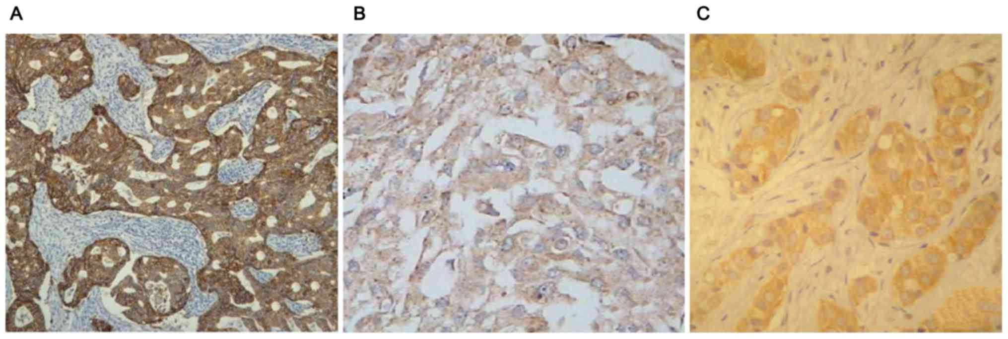
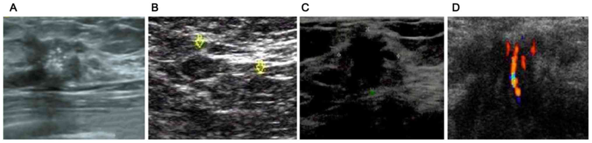
Positive expression of C-erbB-2, VEGF and nm23 was
observed in patients with breast cancer (Fig. 1A-C). Ultrasound examination showed
different signs of breast cancer, including burr sign, abnormal
vascular sign, lymphatic metastasis sign and calcification sign
(Fig. 2A-D).
Analysis of accuracy of ultrasonic
examinations in the diagnosis of breast cancer
There was no significant differences in the accuracy
of diagnosis of breast cancer between the two methods (p>0.05)
(Table II).
| Table II.Comparison of accuracy of ultrasonic
examinations and pathological examinations in the diagnosis of
breast cancer (n, %). |
Table II.
Comparison of accuracy of ultrasonic
examinations and pathological examinations in the diagnosis of
breast cancer (n, %).
| Methods | Diagnosis (%) | Misdiagnosis (%) | Error diagnosis
(%) | Accuracy (%) |
|---|
| Pathological
examinations | 76 (100.00) | 0 (0.00) | 0 (0.00) | 76 (100.00) |
| Enhanced CT | 72 (94.47) | 2 (2.63) | 2 (2.63) | 72 (94.47) |
| χ2 |
|
|
| 2.311 |
| p-value |
|
|
| 0.129 |
Relevance of tumor characteristics of
ultrasonic signs to expression levels of C-erbB-2, VEGF and
nm23
Apparently, the high expression rates of C-erbB-2
and VEGF in the group with tumor diameter ≥3 cm were higher than
those in the group with tumor diameter <3 cm, whereas the high
expression rate of nm23 was higher in the group with tumor diameter
<3 cm. (p<0.05) (Table
III).
| Table III.Correlation between tumor diameter and
high expression rates of C-erbB-2, VEGF and nm23 (n, %). |
Table III.
Correlation between tumor diameter and
high expression rates of C-erbB-2, VEGF and nm23 (n, %).
| Groups | Cases | C-erbB-2 high
expression | VEGF high
expression | nm23 high
expression |
|---|
| Tumor diameter ≥3
cm | 41 | 40 (97.56) | 39 (97.56) | 22 (53.66) |
| Tumor diameter <3
cm | 35 | 19 (54.29) | 19 (54.29) | 34 (97.14) |
| χ2 |
| 17.947 | 15.234 | 16.238 |
| p-value |
| <0.001 | <0.001 | <0.001 |
The high expression rates of C-erbB-2 and VEGF were
higher in the group with spiculated tumor margins than those in the
group with non-spiculated tumor margins, whereas the high
expression rate of nm23 was higher in the group with non-spiculated
tumor margins (p<0.05) (Table
IV). The high expression rates of C-erbB-2 and VEGF were higher
in the group with abnormal tumor vascularity than those in the
group without abnormal tumor vascularity, whereas the high
expression rate of nm23 was higher in the group without abnormal
tumor vascularity (p<0.05) (Table
V).
| Table IV.Correlation between spiculated tumor
margins and high expression rates of C-erbB-2, VEGF and nm23 (n,
%). |
Table IV.
Correlation between spiculated tumor
margins and high expression rates of C-erbB-2, VEGF and nm23 (n,
%).
| Groups | Cases | C-erbB-2 high
expression | VEGF high
expression | nm23 high
expression |
|---|
| Spiculated tumor
margins | 49 | 43 (87.76) | 42 (85.71) | 21 (42.86) |
| Non-spiculated tumor
margins | 27 | 16 (59.25) | 16 (59.25) | 25 (92.59) |
| χ2 |
| 6.582 | 5.356 | 16.001 |
| p-value |
| 0.010 | 0.021 | <0.001 |
| Table V.Correlation between abnormal tumor
vascularity and high expression rates of C-erbB-2, VEGF and nm23
(n, %). |
Table V.
Correlation between abnormal tumor
vascularity and high expression rates of C-erbB-2, VEGF and nm23
(n, %).
| Groups | Cases | C-erbB-2 high
expression | VEGF high
expression | nm23 high
expression |
|---|
| Abnormal tumor
vascularity | 48 | 43 (89.58) | 42 (87.50) | 22 (45.83) |
| Without abnormal
tumor vascularity | 28 | 16 (57.14) | 16 (57.14) | 24 (85.71) |
| χ2 |
| 8.931 | 7.451 | 10.162 |
| p-value |
| 0.003 | 0.007 | 0.001 |
The high expression rates of C-erbB-2 and VEGF were
higher in the group with lymph node metastasis than those in the
group without lymph node metastasis, whereas the high expression
rate of nm23 was higher in the group without lymph node metastasis
(p<0.05) (Table VI). There was no
significant difference in the high expression rates of C-erbB-2,
VEGF and nm23 between the group with calcification and the group
without calcification (p>0.05) (Table VII).
| Table VI.Correlation between lymph node
metastasis and high expression rates of C-erbB-2, VEGF and nm23 (n,
%). |
Table VI.
Correlation between lymph node
metastasis and high expression rates of C-erbB-2, VEGF and nm23 (n,
%).
| Groups | Cases | C-erbB-2 high
expression | VEGF high
expression | nm23 high
expression |
|---|
| Lymph node
metastasis | 30 | 30 (100.00) | 30 (100.00) | 11 (34.38) |
| Without lymph node
metastasis | 46 | 37 (80.43) | 26 (56.52) | 45 (97.83) |
| χ2 |
| 15.304 | 16.500 | 35.664 |
| p-value |
| <0.001 | <0.001 | <0.001 |
| Table VII.Correlation between calcification and
high expression rates of C-erbB-2, VEGF and nm23 (n, %). |
Table VII.
Correlation between calcification and
high expression rates of C-erbB-2, VEGF and nm23 (n, %).
| Groups | Cases | C-erbB-2 high
expression | VEGF high
expression | nm23 high
expression |
|---|
| Calcification | 31 | 21 (63.64) | 21 (63.64) | 23 (69.70) |
| Without
calcification | 45 | 38 (84.44) | 37 (82.22) | 33 (73.33) |
| χ2 |
| 3.416 | 2.543 | 0.009 |
| p-value |
| 0.064 | 0.111 | 0.922 |
Comparison of TIC parameters in
different expression levels of C-erbB-2, VEGF and nm23
The TTP was shorter in patients with high C-erbB-2,
high VEGF and low nm23 expression, the corresponding PI was higher
(p<0.05) (Tables VIII, IX and X).
| Table VIII.Correlation between TIC parameters
and expression level of C-erbB-2. |
Table VIII.
Correlation between TIC parameters
and expression level of C-erbB-2.
| Groups | Cases | TTP (sec) | PI (dB) |
|---|
| C-erbB-2 high
expression | 59 | 6.25±2.04 | 3.57±0.75 |
| C-erbB-2 low
expression | 17 | 9.56±2.52 | 1.69±0.46 |
| t-value |
| 5.585 | 9.790 |
| p-value |
| <0.001 | <0.001 |
| Table IX.Correlation between TIC parameters
and expression level of VEGF. |
Table IX.
Correlation between TIC parameters
and expression level of VEGF.
| Group | Cases | TTP (sec) | PI (dB) |
|---|
| VEGF high
expression | 58 | 6.23±2.03 | 3.57±0.73 |
| VEGF low
expression | 18 | 9.54±2.54 | 1.64±0.47 |
| t-value |
| 5.685 | 10.533 |
| p-value |
| <0.001 | <0.001 |
| Table X.Correlation between TIC parameters
and expression level of nm23. |
Table X.
Correlation between TIC parameters
and expression level of nm23.
| Groups | Cases | TTP (sec) | PI (dB) |
|---|
| nm23 high
expression | 56 | 9.57±2.52 | 1.97±0.32 |
| nm23 low
expression | 20 | 6.26±2.04 | 2.79±0.58 |
| t-value |
| 5.281 | 6.005 |
| p-value |
| <0.001 | <0.001 |
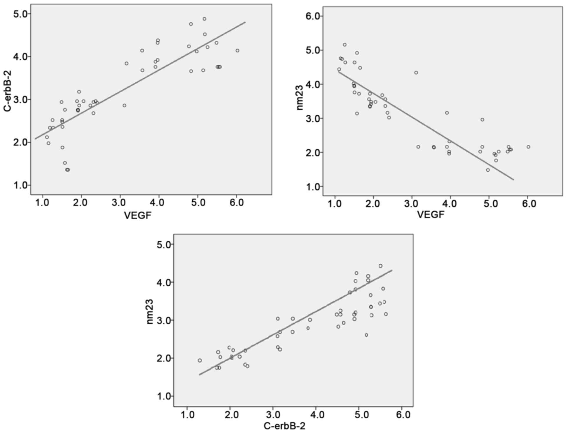
Correlation analysis of C-erbB-2, VEGF
and nm23 expression
Pearson's correlation analysis showed that there was
a positive correlation between C-erbB-2 and VEGF, VEGF and nm23,
nm23 and C-erbB-2 (r=0.463, 0.452, 0.478, P<0.05) (Fig. 3).
Discussion
Breast cancer is the most common cancer among women,
which affects women's physical health as well as their
psychological well-being in a negative way. According to
statistical analysis, breast cancer incidence can be as high as
25%. The current incidence in China is still low, but with the
development of China's society, it has been increasing at a higher
rate than in developed countries (10). Breast cancer ranks the highest in
incidence of cancers diagnosed among Chinese women. The incidence
varies between urban and rural areas in China, which is higher in
urban areas than that in rural areas. The pathogenesis of breast
cancer is currently still under debate. There is consensus that
onset and progression of breast cancer results from multi-step
synergies of various factors, involving mutation and proliferation
of tumor cells, infiltration of the basement membrane, inactivation
of tumor suppressor genes, and imbalance of protease secretion
(11). High risk factors of breast
cancer may include: Age, family history, unmarried late pregnancy,
early menarche, late menopause, frequent chest X-rays, excessive
drinking, long-term use of contraceptives, abortion and inhibition
of breastfeeding (12). Early breast
cancer symptoms are invisible and not noticeable without a
professional screening, thus the disease is easily overlooked and
missed. It can slow down deterioration of breast cancer to a
certain extent and reduce mortality if early screening is performed
and patients receive comprehensive treatments (13).
With continuous improvement of ultrasound
technology, color Doppler ultrasound has become one of the most
important diagnostic methods for breast cancer. The morphology,
size and blood flow of tumor mass can be clearly viewed, thus it
provides a reliable way to effectively distinguish benign and
malignant tumors (14). The C-erbB-2
is an oncogene that indicates tumor prognosis (15). VEGF is one of the most potent and
specific angiogenic factors, and it belongs to the family of
platelet-derived growth factors, including VEGF-A (namely VEGF),
VEGF-B, VEGF-C and VEGF-D. VEGF-A stimulates the vascular
endothelial cells, promoting its division and proliferation, and
ultimately forming new blood vessels. VEGF-B can promote vascular
endothelial growth and migration, tumor growth and metastasis.
VEGF-C and VEGF-D can promote lymphangiogenesis and thus promote
lymphatic metastasis of tumors. VEGF is highly expressed in many
kinds of tumor tissues (16,17). The nm23 gene is a tumor suppressor
gene. It was first found in an animal experiment that the nm23 gene
can inhibit tumor growth and metastasis (18). The results in this study showed that
tumor diameter ≥3 cm, spiculated tumor margin, lymph node
metastasis and abnormal tumor vascularity were all associated with
higher expression of C-erbB-2 and VEGF and lower expression of nm23
(p<0.05). There were no significant differences in expression of
C-erbB-2, VEGF and nm23 between tumor with calcification and tumor
without calcification (p>0.05). Thus, bigger tumor size,
spiculated tumor margins, lymph node metastasis and abnormal tumor
vascularity were specific ultrasonic features of breast cancer.
Under pathological conditions, activation of CerbB-2 led to
expression of VEGF activity. The balance between angiogenic factors
and inhibitory factors was broken, which promoted microvascular
formation, leading to uncontrollable vascular endothelial cell
proliferation. As indicators, expression levels of C-erbB-2 and
VEGF measured by IHC were high. As tumor advanced and grew bigger,
expression of nm23 was hindered. Especially in the lymph nodes
metastasis, the expression level of nm23 was significantly low.
Tumor onset, progression and metastasis all rely on
microvascular formation. Recently, there have been continual
advances in ultrasound technology. Currently ultrasound imaging is
widely used in clinical diagnosis of various cancers due to merits
such as convenience, no radiation exposure and good reproducibility
in evaluation of tumor microvascular network. Ultrasound is
especially highly sensitive in assessing tumor blood flow. Tumor
blood flow pattern can be quantified by analyzing TTP and PI from
the TIC, which may help improve cancer diagnosis (19). The results in this study showed that
the TTP of patients with high expression of C-erbB-2 and VEGF was
shorter than that of patients with low expression of C-erbB-2 and
VEGF, and the corresponding PI was higher. On the contrary, the TTP
of patients with high expression of nm23 was longer than that of
patients with low expression of nm23, and the corresponding PI was
lower (p<0.05). Tumor was in continual progression when
expression levels of C-erbB-2 and VEGF were high. At this stage,
cancer cell proliferation was fast, and the tumor mass became
larger, leading to lymphatic and hematogenous metastasis. In tumor
tissue, blood supply was abundant, and blood flow was fast as well,
thus the average blood volume was large, leading to high PI in the
contrast-enhanced ultrasound imaging. When nm23 expression level
was high, tumor cells were well differentiated, and were less
likely to spread to lymph nodes. At this stage, the tumor blood
flow was slow, leading to low PI in the contrast-enhanced
ultrasound imaging. When nm23 expression level became low,
suppression of tumor metastasis was compromised, which promoted
onset and progression of breast cancer (20).
In summary, ultrasound can show morphological
changes in breast cancer. The ultrasound features and blood flow
parameters were closely associated with expression levels of
C-erbB-2, VEGF and nm23. Their correlations can serve as a
reference for diagnosis and prognosis of breast cancer.
Acknowledgements
Not applicable.
Funding
No funding was received.
Availability of data and materials
The datasets used and/or analyzed during the current
study are available from the corresponding author on reasonable
request.
Authors' contributions
MN analyzed and interpreted the patient data, and
was a major contributor in writing the manuscript. YQ participated
in the analysis and discussion of the data. JZ is responsible for
the collection of the data and the follow-up management of the
patients. YL participated in the experiment and data collection. ZW
was a major contributor in designing the methods. All authors read
and approved the final manuscript.
Ethics approval and consent to
participate
This study was approved by the Ethics Committee of
The Affiliated Hospital of Qingdao University (Qingdao, China).
Signed informed consents were obtained from the patients or
guardians.
Consent for publication
Not applicable.
Competing interests
The authors declare that they have no competing
interests.
References
|
1
|
Shenouda MN, Sadek BT, Goldberg SI,
Keruakous AR, Croft BJ, Abi Raad RF and Taghian AG: Clinical
outcome of isolated locoregional recurrence in patients with breast
cancer according to their primary local treatment. Clin Breast
Cancer. 14:198–204. 2014. View Article : Google Scholar : PubMed/NCBI
|
|
2
|
Akahane K, Tsunoda N, Murata T, Fujii M,
Fuwa Y, Wada K, Oda K and Nagino M: An awareness survey of surgeons
involved in breast cancer treatment regarding their patients
returning to work. Nagoya J Med Sci. 76:315–322. 2014.PubMed/NCBI
|
|
3
|
Barnard ME, Boeke CE and Tamimi RM:
Established breast cancer risk factors and risk of intrinsic tumor
subtypes. Biochim Biophys Acta. 1856:73–85. 2015.PubMed/NCBI
|
|
4
|
Acil H and Cavdar I: Comparison of quality
of life of Turkish breast cancer patients receiving breast
conserving surgery or modified radical mastectomy. Asian Pac J
Cancer Prev. 15:5377–5381. 2014. View Article : Google Scholar : PubMed/NCBI
|
|
5
|
Singleton TP, Perrone T, Oakley G, Niehans
GA, Carson L, Cha SS and Strickler JG: Activation of c-erbB-2 and
prognosis in ovarian carcinoma. Comparison with histologic type,
grade, and stage. Cancer. 73:1460–1466. 1994.
|
|
6
|
Zhao D, Pan C, Sun J, Gilbert C,
Drews-Elger K, Azzam DJ, Picon-Ruiz M, Kim M, Ullmer W, El-Ashry D,
et al: VEGF drives cancer-initiating stem cells through
VEGFR-2/Stat3 signaling to upregulate Myc and Sox2. Oncogene.
34:3107–3119. 2015. View Article : Google Scholar : PubMed/NCBI
|
|
7
|
Sauer T, Furu I, Beraki K, Jebsen PW,
Ormerod E and Naess O: nm23 protein expression in fine-needle
aspirates from breast carcinoma: Inverse correlation with cytologic
grading, lymph node status, and ploidy. Cancer. 84:109–114. 1998.
View Article : Google Scholar : PubMed/NCBI
|
|
8
|
Bao L, Cardiff RD, Steinbach P, Messer KS
and Ellies LG: Multipotent luminal mammary cancer stem cells model
tumor heterogeneity. Breast Cancer Res. 17:1372015. View Article : Google Scholar : PubMed/NCBI
|
|
9
|
Jagadish N, Parashar D, Gupta N, Agarwal
S, Sharma A, Fatima R, Suri V, Kumar R, Gupta A, Lohiya NK, et al:
A novel cancer testis antigen target A-kinase anchor protein
(AKAP4) for the early diagnosis and immunotherapy of colon cancer.
OncoImmunology. 5:e10789652016. View Article : Google Scholar : PubMed/NCBI
|
|
10
|
Youlden DR, Cramb SM, Dunn NA, Muller JM,
Pyke CM and Baade PD: The descriptive epidemiology of female breast
cancer: An international comparison of screening, incidence,
survival and mortality. Cancer Epidemiol. 36:237–248. 2012.
View Article : Google Scholar : PubMed/NCBI
|
|
11
|
Khan MA: Effect of preoperative
intravenous steroids on seroma formation after modified radical
mastectomy. J Ayub Med Coll Abbottabad. 29:207–210. 2017.PubMed/NCBI
|
|
12
|
Memmi EM, Sanarico AG, Giacobbe A,
Peschiaroli A, Frezza V, Cicalese A, Pisati F, Tosoni D, Zhou H,
Tonon G, et al: p63 sustains self-renewal of mammary cancer stem
cells through regulation of Sonic Hedgehog signaling. Proc Natl
Acad Sci USA. 112:3499–3504. 2015. View Article : Google Scholar : PubMed/NCBI
|
|
13
|
Thorsen LB, Offersen BV, Danø H, Berg M,
Jensen I, Pedersen AN, Zimmermann SJ, Brodersen HJ, Overgaard M and
Overgaard J: DBCG-IMN: A population-based cohort study on the
effect of internal mammary node irradiation in early node-positive
breast cancer. J Clin Oncol. 34:314–320. 2016. View Article : Google Scholar : PubMed/NCBI
|
|
14
|
Faustino-Rocha AI, Gama A, Oliveira PA,
Alvarado A, Fidalgo-Gonçalves L, Ferreira R and Ginja M:
Ultrasonography as the gold standard for in vivo volumetric
determination of chemically-induced mammary tumors. In Vivo.
30:465–472. 2016.PubMed/NCBI
|
|
15
|
Farabegoli F, Ceccarelli C, Santini D,
Baldini N, Taffurelli M, Marrano D, Treré D and Derenzini M:
c-erbB-2 over-expression in amplified and non-amplified breast
carcinoma samples. Int J Cancer. 84:273–277. 1999. View Article : Google Scholar : PubMed/NCBI
|
|
16
|
Hamnvik OP, Choueiri TK, Turchin A, McKay
RR, Goyal L, Davis M, Kaymakcalan MD and Williams JS: Clinical risk
factors for the development of hypertension in patients treated
with inhibitors of the VEGF signaling pathway. Cancer. 121:311–319.
2015. View Article : Google Scholar : PubMed/NCBI
|
|
17
|
Ciamporcero E, Miles KM, Adelaiye R,
Ramakrishnan S, Shen L, Ku S, Pizzimenti S, Sennino B, Barrera G
and Pili R: Combination strategy targeting VEGF and HGF/c-met in
human renal cell carcinoma models. Mol Cancer Ther. 14:101–110.
2015. View Article : Google Scholar : PubMed/NCBI
|
|
18
|
Lin CH, Dammai V, Adryan B and Hsu T:
Interaction between Nm23 and the tumor suppressor VHL. Naunyn
Schmiedebergs Arch Pharmacol. 388:143–152. 2015. View Article : Google Scholar : PubMed/NCBI
|
|
19
|
Ahmed M: Image-guided tumor ablation:
Standardization of terminology and reporting criteria - a 10-year
update: Supplement to the consensus document. J Vasc Interv Radiol.
25:1706–1708. 2014. View Article : Google Scholar : PubMed/NCBI
|
|
20
|
Simpson JF, O'Malley F, Dupont WD and Page
DL: Heterogeneous expression of nm23 gene product in noninvasive
breast carcinoma. Cancer. 73:2352–2358. 1994. View Article : Google Scholar : PubMed/NCBI
|