Introduction
With the progress in risk stratification,
chemotherapy, hematopoietic stem cell transplantation (HSCT) and
supportive treatment, the current overall survival (OS) of acute
lymphoblastic leukemia (ALL), as the most common tumor in children,
has been more than 80%, but there are still approximately 20%
(1) children with relapse. Most
pediatric patients will suffer from relapse again even after the
relapse is treated with high-intensity combined chemotherapy and/or
allogeneic HSCT, and only approximately one-third of pediatric
patients can be cured, so the overall prognosis is not
significantly improved (2), and the
relapse is still an important risk factor threatening the long-term
survival and healing of ALL pediatric patients. The age in the
initial diagnosis, leukocyte count, immunophenotyping,
cytogenetics, gene characteristics, minimal residual disease (MRD)
and early therapeutic response are the main risk factors for
assessing the relapse of ALL pediatric patients (3). According to Chinese Children's Leukemia
Group (CCLG)-2008-ALL protocol, ALL pediatric patients can be
divided into standard risk group, intermediate risk group and high
risk group. The relapse rates of pediatric patients in moderate and
high risk groups are relatively higher, and the long-term
event-free survival (EFS) rates are lower, especially pediatric
patients in high risk group, which may be related to the higher age
in the initial diagnosis, high leukocyte count, cytogenetic changes
and poor early therapeutic response. However, some pediatric
patients in standard risk group also suffer from relapse in the
medium and late stage, indicating that the current risk
stratification cannot predict the prognosis very accurately, and it
is needed to find new poor prognosis-related factors as soon as
possible and use it in the guidance of clinical stratification and
treatment.
Relapse occurs most frequently in bone marrow of ALL
pediatric patients. According to the research of Children's
Oncology Group (COG), the 5-year EFS rate of intramedullary relapse
is approximately 30% (4), and the
5-year EFS rates of simple intramedullary relapse, simple
extramedullary relapse and intra-extramedullary relapse are 24%,
59% and 39%, respectively (5,6). The results show that the prognosis of
simple extramedullary (central nervous leukemia and testicular
leukemia) relapse is the best, and the prognosis of simple bone
marrow relapse is inferior to that of intra-extramedullary relapse
(6,7).
An AAL LOIP2 study performed by COG recently made a conclusion that
the relapse time can predict the prognosis; only less than 1/3
pediatric patients with early relapse (relapse within 36 months
after the initial diagnosis according to COG standard) can survive,
while nearly 1/2 pediatric patients with late relapse (relapse at
more than 36 months after the initial diagnosis according to COG
standard) can survive (8,9). Drug-resistant mutation has occurred at
the onset in T-cell ALL (T-ALL) pediatric patients due to rapid
cell mutation and low apoptosis rate (10), and there is much residual leukemia
cloning; thus early relapse occurs more easily in central nervous
system in T-ALL than B-cell ALL (B-ALL). A large number of clinical
studies in China and foreign countries have confirmed that the MRD
levels at different time points of chemotherapy are closely related
to the relapse and long-term EFS, and monitoring the MRD level is
conducive to the evaluation of curative effect and adjustment of
chemotherapy regimens of ALL (11,12).
In this study, the clinical characteristics and
curative effect of 390 pediatric patients initially diagnosed as
B-ALL in Jining First People's Hospital (Jining, China) in the past
6 years were analyzed, and the risk factors related to ALL relapse
were searched, so as to lay a foundation for reducing relapse and
studying its related gene molecules in the future.
Materials and methods
Objects of study
A total of 390 pediatric patients admitted and
initially diagnosed as B-ALL in Jining First People's Hospital from
August 2010 to May 2016 were selected, and they were diagnosed via
bone marrow morphology, immunology, cytogenetics and molecular
biology (MICM) classification. All pediatric patients were followed
up; this study followed the ethical standards set by the
Institutional Review Board and was approved by The Ethics Committee
of Jining First People's Hospital. Written, informed consent was
obtained from all patients or their parents prior to their
inclusion within the study.
Risk stratification
The risk of leukemia was stratified according to the
age of pediatric patients in the initial diagnosis, peripheral
leukocyte count, abnormalities in cytogenetics and molecular
biology, whether they were sensitive to prednisone, bone marrow
remission status at 33 days and MRD at 33 days; the therapeutic
regimen was based on the CCLG-2008 protocol (modified BFM ALL-95
protocol) (13), and the diagnosis
and efficacy were based on ‘Diagnosis and Treatment Recommendations
of Pediatric Acute Leukemia (Revised Draft)’.
Criteria of risk stratification
The risk was stratified according to the age of
pediatric patients in the initial diagnosis, peripheral leukocyte
count, bone marrow puncture results (immunophenotyping, chromosome
and 29 fusion genes) and treatment effects, and ALL pediatric
patients could be divided into standard risk (SR) group,
intermediate risk (IR) group and high risk (HR) group.
Immunophenotyping
Bone marrow (2–3 ml) was taken from pediatric
patients for anticoagulation to separate the karyocytes, followed
by immunofluorescence labeling using the living-cell index method
(FITC or PE). The antibodies used were all mouse monoclonal
antibodies and the dilution was 1/100: CD2 (cat. no. 563820); CD3
(cat. no. 565066); CD5 (cat. no. 564648); CD7 (cat. no. 566119);
CD10 (cat. no. 564959); CD19 (cat. no. 564978); CD20 (cat. no.
564918); CD22 (cat. no. 563940); cCD79 (cat. no. 555302); cCD3
(cat. no. 565065); HLA-DR (cat. no. 564516); TdT (cat. no. 565229);
MPO (cat. no. 556035) and Smlg (cat. no. 564555). All were
purchased from BD Biosciences (San Diego, CA, USA). Flow cytometer
(Beckman Coulter, Brea, CA, USA) was used; antigen-expressing cells
in juvenile cell group >20% indicated the positive.
Chromosome and fusion gene
detection
The chromosomes were taken from the bone marrow of
pediatric patients, and the chromosome samples were prepared using
the direct method or 24 h short-term culture. The karyotype was
analyzed using the conventional R-banding technique. The chromosome
abnormality was explained based on the International System for
Human Cytogenetic Nomenclature (ISCN1985) (14). Bone marrow (2–3 ml) with heparin
anticoagulation was taken in the initial diagnosis, and the
mononuclear cells were extracted using lymphocyte analytic liquid
(Ficoll). Twenty-nine fusion genes [including ETV6/RUNXI
(TEL/AML1), MLL rearrangement, HOX11, E2A/PBX1, and BCR/ABL] were
detected via nested reverse transcription polymerase chain reaction
(RT-PCR).
MRD screening and monitoring
Bone marrow (2–3 ml) with heparin anticoagulation
was taken from pediatric patients in the initial diagnosis before
chemotherapy, and the karyocytes were separated routinely and
counted. Then the karyocytes were stained using CD45-FITC grading,
CD10-PE, CD34-Percp and CD19-APC monoclonal antibodies; the
antibody combination was effective if the leukemia cells and normal
bone marrow cells could be distinguished. At 33 days and 12 weeks
after induction chemotherapy, the bone marrow samples were taken
from pediatric patients, and the mononuclear cells were separated
using Ficoll, followed by four-color fluorescence labeling
detection (CD58, CD66c, CD38, CD123 and TdT were added based on
CD34/CD19/CD10); the original data in the initial diagnosis and
screening were called up; in the two-parameter diagram, if there
were cells in the original area of leukemia cells, it indicated the
minimal residual leukemia; the proportion of these residual cells
in total bone marrow mononuclear cells was the result of monitoring
MRD.
Therapeutic regimen
Among the newly-diagnosed pediatric patients
selected, except those with mature-type B-ALL (n=12) who were
treated as non-Hodgkin's lymphoma, all the other pediatric patients
were treated according to CCLG-2008-ALL protocol and chemotherapy
regimens with different intensities according to different risk
degrees as follows: i) remission induction: pretreatment with
prednisone for 7 days, VDLD (vincristine, daunorubicin,
L-asparaginase or pegaspargase, dexamethasone) regimen in
induction; ii) early intensive treatment: CAM (cyclophosphamide,
cytarabine, 6-mercaptopurine or thioguanine) program (SR group ×
one round; IR and HR groups × two rounds); iii) consolidation
therapy: SR and IR groups: HD-methotrexate (MTX) + 6-MP program
(2.0 g/m2 MTX in SR group, 5.0 g/m2 MTX in IR
group); HR group: HR-1, HR-2 and HR-3 (a total of two rounds); iv)
delayed enhancement: the same VDLD and CAM programs as above [two
rounds of delayed enhancement for IR group (one round of
maintenance chemotherapy between the two rounds of delayed
enhancement)]; v) maintenance treatment: 6-MP + MTX. The single or
triple chemotherapy drugs were regularly injected intrathecally to
prevent the central nervous system leukemia.
Therapeutic regimen of relapse
(15)
Re-induction therapy: VMDP (vincristine,
mitoxantrone, dexamethasone, pegaspargase). Intensive consolidation
therapy: FLAG (fludarabine, HD-cytarabine, granulocyte-stimulating
factor) or CAM + 6-MP. Transplantation or continuous treatment
[delayed enhancement (vincristine, dexamethasone, methotrexate,
6-MP, cyclophosphamide, cytarabine, etoposide) + maintenance
chemotherapy (vincristine, dexamethasone, methotrexate, 6-MP)].
Extramedullary relapse: local radiotherapy after chemotherapy.
CNSL: head radiotherapy 1800 cGy, 150 cGy/day, 12 days; TL:
testicular local radiotherapy 2400 cGy, 200 cGy/day, 12 days.
Efficacy evaluation
Complete remission (CR): the primitive and juvenile
lymphocytes <5% on the bone marrow smear after induction
chemotherapy. Relapse: including simple intramedullary relapse,
extramedullary relapse (central nervous system or testicular
relapse) and intra-extramedullary relapse.
Statistical analysis
Patients were followed up until December 31, 2015,
the median follow-up time was 22.7 months (2.8–62.4 months), and
there were a total of 80 cases of relapse. EFS: the time from
relapse to relapse again or death. Statistical Product and Service
Solutions (SPSS, Chicago, IL, USA) 17.0 Software Package was used
for the comparison of 3-year EFS rate of B-ALL pediatric patients
in intramedullary relapse group at different time points;
Kaplan-Meier survival curve analysis method was used for the
comparison of 3-year EFS rate in different risk degrees; bilateral
log-rank test was used for the survival situations in different
groups. The independent-samples t-test was used for the clinical
characteristics between B-ALL relapse group and non-relapse group,
and Chi-square test was used for the comparison of constituent
ratio of clinical characteristics between the two groups. P<0.05
was considered to indicate a statistically significant
difference.
Results
General data
The general data of subjects in the two groups are
shown in Table I. There was a
significant difference in the proportion of B-ALL pediatric
patients with different risk stratification in relapse group and
non-relapse group; the pairwise comparison among SR group, IR group
and HR group showed that the difference in the proportion of
relapse in B-ALL pediatric patients was not statistically
significant between SR group and IR group, but the difference was
statistically significant compared with that in HR group (P=0.013).
The comparison of pediatric patients in B-ALL relapse group and
non-relapse group revealed that there was no statistically
significant difference in the sex composition between the two
groups (P=0.421, P>0.05), but there were statistically
significant differences in the age and ratio of white blood cell
(WBC) ≥50 × 109/l in the initial diagnosis (P=0.031,
P=0.032, P<0.05).
 | Table I.Comparison of clinical features of
relapse and non-relapse of B-ALL. |
Table I.
Comparison of clinical features of
relapse and non-relapse of B-ALL.
| Clinical
features | Relapse group | Non-relapse
group | P-value |
|---|
| Number (n) | 80 | 310 |
|
| Sex |
|
| 0.421 |
| Male | 54 (67.5%) | 173 (55.8%) |
|
|
Female | 26 (32.5%) | 137 (44.2%) |
|
| Age (years) |
|
| 0.031 |
|
<1 | 0 | 2 (0.6%) |
|
| 1–10 | 68 (85%) | 280 (90.3%) |
|
| ≥10 | 12 (15%) | 28 (9.1%) |
|
| WBC in the initial
diagnosis |
|
| 0.032 |
|
<50×109/l | 51 (63.8%) | 264 (85.2%) |
|
|
≥50×109/l | 29 (36.2%) | 46 (14.8%) |
|
| Response of
prednisone pretreatment |
|
| 0.01 |
|
Sensitive | 60 (75%) | 287 (92.6%) |
|
|
Non-sensitive | 20 (25%) | 23 (7.4%) |
|
| MRD (33 days) |
|
| 0.015 |
|
<10−2 | 65 (81.2%) | 290 (93.5%) |
|
|
≥10−2 | 15 (18.8%) | 20 (6.5%) |
|
| MRD (12 weeks) |
|
| 0.001 |
|
<10−3 | 56 (70%) | 282 (91.0%) |
|
|
≥10−3 | 24 (30%) | 28 (9%) |
|
| Abnormal
chromosomes | 36 (45%) | 162 (52.2%) | 0.890 |
| Fusion genes | 21 (26.3%) | 108 (34.8%) | 0.045 |
| Risk
stratification |
|
| 0.013 |
| SR | 21 (26.3%) | 98 (31.6%) |
|
| IR | 26 (32.5%) | 121 (39.0%) |
|
| HR | 33 (41.2%) | 91 (29.4%) |
|
| After the first
course of treatment |
|
| 0.654 |
|
Remission | 69 (86.3%) | 273 (88.1%) |
|
|
Non-remission | 11 (13.7%) | 37 (11.9%) |
|
Cytogenetics and molecular biology
results
The cytogenetics and molecular biology results of
pediatric patients with relapse and non-relapse in B-ALL group are
shown in Tables II and III. For chromosome analysis, we did not
detect significant difference between two groups while for gene
analysis, fusion gene rate in the relapse group was significantly
higher than that in the non-relapse group.
| Table II.Comparisons of chromosomes between
relapse group and non-relapse group with B-ALL. |
Table II.
Comparisons of chromosomes between
relapse group and non-relapse group with B-ALL.
| Items | Normal
chromosome | Abnormal chromosome
quality | Abnormal chromosome
counts |
|---|
| Relapse group | 54.9% (44/80) | 21.3% (17/80) | 23.8% (19/80) |
| Non-relapse
group | 52.9%
(164/310) | 21.9% (68/310) | 25.2% (78/310) |
| P-value | 0.483 | 0.873 | 0.678 |
| Table III.Comparisons of fusion genes between
relapse group and non-relapse group of B-ALL. |
Table III.
Comparisons of fusion genes between
relapse group and non-relapse group of B-ALL.
| Items | Fusion genes | Normal genes |
|---|
| Relapse group | 83.8% (67/80) | 16.2% (13/80) |
| Non-relapse
group | 61.0%
(189/310) | 39% (121/310) |
| P-value | 0.032 |
Analysis of MRD monitoring at
different time points
MRD of pediatric patients with B-ALL relapse and
non-relapse was monitored at 33 days and 12 weeks after
chemotherapy, and they were divided according to different MRD
levels for analysis and comparison (Tables IV and V). At day 33, there were more children
relapsed in the MRD ≥10−2 group than that in MRD
<10−2 group (P<0.05). At week 12, there were more
children relapsed in the MRD ≥10−3 group than that in
MRD <10−3 group (P<0.05). The relapse rate in the
<10−4 group was lower than non-relapse rate
(P<0.05). Moreover, the ratio in
10−4−10−3, 10−3−10−2
and ≥10−2 in relapse group was higher than that in
non-relapse group. As a result, MRD <10−4 could be
thought as a cut-off point of low relapse rate.
| Table IV.Comparisons of MRD at 33 days between
relapse and non-relapse group with B-ALL. |
Table IV.
Comparisons of MRD at 33 days between
relapse and non-relapse group with B-ALL.
|
|
|
| MRD at 33 days |
|---|
|
|
|
|
|
|---|
| Items | CR | Non-CR |
<10−4 |
10−4−10−3 |
10−3−10−2 |
≥10−2 |
|---|
| Relapse group | 70 | 10 | 32 (40%) | 24 (30.0%) | 12 (15%) | 12 (15%) |
| Non-relapse
group | 243 | 67 | 173 (55.8%) | 88 (28.4%) | 45 (14.5%) | 4 (1.3%) |
| P-value | P>0.05 | 0.043 | 0.122 | 0.435 | 0.001 |
| Table V.Comparisons of MRD at 12 weeks
between relapse and non-relapse group with B-ALL. |
Table V.
Comparisons of MRD at 12 weeks
between relapse and non-relapse group with B-ALL.
|
|
|
| MRD at 12
weeks |
|---|
|
|
|
|
|
|---|
| Items | CR | Non-CR |
<10−4 |
10−4−10−3 |
10−3−10−2 |
≥10−2 |
|---|
| Relapse group | 68 | 12 | 24 (30%) | 29 (36.3%) | 18 (22.5%) | 9 (11.2%) |
| Non-relapse
group | 233 | 77 | 162 (52.3%) | 102 (32.9%) | 25 (8.1%) | 21 (6.7%) |
| P-value | P>0.05 | 0.032 | 0.546 | 0.001 | 0.002 |
Relapse site and time
The relapse site and time of pediatric patients in
the relapse group and their relationship with risk typing are shown
in Tables VI and VII. As defined by
Berlin-Frankfurt-Munster-ALL Group, early relapse: the time from
initial diagnosis to relapse was ≤18 months; medium relapse: the
time from initial diagnosis to relapse was >18 months and CR
<30 months; late relapse: CR >30 months.
| Table VI.The relapse site of 80 B-ALL
cases. |
Table VI.
The relapse site of 80 B-ALL
cases.
| Bone marrow | Central nervous
system | Testis | Combining
relapse |
|---|
| 88.8% (71/80) | 5% (4/80) | 5% (4/80) | 3.8% (3/80) |
| Table VII.Relapse stages of different risk
stratifications of B-ALL. |
Table VII.
Relapse stages of different risk
stratifications of B-ALL.
|
| Risk
stratification |
|---|
|
|
|
|---|
| Stage of
relapse | Standard risk | Intermediate
risk | High risk | Total number |
|---|
| Early stage | 2 | 12 | 20 | 34 |
| Medium stage | 8 | 11 | 19 | 38 |
| Late stage | 5 | 2 | 1 | 8 |
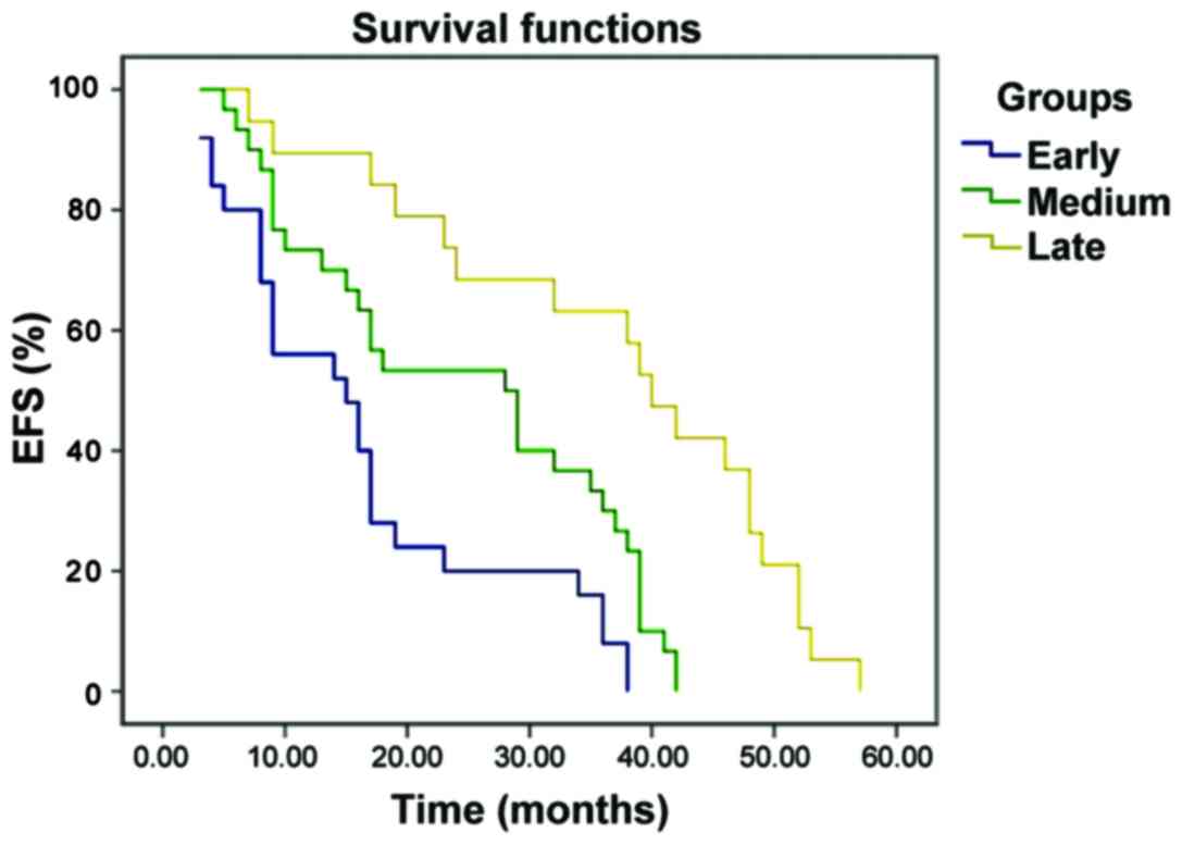
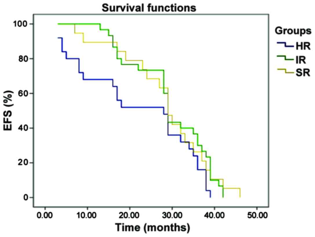
Long-term survival analysis
The EFS curve of B-ALL pediatric patients with
early, medium and late bone marrow relapse, the EFS curve of B-ALL
pediatric patients with bone marrow relapse in SR group, IR group
and HR group are shown in Figs. 1 and
2. The 3-year event-free survival
(EFS) rates of pediatric patients with early, medium and late B-ALL
relapse were 12.5±7.8, 33.1±9.8 and 63.6±6.1%, respectively, and
the prognosis of late relapse was significantly better than that of
early relapse (P<0.001). The 3-year EFS rates of pediatric
patients with bone marrow relapse in standard risk group,
intermediate risk group and high risk group were 29.1±6.9, 31.3±6.5
and 28.3±6.3%, respectively; there were no statistically
significant differences (P=0.387, P>0.05).
Discussion
ALL is the most common type of AL in children,
accounting for approximately 80% (15,16), whose
immunophenotyping is mostly B-lineage expression. In recent years,
with the improvement of risk stratification, chemotherapy regimens
and HSCT, the cure rate of ALL pediatric patients has been up to
70–80% (17), and the long-term
survival rate has reached approximately 80% (18), but approximately 20% cases still
suffer from relapse. Only 30–40% pediatric patients with ALL
relapse are cured (19,20) and its mortality rate is high, so it is
known as one of the four major tumors in children (the other three
are acute leukemia, lymphoma and brain tumor) (21). Therefore, the key to improving the
long-term survival of ALL pediatric patients is to reduce its
relapse rate, master the major risk factors for ALL relapse and
give appropriate treatment.
Intramedullary relapse is the most common in ALL
pediatric patients, and the curative effect on it is poor compared
with that on extramedullary relapse and intra-extramedullary
relapse, and the prognosis is positively correlated with relapse
time. Only 10% pediatric patients with early relapse and 50%
pediatric patients with late relapse can be cured. In this study,
there were a total of 71 cases of B-ALL intramedullary relapse. The
3-year EFS rates in early, medium and late stage were 12.5±7.8%,
33.1±9.8% and 63.6±6.1%, respectively. The bilateral log-rank test
showed that the prognosis of early relapse was worse than that of
late relapse, and the difference was statistically significant
(P<0.001). The MRD level is an independent factor of predicting
relapse in chemotherapy (22), which
is closely related to the long-term EFS of pediatric patients.
Moreover, monitoring the MRD level has an important value in the
evaluation of curative effect and adjustment of chemotherapy
intensity of leukemia.
ALL relapse was more common in children in IR group
and HR group, but the clinical observation found that some SI
pediatric patients also suffered from relapse, mostly the medium
and late relapse. Therefore, the current risk stratification cannot
accurately predict the prognosis yet, and the key to reducing ALL
relapse in the future is to find new poor prognosis-related genes
used to assess the prognosis and guide the treatment.
Acknowledgements
Not applicable.
Funding
No funding was received.
Availability of data and materials
All data generated or analyzed during this study are
included in this published article.
Authors' contributions
XZ and HW designed the present study and prepared
the manuscript; HF, XZ and BS collected all data; HF and BS
performed the flow cytometry; GZ, HW and LD analyzed the data; XZ
and HW interpreted the data. All authors read and approved the
final manuscript.
Ethics approval and consent to
participate
The present study was approved by the Ethics
Committee of Jining First People's Hospital (Jining, China).
Written, informed consent was obtained from all patients or their
parents prior to their inclusion within the study.
Patient consent for publication
Not applicable.
Competing interests
The authors declare that they have no competing
interests.
References
|
1
|
Hunger SP and Mullighan CG: Acute
lymphoblastic leukemia in children. N Engl J Med. 373:1541–1552.
2015. View Article : Google Scholar : PubMed/NCBI
|
|
2
|
Roy A, Cargill A, Love S, Moorman AV,
Stoneham S, Lim A, Darbyshire PJ, Lancaster D, Hann I, Eden T, et
al: Outcome after first relapse in childhood acute lymphoblastic
leukaemia - lessons from the United Kingdom R2 trial. Br J
Haematol. 130:67–75. 2005. View Article : Google Scholar : PubMed/NCBI
|
|
3
|
Locatelli F, Schrappe M, Bernardo ME and
Rutella S: How I treat relapsed childhood acute lymphoblastic
leukemia. Blood. 120:2807–2816. 2012. View Article : Google Scholar : PubMed/NCBI
|
|
4
|
Raetz EA, Borowitz MJ, Devidas M, Linda
SB, Hunger SP, Winick NJ, Camitta BM, Gaynon PS and Carroll WL:
Reinduction platform for children with first marrow relapse of
acute lymphoblastic Leukemia: A Children's Oncology Group Study. J
Clin Oncol. 26:3971–3978. 2008. View Article : Google Scholar : PubMed/NCBI
|
|
5
|
Nguyen K, Devidas M, Cheng SC, La M, Raetz
EA, Carroll WL, Winick NJ, Hunger SP, Gaynon PS and Loh ML:
Children's Oncology Group: Factors influencing survival after
relapse from acute lymphoblastic leukemia: A Children's Oncology
Group study. Leukemia. 22:2142–2150. 2008. View Article : Google Scholar : PubMed/NCBI
|
|
6
|
Einsiedel HG, von Stackelberg A, Hartmann
R, Fengler R, Schrappe M, Janka-Schaub G, Mann G, Hählen K, Göbel
U, Klingebiel T, et al: Long-term outcome in children with relapsed
ALL by risk-stratified salvage therapy: Results of trial acute
lymphoblastic leukemia-relapse study of the
Berlin-Frankfurt-Münster Group 87. J Clin Oncol. 23:7942–7950.
2005. View Article : Google Scholar : PubMed/NCBI
|
|
7
|
Lawson SE, Harrison G, Richards S, Oakhill
A, Stevens R, Eden OB and Darbyshire PJ: The UK experience in
treating relapsed childhood acute lymphoblastic leukaemia: A report
on the medical research council UKALLR1 study. Br J Haematol.
108:531–543. 2000. View Article : Google Scholar : PubMed/NCBI
|
|
8
|
Bailey LC, Lange BJ, Rheingold SR and
Bunin NJ: Bone-marrow relapse in paediatric acute lymphoblastic
leukaemia. Lancet Oncol. 9:873–883. 2008. View Article : Google Scholar : PubMed/NCBI
|
|
9
|
Hogan LE, Meyer JA, Yang J, Wang J, Wong
N, Yang W, Condos G, Hunger SP, Raetz E, Saffery R, et al:
Integrated genomic analysis of relapsed childhood acute
lymphoblastic leukemia reveals therapeutic strategies. Blood.
118:5218–5226. 2011. View Article : Google Scholar : PubMed/NCBI
|
|
10
|
Pieters R, den Boer ML, Durian M, Janka G,
Schmiegelow K, Kaspers GJ, van Wering ER and Veerman AJ: Relation
between age, immunophenotype and in vitro drug resistance in 395
children with acute lymphoblastic leukemia - implications for
treatment of infants. Leukemia. 12:1344–1348. 1998. View Article : Google Scholar : PubMed/NCBI
|
|
11
|
Borowitz MJ, Devidas M, Hunger SP, Bowman
WP, Carroll AJ, Carroll WL, Linda S, Martin PL, Pullen DJ,
Viswanatha D, et al: Children's Oncology Group: Clinical
significance of minimal residual disease in childhood acute
lymphoblastic leukemia and its relationship to other prognostic
factors: A Children's Oncology Group study. Blood. 111:5477–5485.
2008. View Article : Google Scholar : PubMed/NCBI
|
|
12
|
Zhang R, Yang JY, Sun HQ, Jia H, Liao J,
Shi YJ and Li G: Comparison of minimal residual disease (MRD)
monitoring by WT1 quantification between childhood acute myeloid
leukemia and acute lymphoblastic leukemia. Eur Rev Med Pharmacol
Sci. 19:2679–2688. 2015.PubMed/NCBI
|
|
13
|
Möricke A, Reiter A, Zimmermann M, Gadner
H, Stanulla M, Dördelmann M, Löning L, Beier R, Ludwig WD, Ratei R,
et al: German-Austrian-Swiss ALL-BFM Study Group: Risk-adjusted
therapy of acute lymphoblastic leukemia can decrease treatment
burden and improve survival: Treatment results of 2169 unselected
pediatric and adolescent patients enrolled in the trial ALL-BFM 95.
Blood. 111:4477–4489. 2008. View Article : Google Scholar : PubMed/NCBI
|
|
14
|
No authors listed: An International System
for Human Cytogenetic Nomenclature (1985) ISCN 1985. Report of the
Standing Committee on Human Cytogenetic Nomenclature. Birth Defects
Orig Artic Ser. 21:1–117. 1985.
|
|
15
|
Parker C, Waters R, Leighton C, Hancock J,
Sutton R, Moorman AV, Ancliff P, Morgan M, Masurekar A, Goulden N,
et al: Effect of mitoxantrone on outcome of children with first
relapse of acute lymphoblastic leukaemia (ALL R3): An open-label
randomised trial. Lancet. 376:2009–2017. 2010. View Article : Google Scholar : PubMed/NCBI
|
|
16
|
Gao YJ, Zhu XH, Yang Y, Wu Y, Lu FJ, Zhai
XW and Wang HS: Prevalence of ETV6-RUNX1 fusion gene in children
with acute lymphoblastic leukemia in China. Cancer Genet Cytogenet.
178:57–60. 2007. View Article : Google Scholar : PubMed/NCBI
|
|
17
|
Mariottini P and Amaldi F: The 5′
untranslated region of mRNA for ribosomal protein S19 is involved
in its translational regulation during Xenopus development. Mol
Cell Biol. 10:816–822. 1990. View Article : Google Scholar : PubMed/NCBI
|
|
18
|
Pui CH, Carroll WL, Meshinchi S and Arceci
RJ: Biology, risk stratification, and therapy of pediatric acute
leukemias: An update. J Clin Oncol. 29:551–565. 2011. View Article : Google Scholar : PubMed/NCBI
|
|
19
|
Gorman MF, Ji L, Ko RH, Barnette P,
Bostrom B, Hutchinson R, Raetz E, Seibel NL, Twist CJ, Eckroth E,
et al: Outcome for children treated for relapsed or refractory
acute myelogenous leukemia (rAML): A Therapeutic Advances in
Childhood Leukemia (TACL) Consortium study. Pediatr Blood Cancer.
55:421–429. 2010. View Article : Google Scholar : PubMed/NCBI
|
|
20
|
van den Berg H, de Groot-Kruseman HA,
Damen-Korbijn CM, de Bont ES, Schouten-van Meeteren AY and
Hoogerbrugge PM: Outcome after first relapse in children with acute
lymphoblastic leukemia: A report based on the Dutch Childhood
Oncology Group (DCOG) relapse all 98 protocol. Pediatr Blood
Cancer. 57:210–216. 2011. View Article : Google Scholar : PubMed/NCBI
|
|
21
|
Gaynon PS: Childhood acute lymphoblastic
leukaemia and relapse. Br J Haematol. 131:579–587. 2005. View Article : Google Scholar : PubMed/NCBI
|
|
22
|
von Stackelberg A, Seeger K, Henze G and
Eckert C: Clinical significance of minimal residual disease in
childhood acute lymphoblastic leukemia after first relapse.
Leukemia. 18:1727–1729. 2004. View Article : Google Scholar : PubMed/NCBI
|