Introduction
Hepatocellular carcinoma (HCC) is one of the five
most common cancer types globally and is an aggressive malignancy
with poor prognosis (1). The majority
of HCC cases occur in eastern Asia and sub-Saharan Africa,
particularly in China, which accounts for >50% of HCC cases
globally (2). The definition of small
HCC (SHCC) is a single HCC nodule <5 cm or ≤3 nodules and a
maximum diameter of each nodule <3 cm (3). A single HCC nodule of <3 cm is
generally considered to be early stage HCC, according to the
Barcelona Clinic Liver Cancer stage (BCLC) criteria (4).
Currently, several methods are used for the
treatment of SHCC. Surgical resection (SR) is still regarded as the
‘gold standard’ treatment for HCC, particularly for SHCC (5). However, the long-term clinical outcomes
remain frustrating due to a high recurrence rate (4,5). In
addition, a number of patients cannot undergo the SR treatment by
the time the diagnosis of SHCC is confirmed (6). A number of patients may worry about the
high risk of hepatolobectomy and refuse surgical treatment
(7). A number of alternative, local,
mini-invasive therapies for SHCC have been produced and have become
popular treatments with less surgical intervention and fewer
complications (8–10). Several previous studies have reported
that the local mini-invasive therapies (monotherapy or combined
therapy) were safe and effective for patients with SHCC in the
short term (11,12). However, little attention has been paid
to whether local mini-invasive therapies were effective for
patients with SHCC in the long term (13). Furthermore, no data have been reported
concerning the appropriate treatment for patients with SHCC,
particularly for patients with SHCC and variousα-fetoprotein (AFP)
levels (14,15).
A number of researchers have drawn different
conclusions regarding the value of features, including tumor size
and margin, in the prognosis of postoperative outcomes of patients
with SHCC (16–19). Preoperative indicators were
significantly important for the prognosis of patients with SHCC
(20,21). However, to the best of our knowledge,
there are still only scarce reports regarding the preoperative
indicators in clinical and laboratory testing as predictive factors
for patients with SHCC. Consequently, the present study sought to
explore the influence predictive factors for patients with SHCC by
comprehensively analyzing medical histories, imaging features and
laboratory results.
Treatment programs for 206 patients with SHCC were
determined through the BCLC proposal and patients' informed consent
(22,23). By comparing three local tumor lesion
treatments, the appropriate treatment for patients with SHCC was
elucidated. In addition, preoperative indicators as predictors of
HCC prognosis were determined.
Materials and methods
Patients
According to the inclusion and exclusion criteria
for the present study, 206 patients were enrolled, including 159
males (77.18%) and 47 females (22.82%), aged 13–87 years with a
mean age of 55.68±11.61 years. The inclusion criteria for the study
population was as follows: i) Patients aged between 13–87 years;
ii) single SHCC (≤3 cm) or multifocal HCC <3.0 cm in the
greatest dimension; iii) diabetes mellitus (DM) and hypertension,
if present, were controlled with medication; and iv) no multiple
organ failure and no severe underlying diseases. Patients were
excluded from the study if they: i) Received other treatments in
another hospital; ii) were missing data; iii) were not tracked
adequately; iv) received a liver transplant; v) received systemic
chemotherapy; vi) received sorafenib; or vii) had another type of
malignant tumor.
Collection of data and primary
end-point assessment
Clinical data were collected from each patient at
the time of SHCC diagnosis, including sex, age, other chronic
diseases [including hypertension and type 2 DM (T2DM)],
Child-Pugh grade and BCLC stage. Imaging features were also
collected, including tumor size, tumor number, cirrhosis, portal
vein tumor thrombus (PVTT) and intrahepatic metastasis (IM). All
laboratory indicators were collected in the week prior to surgery.
Laboratory results determined the AFP, hepatitis B virus surface
antigen and hepatitis C antibody (HCV-Ab) levels. The main endpoint
was survival time, which was defined as the duration from the time
of primary treatment for SHCC to mortality or August 2016,
whichever was earlier. The secondary endpoint was outcomes during
follow-up, including survival and mortality.
Treatments and follow-up
The SR was performed as a strictly standardized
procedure for hepatobiliary surgery. A partial hepatectomy with 1–2
cm tumor-free margin was performed in these patients.
Intra-operative ultrasonography was routinely used to estimate the
number of tumors, and tumor size(s), location(s) and border.
Transarterial chemoembolization (TACE) was performed
using the following procedures. Following using 5-French catheter
selection to perform arteriography of the superior mesenteric,
celiac and common hepatic arteries, the hepatic artery was
catheterized with a coaxial microcatheter. The microcatheter was
positioned into or as close as possible to the tumor feeding
branch; then, an emulsion of doxorubicin hydrochloride (Adriamycin)
and iodized oil (Lipiodol; Guerbet, Aulnay-sous-Bois, France) was
slowly infused through the catheter. Oily TACE was performed as
selectively as possible and a microcatheter was routinely used. The
doses of iodized oil and doxorubicin were determined according to
the tumor size and tumor vascularity. The maximum doses of iodized
oil and doxorubicin for a single session of TACE was 25 ml and 70
mg, respectively. Infusion of the Lipiodol® mixture was
followed by particulate embolization with 1–2 mm diameter gelatin
sponge pledgets (Cutanplast; Mascia Brunelli, Milan, Italy).
Percutaneous microwave coagulation therapy (PMCT)
was performed using the KY-2000 microwave therapy instruments
(Xuzhou Hengda Electronic Co., Ltd., Xuzhou, China). The PMCT
procedure was performed by an experienced hepatobiliary surgeon
following local anesthesia using 2% lidocaine. The entire procedure
was guided and constantly monitored using real-time ultrasound
(MyLab™ Twice; Esaote Co., Ltd., Genoa, Italy).
Following anesthesia was achieved, a 15-cm 16-gauge cooling
unipolar was inserted into the center of the nodule, and
coagulation therapy was performed at 2,450 MHz with 60–80 W output
for 8–10 min/ablation. The ablation was performed repeatedly until
the tumors attained completed necrosis as monitored by real-time
ultrasound, and the hyperechoic area overlapped the area of the
tumor with a surrounding ≥1 cm safety margin.
Following TACE, PMCT or SR, patients were followed
up every 1–3 months during the first 2 years, and at 3–6 months
intervals thereafter. Following treatment, patients with SHCC were
evaluated for treatment response by combining contrast-enhanced
computer tomography with liver function. Patients who experienced
recurrence were given subsequent treatment by physicians if
clinically feasible.
Statistical analysis
The Kruskal-Wallis test was performed to analyze
continuous variables; the results are expressed as the mean ±
standard deviation for normal distributions and as median and
interquartile range (Q1-Q3) for skewed distributions. For
categorical variables, the χ2 test and Fisher's exact
test were utilized. Analyses regarding survival time were generated
using the Kaplan-Meier method and COX regression analysis. For
Fig. 1, the Spearman's rank
correlation coefficient was used. All statistical tests were
two-sided, and P<0.05 was considered to indicate a statistically
significant difference. All statistical analyses were performed
using the SPSS v.22.0 software (IBM Corp., Armonk, NY, USA).
Ethical approval
All procedures in the current study were in
accordance with the ethical standards of the Institutional Research
Committee and with The Declaration of Helsinki. This type of study
was a retrospective data analysis, so formal consent was not
required.
Results
Patient population
The sample was predominantly male (159/202, 77.2%),
full-grown adults with a long period of HBV infection (189/202,
91.7%). As depicted in Table I, the
majority of the patients had cirrhosis (88.8%). The majority of
patients (154/202, 74.8%) had well-preserved liver function
(Child-Pugh A), whereas 49 (23.8%) and 3 patients (1.5%) had
Child-Pugh B and C functional status, respectively. The majority of
patients (182/202, 88.3%) had an early-stage tumor (BCLC stage A);
whilst, 9 (4.4%) and 15 patients (7.3%) had tumors classified as
BCLC stage B and C, respectively. The mean number of tumors was
1.31±0.62 (range, 1–3), the mean tumor length was 2.07±0.63 cm
(range, 0.5–3 cm) and the mean tumor width was 1.61±0.56 cm (range,
0.5–2.9 cm). Of these patients, 29 (14.1%) had IM, 19 (9.2%) had
pathological vascular invasion and 51 (24.75%) had ≥400 ng/ml
AFP.
 | Table I.Demographic and clinical data of 206
patients with SHCC. |
Table I.
Demographic and clinical data of 206
patients with SHCC.
| Variables | Value | Percentage (%) |
|---|
| Sex,
male/female | 159/47 | 77.2/22.8 |
| Age, years | 55.68±11.61 |
|
| Cirrhosis,
negative/positive | 29/177 | 14.1/85.9 |
| Tumor length,
cm | 2.07±0.63 |
|
| Tumor width,
cm | 1.61±0.56 |
|
| Tumor number | 1.31±0.62 |
|
| IM,
negative/positive | 177/29 | 85.9/14.1 |
| PVTT,
negative/positive | 187/19 | 90.8/9.2 |
| Child-Pugh,
A/B/C | 154/49/3 | 74.8/23.8/1.5 |
| BCLC stage,
A/B/C | 182/9/15 | 88.3/4.4/7.3 |
| Hypertension,
negative/positive | 168/24 | 87.5/12.5 |
| Diabetes mellitus
type 2, negative/positive | 167/25 | 87/13 |
|
TACE/TACE-PMCT/SR | 68/82/56 | 33/39.8/27.2 |
| <400 ng/ml
AFP/≥400 ng/ml AFP | 117/89 | 56.8/43.2 |
General characteristics of subjects in
the three groups
According to the BCLC proposal and with patients'
informed consent, a total of 206 patients were included in the
present study. A total of 68 patients were initially treated with
TACE, 82 patients were treated with TACE-PMCT and 56 patientswere
treated with SR. The demographic and clinicopathological
characteristics of the three groups are summarized in Table II. The mean patient age in the SR
group was younger than the other two groups (P=0.019), and the mean
number of tumors was also less than in the other two groups
(P=0.001). All patients in the SR group were BCLC stage A. The
majority of patients (55/56) were infected with the hepatitis B
virus. The proportion of patients with hypertension in the SR group
was lower than that of the other two groups (P=0.032). Other
laboratory and imaging parameters were not significantly different
among three groups including AFP, HCV-Ab, tumor length, tumor
width, cirrhosis, PVTT and IM (all of them, P>0.05). Sex,
Child-Pugh grade and T2DM were also not significantly
different among the three groups (all, P>0.05).
| Table II.Baseline character of TACE, TACE-PMCT
and SR group. |
Table II.
Baseline character of TACE, TACE-PMCT
and SR group.
| Variables | TACE (n=68) | TACE-PMCT
(n=82) | SR (n=56) | P-value |
|---|
| Age, years | 56.96±9.20 | 57.15±13.00 | 51.99±11.45 | 0.019a |
| Sex,
male/female | 50/17 | 59/23 | 49/7 | 0.139 |
| <400 ng/ml
AFP | 52 | 57 | 46 | 0.307 |
| ≥400 ng/ml AFP | 16 | 18 | 17 |
|
| HCV-Ab, N/P | 67/1 | 77/5 | 52/4 | 0.273 |
| HBsAg, N/P | 5/63 | 13/69 | 1/54 | 0.017a |
| Tumor length,
cm | 1.96±0.58 | 2.07±0.65 | 2.20±0.62 | 0.102 |
| Tumor width,
cm | 1.59±0.55 | 1.68±0.57 | 1.56±0.54 | 0.427 |
| Tumor number | 1.32±0.66 | 1.46±0.72 | 1.05±0.23 | 0.001a |
| Cirrhosis,
negative/positive | 8/60 | 9/73 | 6/50 | 0.981 |
| IM,
negative/positive | 57/11 | 71/11 | 49/7 | 0.822 |
| PVTT,
negative/positive | 61/7 | 75/7 | 51/5 | 0.930 |
| Child-Pugh A | 45 | 64 | 45 | 0.281 |
| Child-Pugh B | 21 | 17 | 17 |
|
| Child-Pugh C | 2 | 1 | 0 |
|
| BCLC stage A | 57 | 69 | 56 | 0.022a |
| BCLC stage B | 3 | 6 | 0 |
|
| BCLC stage C | 8 | 7 | 0 |
|
| Hypertension,
negative/positive | 55/13 | 69/13 | 54/2 | 0.032a |
| T2DM,
negative/positive | 57/11 | 69/13 | 51/5 | 0.430 |
Associations between survival time and
local lesion treatment strategies
No fatal treatment-associated complications were
recorded for these patients. This indicates that the local invasive
treatments were safe and effective in the short term. The median
survival time was 27 (range, 14–49) months in the TACE group, 29.5
(range, 16–52) months in the TACE-PMCT group and 36.5 (range,
26–52) months in the SR group. The results demonstrated that there
were no significant differences in the survival time of patients
with SHCC among the three groups (P=0.091). In addition, the 1, 3
and 5-year survival rates of patients with SHCC were 82.4, 64.9 and
46.8% in the TACE group; 89, 72.6 and 58.3% in the TACE-PMCT group;
and 88.8, 72.3 and 58.6% in the SR group, respectively. This
indicated that there were no significant differences among the
three groups (P=0.181) for survival time.
Results of the association between
survival time and number of TACE sessions
The proportion of patients receiving TACE treatment
was the highest in the present study, including the TACE and
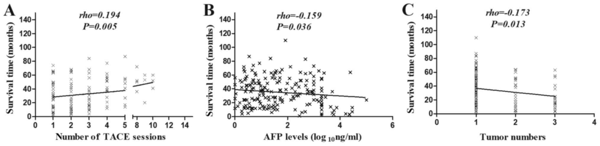
TACE-PMCT group. Fig. 1A depicts that
the number of TACE sessions (r=0.194; P=0.005) was positively
correlated with the survival time for patients with SHCC in the
TACE and TACE-PMCT group. In addition, there were 24 patients in
this group with BCLC stage B or C tumors. They could not undergo
the SR treatment and so had received TACE or TACE-PMCT treatment.
The median survival time was 13.5 (range, 4.25–27) months for these
patients. Therefore, TACE treatment was one of alternative
treatment therapies for patients with SHCC, particularly for
unresectable patients with SHCC.
Results of the association between
survival time and other factors
Fig. 1B and 1C
depicted that AFP level (r=−0.159 and P=0.036) and tumor numbers
were negatively correlated with survival time (r=−0.173 and
P=0.013), which indicated that the patients with higher AFP level
and multiple tumor lesions had reduced survival time, compared with
patients with lower AFP level and a single tumor lesion.
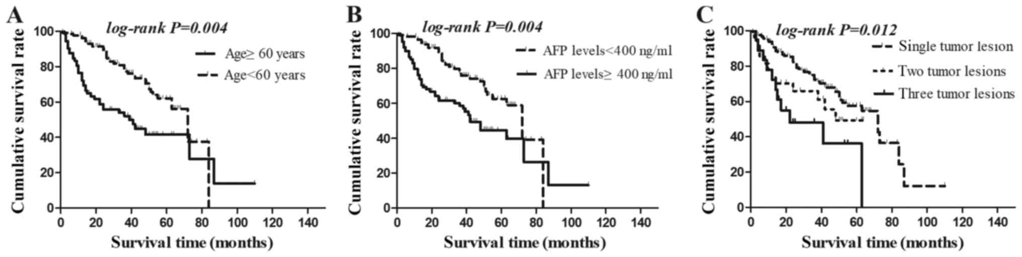
Cumulative survival rate
Kaplan-Meier survival analyses were used to analyze
the cumulative survival rate among the subgroups. The patients' age
and preoperative AFP level were divided into two groups. Fig. 2A depicted that the cumulative survival
rate of patients with SHCC >60 years of age was significantly
lower than that of patients with SHCC <60 years of age (log-rank
test P=0.005). Similarly, Fig. 2B
demonstrates that the cumulative survival rate of patients with
SHCC with preoperative AFP levels of ≥400 ng/ml was significantly
lower than that of patients with SHCC with preoperative AFP level
of <400 ng/ml (log-rank test P=0.012). Fig. 2C depicts that the cumulative survival
rate classifying by tumor numbers was also significantly different
in patients with SHCC (log-rank test P=0.004). The data indicates
that patients with SHCC, a single tumor lesion had higher
cumulative survival rate than patients with multiple tumor lesions.
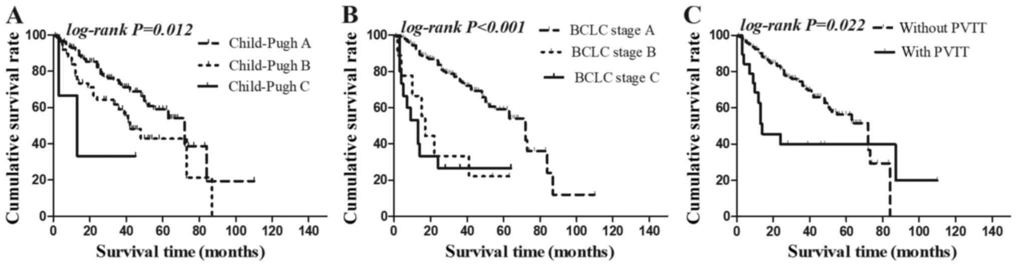
In addition, Fig. 3A and B
illustrated that the cumulative survival rates, considering
Child-Pugh grade (log-rank test P=0.012) and BCLC stage, were
significantly different in patients with SHCC (log-rank test,
P<0.001). The Child-Pugh A and BCLC stage A patients had higher
cumulative survival rate than patients classified as Child-Pugh B
and BCLC stage B or classified as Child-Pugh C and BCLC stage C.
Similarly, Fig. 3C depicts that the
cumulative survival rate classifying by PVTT were also
significantly different in patients with SHCC (log-rank test,
P=0.02). The data indicated that SHCC patients without PVTT had
higher cumulative survival rate than patients with multiple tumor
lesions and PVTT. However, there were no statistically significant
differences in the cumulative survival rate of patients with SHCC
when classifying by sex (log-rank test, P=0.227), age (log-rank
test, P=0.87) or T2DM (log-rank test, P=0.52).
Multivariate analyses
The Cox regression model was used to calculate
hazard ratios (HRS). Table III
demonstrated that patient age [<60 years vs. ≥60 years;
HR=0.548; 95% confidence interval (CI): 0.331–0.908; P=0.020],
preoperative AFP level (<400 ng/ml vs. ≥400 ng/ml; HR=0.548; 95%
CI: 0.306–0.961; P=0.036) and BCLC stage (A vs. C; HR=0.212; 95%
CI: 0.098–0.461; P<0.001) were independent prognostic factors
for the survival time of patients with SHCC. Therefore, these
results indicate that BCLC stage A was a protective factor, whist
older age and higher preoperative AFP levels were risk factors for
the survival time of patients with SHCC.
| Table III.Risk factors for the mortality of
patients with SHCC. |
Table III.
Risk factors for the mortality of
patients with SHCC.
| Factor | β | SE | Wald
χ2 | HR | 95% CI | P-values |
|---|
| Age (<60 vs. ≥60
years) | −0.602 | 0.258 | 5.452 | 0.548 | 0.331–0.908 | 0.020a |
| AFP (<400 vs.
≥400 ng/ml) | −0.612 | 0.292 | 4.394 | 0.542 | 0.306–0.961 | 0.036a |
| BCLC stage, A vs.
C | −1.550 | 0.395 | 15.363 | 0.212 | 0.098–0.461 |
<0.001a |
| BCLC stage, B vs.
C | −0.628 | 0.573 | 1.203 | 0.534 | 0.106–8.609 | 0.405 |
Subgroup analysis based on
preoperative AFP level
Multivariate survival analysis revealed that
preoperative AFP levels were an independent prognostic factor for
patients with SHCC. Subsequently, subgroup survival analysis was
performed based on preoperative AFP level. The analysis of the
patients with ≥400 ng/ml AFP revealed that the median survival time
was 17 (range, 12–44) months in the TACE group, 27 (range, 14–55)
months in the TACE-PMCT group and 36 (range, 28.25–52) months in
the SR group. Thus, the median survival time of the patients in the
SR group was significantly longer than that of the patients in the
TACE or TACE-PMCT groups (P=0.035). By contrast, in the analysis of
the patients with <400 ng/ml preoperative AFP, the median
survival time was 34.5 (range, 15.25–51.5), 38 (range, 22–52) and
38.5 (range, 24–52) months in the TACE group, TACE-PMCT group and
SR group, respectively. Therefore, there was no significant
difference in the survival time of patients with SHCC with <400
ng/ml preoperative AFP among the three groups (P=0.697). A
multivariate analysis of the patients with ≥400 ng/ml preoperative
AFP demonstrated that the number of tumors (HR=0.205; P=0.041),
hypertension (HR=0.283; P=0.048) and BCLC stage (HR=1.96;
P<0.001) were independent prognostic factors for patients with
SHCC with preoperative AFP levels of ≥400 ng/ml. The results of the
multivariate analysis are depicted in Table IV. Multivariate analysis of the
patients with <400 ng/ml preoperative AFP demonstrated that age
(HR=0.498; P=0.043) was an independent prognostic factor for those
patients with SHCC.
| Table IV.Risk factors for mortality of
patients with SHCC and ≥400 ng/ml AFP. |
Table IV.
Risk factors for mortality of
patients with SHCC and ≥400 ng/ml AFP.
| Factor | β | SE | Wald
χ2 | HR | 95% CI | P-values |
|---|
| Tumor number,
single lesion vs. three tumor lesions | −1.587 | 0.628 | 6.387 | 0.205 | 0.606–0.701 | 0.011a |
| BCLC stage, A vs.
C | −2.647 | 0.685 | 14.939 | 0.071 | 0.104–0.464 |
<0.001a |
| BCLC stage, B vs.
C | −0.551 | 0.228 | 5.837 | 0.576 | 0.019–0.271 | 0.012a |
| Hypertension, no
vs. yes | −1.263 | 0.640 | 3.897 | 0.283 | 0.081–0.991 | 0.048a |
Discussion
HCC affects millions of individuals globally
(24). The observation of high-risk
patients increases the early detection of SHCC but there are still
a number of patients with SHCC who cannot undergo surgery by the
time SHCC is diagnosed (25). To
date, the treatment of SHCC remains a critical issue (26,27). Three
aspects of this problem have been addressed in the present study.
The first question involved the comparison of the TACE, TACE-PMCT
and SR treatment modalities for patients with SHCC. The second
question associated with the exploration of predictive factors for
patients with SHCC. The third aspect was subgroup analysis based on
preoperative AFP level. It was confirmed that there was no
significant difference in the survival time of patients with SHCC
receiving TACE, TACE-PMCT or SR treatments; however, it was
determined that the number of TACE sessions was positively
correlated with the survival time of patients with SHCC. Then, COX
regression analysis indicated that age, BCLC stage and preoperative
AFP levels were independent predictors for patients with SHCC.
Additionally, according to the preoperative AFP level subgroup
analysis, there was a significant difference in survival time for
patients with SHCC with preoperative AFP ≥400 ng/ml among the three
groups. Furthermore, tumor numbers, hypertension and BCLC stage
were independent prognostic factors for patients with SHCC and ≥400
ng/ml preoperative AFP.
A 3 cm cutoff was selected to define SHCC in the
present study as a result of this threshold being accepted for
curative treatment by the Asian Pacific Association for the Study
of the Liver (28,29). SR is regarded as the ‘gold standard’
in SHCC treatment (30). However,
according to Ochiai et al (6),
it appears that if there are preoperative risk factors for patients
with SHCC, they should not receive SR and should be considered for
other treatments as therapeutic options for SHCC. Good alternatives
are available since the efficacy and safety of TACE have been
demonstrated numerous times in patients with SHCC (9–11).
Furthermore, TACE alone is an effective treatment option for
patients with single HCC (31).
However, it remains controversial whether SR or local mini-invasive
therapies are the improved treatment option for patients with
single nodules ≤3 cm or multifocal HCC <3.0 cm in the greatest
dimension (32–34). Additionally, the impact of TACE,
TACE-PMCT and SR treatments on the survival time of patients with
SHCC had not been reported (12,33,35). In
the present study, it was determined that TACE alone or TACE-PMCT
treatment was safe and effective in the short-term and long-term
observation. In addition, there was no significant difference in
the survival time of the patients receiving the aforementioned
treatment strategies. It was indicated that the survival time in
the SR group had a notable tendency of being longer than that of
the TACE group and TACE-PMCT group, although there was no
significant difference (P=0.091), which might be caused by a number
of potential factors, including: Although SR was a more radical
therapy, as well as a higher risk therapy, it was prone to decrease
postoperative residual liver function and increase serious
postoperative complication, particularly for cirrhotic liver
patients; PMCT with the cooling electrode can produce higher local
temperature (36), However, it is
difficult to accurately cover every melting zone in the
three-dimensional liver under two-dimensional ultrasonography
guidance, particularly for SHCC with irregular shapes (32); there were no evidence that indicated
TACE treatment to be superior, in terms of survival time or
survival rate, to SR treatment; there were no large-scale studies
in the comparisons of survival time and survival rate for
unresectable patients with SHCC receiving different tumor lesions
treatments. Only a few teams reported the comparison of treatment
protocols and prognosis of SHCC (12,37). Tamai
et al (37) reported that
RFA-TACE should be considered for the treatment of single
hypervascular HCC rather than RFA alone. Kim et al (35) reported that SR provided a survival
benefit over TACE in intermediate-stage HCC. However, it was
determined that TACE sessions were positively correlated with
survival time of patients with SHCC in the TACE group and TACE-PMCT
group (excluding SR group). The main reasons for TACE leading to
the longer survival time of patients with SHCC were higher tumor
necrosis rates and less hepatic function damage. Accordingly,
although the data indicated that SR is not significantly different
with regard to the overall survival time in the patients with SHCC
between the two groups, this method can be employed for SHCC. This
was based on the median survival time of patients in the SR group
[36 (range, 28.25–52) months] that was significantly longer than
the TACE [17 (range, 12–44) months] and TACE-PMCT groups [27
(range, 14–55) months] (P=0.035). Furthermore, the 1, 2 and 3-year
survival rates for patients with SHCC and ≥400 ng/ml AFP in SR
group (90.9, 81.8 and 61.0%) was mostly higher than in TACE group
(70.8, 55.5 and 49.9%) and TACE-PMCT group (83.7, 68.0 and 60.8%)
(log-rank test, P=0.664). In addition, the present study indicated
that TACE treatment was one of alternative treatment therapies for
the unresectable small tumor lesions.
Serum AFP was an important tumor biomarker of HCC.
Nomura et al (38) analyzed
606 patients with HCC and indicated that that serum AFP levels
could be used as an indicator to assess the clinical features and
prognosis of HCC (28). Additionally,
Choi et al (39) further
demonstrated that serum AFP levels and tumor size prior to RFA were
important predictors of long-term outcomes in HCC. In addition,
Carr et al (40) reported that
elevated AFP levels are associated with reduced survival time of
patients with large tumors. More recent studies by Blank et
al (41), and Terentiev and
Moldogazievain (42), reported that
preoperative serum AFP was an independent predictive factor among
patients with HBV-HCC following surgical resection (41,42);
however, the literature contained only scarce research about the
impact of the preoperative AFP level on the survival time of
patients with SHCC. In the present study, higher preoperative AFP
level was identified as an independent risk factor for the survival
time of patients with SHCC. This result was comparative with a 2012
study by Giannini et al (43),
in which they collected a large amount of sample data and failed to
indicate a prognostic value of AFP on the survival time of patients
with compensated cirrhosis and SHCC. The reason for the different
results may be due to different treatment modalities and the
setting of AFP subgroup boundaries.
In the present study, it was demonstrated that
preoperative AFP levels have a prognostic relevance for patients
with SHCC. Then, considering patients with ≥400 ng/ml preoperative
AFP, the analysis demonstrated that patients receiving SR treatment
had significantly longer survival time than TACE and TACE-PMCT
groups. Further analysis determined that tumor numbers,
hypertension and BCLC stage were independent predictive factors for
patients with SHCC, while the treatment strategies were not
predictive factors for the survival time of patients with SHCC with
preoperative AFP levels of <400 ng/ml. To the best of our
knowledge, the current study is the first to identify hypertension
as an independent predictive factor for patients with SHCC with
≥400 ng/ml AFP, but a larger sample is desirable to confirm this
correlation. A number of other results of the present study are
inconsistent with the results of previous studies (34). Nagashima et al (30) reported that AFP level was not
significantly associated with survival rate in treating patients
with SHCC by surgical resection. Then, Graham et al
(44) reported that patients with
single HCC using the Milan criteria and AFP-positive status should
not undergo resection but rather receive orthotropic liver
transplantation. In combination, the differences in results may be
due to a lack of subgroup analysis based on preoperative AFP levels
in the study by Nagashima et al (30). In addition, due to the lack of liver
donors, SR treatment is a preferable option for patients with SHCC
with AFP levels of ≥400 ng/ml.
Kaplan Meier survival analysis demonstrated that
Child-Pugh grade and BCLC stage are beneficial factors for the
survival time of patients with SHCC. Conversely, age, preoperative
AFP levels, tumor size and PVTT were identified as adverse factors.
Those results were similar to other associated HCC types (45–47). Cox
regression analysis demonstrated that BCLC stage was a protective
factor for survivaltime and patients with SHCC who classified as
BCLC stage A have higher survival rates than those grouped in BCLC
stage B or C. For older patients with higher preoperative AFP
levels, a poor prognosis was predicted. Previous publications have
reported that T2DM and impaired glucose tolerance are
predictors of poor prognosis for patients with SHCC (≤5 cm)
(48,49). However, it was not determined in the
present study that there was any association between
T2DM and the prognosis of SHCC (≤3 cm).
In conclusion, although the present study indicated
that SR is not significantly different with regard to the overall
survival time in the patients with SHCC between the 2 groups, this
treatment therapy can be employed for patients with SHCC. This was
based on the patients with SHCC with ≥400 ng/ml AFP in the SR group
had longer survival time and a higher survival rate than the TACE
and TACE-PMCT group. In addition, the present study indicated that
TACE treatment was one of alternative treatment therapies for the
unresectable small tumor lesions. However, the results of this
retrospective study need to amplify the sample to identify the
benefits from TACE treatments and be validated by prospective
clinical trial.
Acknowledgements
Not applicable.
Funding
This research was partially supported by grants from
Jiangsu Provincial Special Program of Medical Science (grant no.
BL2014005 to Yongxiang Yi), the Science and Technology Commission
of Nanjing (No.201605033 to Wei Ye), the Project of Six Talent
Peaks of Jiangsu Province (No.WSN-177 to Wei Ye) and the Project of
Jiangsu Provincial Medical Youth Talent (Wei Ye), Nanjing Medical
Science and Technology Development Foundation (grant no. YKK-17173
to Wei Ye).
Availability of data and materials
The datasets used or analyzed during the current
study are available from the corresponding author on reasonable
request.
Authors' contributions
YW collected and analyzed the patient data,
contributed to the discussion, wrote the manuscript, reviewed and
edited the manuscript. YY, FD, WY and WZ collected and analyzed the
patient data, contributed to the discussion, and reviewed and
edited the manuscript. YW is the guarantor of this work and, as
such, had full access to all the data in the study and took
responsibility for the integrity of the data and the accuracy of
the data analysis. All authors read and approved the final
manuscript.
Ethics approval and consent to
participate
All procedures in the current study were in
accordance with the ethical standards of the Institutional Research
Committee and with The Declaration of Helsinki. The study was
approved by the Ethics Committee of the Second Hospital of Nanjing
and written informed consent for participation was obtained. This
study had no influence on the subsequent management of
patients.
Patient consent for publication
All patients, patients' parents or next of kin (if
patients have deceased) have provided written informed consent for
the publication of any associated data.
Competing interests
The authors declare that they have no competing
interests.
Authors' information
YW, WY and WZ have extensive experience in the
treatment of liver diseases. WZ is an academic leader of department
of the liver disease in The Second Hospital of Nanjing. YY is the
leader of the department of hepatobiliary surgery in The Second
Hospital of Nanjing. FD is experienced at interventional therapy
for hepatocellular carcinoma in The Second Hospital of Nanjing.
References
|
1
|
Wang X, Wang ZS and Wu LQ: Combined
measurements of tumor number and size helps estimate the outcome of
resection of Barcelona clinic liver cancer stage B hepatocellular
carcinoma. Bmc Surg. 16:2016. View Article : Google Scholar
|
|
2
|
Qin SK, Bai YX, Lim HY, Thongprasert S,
Chao Y, Fan J, Yang TS, Bhudhisawasdi V, Kang WK, Zhou Y, et al:
Randomized, multicenter, open-label study of oxaliplatin plus
fluorouracil/leucovorin versus doxorubicin as palliative
chemotherapy in patients with advanced hepatocellular carcinoma
from Asia. J Clin Oncol. 31:3501–3508. 2013. View Article : Google Scholar : PubMed/NCBI
|
|
3
|
França AV, Elias Junior J, Lima BL,
Martinelli AL and Carrilho FJ: Diagnosis, staging and treatment of
hepatocellular carcinoma. Braz J Med Biol Res. 37:1689–1705. 2004.
View Article : Google Scholar : PubMed/NCBI
|
|
4
|
Llovet JM, Fuster J and Bruix J;
Barcelona-Clinic Liver Cancer G, : The Barcelona approach:
Diagnosis, staging, and treatment of hepatocellular carcinoma.
Liver Transpl. 10:S115–S120. 2004. View
Article : Google Scholar : PubMed/NCBI
|
|
5
|
Yang LY, Fang F, Ou DP, Wu W, Zeng ZJ and
Wu F: Solitary large hepatocellular carcinoma a specific subtype of
hepatocellular carcinoma with good outcome after hepatic resection.
Ann Surg. 249:118–123. 2009. View Article : Google Scholar : PubMed/NCBI
|
|
6
|
Ochiai T, Sonoyama T, Ichikawa D, Fujiwara
H, Okamoto K, Sakakura C, Ueda Y, Otsuji E, Itoi H, Hagiwara A and
Yamagishi H: Poor prognostic factors of hepatectomy in patients
with resectable small hepatocellular carcinoma and cirrhosis. J
Cancer Res Clin Oncol. 130:197–202. 2004. View Article : Google Scholar : PubMed/NCBI
|
|
7
|
Cong WM and Wu MC: Small hepatocellular
carcinoma: Current and future approaches. Hepatol Int. 7:805–812.
2013. View Article : Google Scholar : PubMed/NCBI
|
|
8
|
Kaneko H, Takagi S and Shiba T:
Laparoscopic partial hepatectomy and left lateral segmentectomy:
Technique and results of a clinical series. Surgery. 120:468–475.
1996. View Article : Google Scholar : PubMed/NCBI
|
|
9
|
Seki T, Tamai T, Nakagawa T, Imamura M,
Nishimura A, Yamashiki N, Ikeda K and Inoue K: Combination therapy
with transcatheter arterial chemoembolization and percutaneous
microwave coagulation therapy for hepatocellular carcinoma. Cancer.
89:1245–1251. 2000. View Article : Google Scholar : PubMed/NCBI
|
|
10
|
Shiozawa K, Watanabe M, Wakui N, Ikehara
T, Iida K and Sumino Y: Risk factors for the local recurrence of
hepatocellular carcinoma after single-session percutaneous
radiofrequency ablation with a single electrode insertion. Mol Med
Rep. 2:89–95. 2009.PubMed/NCBI
|
|
11
|
Terzi E, Piscaglia F, Forlani L, Mosconi
C, Renzulli M, Bolondi L and Golfieri R; BLOG-Bologna Liver
Oncology Group, ; S. Orsola-Malpighi Hospital, University of
Bologna, Bologna, Italy, : TACE performed in patients with a single
nodule of hepatocellular carcinoma. BMC Cancer. 14:6012014.
View Article : Google Scholar : PubMed/NCBI
|
|
12
|
Yang WZ, Jiang N, Huang N, Huang JY, Zheng
QB and Shen Q: Combined therapy with transcatheter arterial
chemoembolization and percutaneous microwave coagulation for small
hepatocellular carcinoma. World J Gastroenterol. 15:748–752. 2009.
View Article : Google Scholar : PubMed/NCBI
|
|
13
|
Fu CC, Liu NZ, Deng QS, Li XW, Ma KS and
Bie P: Radiofrequency ablation vs. surgical resection on the
treatment of patients with small hepatocellular carcinoma: A system
review and meta-analysis sf five randomized controlled trials.
Hepato-Gastroenterol. 61:1722–1729. 2014.
|
|
14
|
De Carlis L, Giacomom A, Pirotta V,
Lauterio A, Slim AO, Sammartino C, Cardillo M and Forti D: Surgical
treatment of hepatocellular cancer in the era of hepatic
transplantation. J Am Coll Surgeons. 196:887–897. 2003. View Article : Google Scholar
|
|
15
|
Kamo N, Kaido T, Yagi S, Okajima H and
Uemoto S: Liver transplantation for small hepatocellular carcinoma.
Hepatol Surg Nutr. 5:391–398. 2016. View Article : Google Scholar
|
|
16
|
Hsu HC, Wu TT, Wu MZ, Sheu JC, Lee CS and
Chen DS: Tumor invasiveness and prognosis in resected
hepatocellular carcinoma. Clinical and pathogenetic implications.
Cancer. 61:2095–2099. 1988. View Article : Google Scholar : PubMed/NCBI
|
|
17
|
Huang G, Lau WY, Wang ZG, Pan ZY, Yuan SX,
Shen F, Zhou WP and Wu MC: Antiviral therapy improves postoperative
survival in patients with hepatocellular carcinoma: A randomized
controlled trial. Ann Surg. 261:56–66. 2015. View Article : Google Scholar : PubMed/NCBI
|
|
18
|
Ko CJ, Chien SY, Chou CT, Chen LS, Chen ML
and Chen YL: Factors affecting prognosis of small hepatocellular
carcinoma in Taiwanese patients following hepatic resection. Can J
Gastroenterol. 25:485–491. 2011. View Article : Google Scholar : PubMed/NCBI
|
|
19
|
Shimozawa N and Hanazaki K: Longterm
prognosis after hepatic resection for small hepatocellular
carcinoma. J Am Coll Surg. 198:356–365. 2004. View Article : Google Scholar : PubMed/NCBI
|
|
20
|
Sugimachi K, Shirabe K, Taketomi A,
Soejima Y, Iguchi T, Takeishi K, Toshima T, Aishima S, Tajima T and
Maehara Y: Prognostic significance of preoperative imaging in
recipients of living donor liver transplantation for hepatocellular
carcinoma. Transplantation. 91:570–574. 2011. View Article : Google Scholar : PubMed/NCBI
|
|
21
|
Hakamada K, Kimura N, Miura T, Morohashi
H, Ishido K, Nara M, Toyoki Y, Narumi S and Sasaki M:
Des-gamma-carboxy prothrombin as an important prognostic indicator
in patients with small hepatocellular carcinoma. World J
Gastroentero. 14:1370–1377. 2008. View Article : Google Scholar
|
|
22
|
Hernández-Guerra M, Hernández-Camba A,
Turnes J, Ramos LM, Arranz L, Mera J, Crespo J and Quintero E:
Application of the Barcelona clinic liver cancer therapeutic
strategy and impact on survival. United European Gastroenterol J.
3:284–293. 2015. View Article : Google Scholar : PubMed/NCBI
|
|
23
|
Prajapati HJ and Kim HS: Treatment
algorithm based on the multivariate survival analyses in patients
with advanced hepatocellular carcinoma treated with trans-arterial
chemoembolization. PLoS One. 12:e01707502017. View Article : Google Scholar : PubMed/NCBI
|
|
24
|
Lok AS: Prevention of hepatitis B
virus-related hepatocellular carcinoma. Gastroenterology.
127:S303–S309. 2004. View Article : Google Scholar : PubMed/NCBI
|
|
25
|
Zhou XD, Tang ZY, Yang BH, Lin ZY, Ma ZC,
Ye SL, Wu ZQ, Fan J, Qin LX and Zheng BH: Experience of 1000
patients who underwent hepatectomy for small hepatocellular
carcinoma. Cancer. 91:1479–1486. 2001. View Article : Google Scholar : PubMed/NCBI
|
|
26
|
Zhao JH, Zhang H, Wei LS, Xie SP and Suo
ZM: Comparing the long-term efficacy of standard and combined
minimally invasive procedures for unresectable HCC: A mixed
treatment comparison. Oncotarget. 8:15101–15113. 2017.PubMed/NCBI
|
|
27
|
Takuma Y, Takabatake H, Morimoto Y,
Toshikuni N, Kayahara T, Makino Y and Yamamoto H: Comparison of
combined transcatheter arterial chemoembolization and
radiofrequency ablation with surgical resection by using propensity
score matching in patients with hepatocellular carcinoma within
milan criteria. Radiology. 269:927–937. 2013. View Article : Google Scholar : PubMed/NCBI
|
|
28
|
Omata M, Lesmana LA, Tateishi R, Chen PJ,
Lin SM, Yoshida H, Kudo M, Lee JM, Choi BI, Poon RT, et al: Asian
Pacific Association for the study of the liver consensus
recommendations on hepatocellular carcinoma. Hepatol Int.
4:439–474. 2010. View Article : Google Scholar : PubMed/NCBI
|
|
29
|
Poon RT, Fan ST, Lo CM, Liu CL and Wong J:
Difference in tumor invasiveness in cirrhotic patients with
hepatocellular carcinoma fulfilling the Milan criteria treated by
resection and transplantation: Impact on long-term survival. Ann
Surg. 245:51–58. 2007. View Article : Google Scholar : PubMed/NCBI
|
|
30
|
Nagashima I, Hamada C, Naruse K, Osada T,
Nagao T, Kawano N and Muto T: Surgical resection for small
hepatocellular carcinoma. Surgery. 119:40–45. 1996. View Article : Google Scholar : PubMed/NCBI
|
|
31
|
Segawa T, Izawa K, Tsunoda T, Kanematsu T,
Shima M, Matsunaga N and Hayashi K: Evaluation of hepatectomy in
small hepatocellular carcinoma-comparison with transcatheter
arterial embolization therapy. Nihon Geka Gakkai Zasshi.
93:1095–1099. 1992.(In Japanese). PubMed/NCBI
|
|
32
|
Huang J, Yan L, Cheng Z, Wu H, Du L, Wang
J, Xu Y and Zeng Y: A randomized trial comparing radiofrequency
ablation and surgical resection for HCC conforming to the Milan
criteria. Ann Surg. 252:903–912. 2010. View Article : Google Scholar : PubMed/NCBI
|
|
33
|
Shi J, Sun Q, Wang Y, Jing X, Ding J, Yuan
Q, Ren C, Shan S, Wang Y and Du Z: Comparison of microwave ablation
and surgical resection for treatment of hepatocellular carcinomas
conforming to Milan criteria. J Gastroenterol Hepatol.
29:1500–1507. 2014. View Article : Google Scholar : PubMed/NCBI
|
|
34
|
Maluccio M and Covey A: Recent progress in
understanding, diagnosing, and treating hepatocellular carcinoma.
CA Cancer J Clin. 62:394–399. 2012. View Article : Google Scholar : PubMed/NCBI
|
|
35
|
Kim JY, Sinn DH, Gwak GY, Choi GS, Saleh
AM, Joh JW, Cho SK, Shin SW, Carriere KC, Ahn JH, et al:
Transarterial chemoembolization versus resection for
intermediate-stage (BCLC B) hepatocellular carcinoma. Clin Mol
Hepatol. 22:250–258. 2016. View Article : Google Scholar : PubMed/NCBI
|
|
36
|
Boutros C, Somasundar P, Garrean S, Saied
A and Espat NJ: Microwave coagulation therapy for hepatic tumors:
Review of the literature and critical analysis. Surg Oncol.
19:e22–e32. 2010. View Article : Google Scholar : PubMed/NCBI
|
|
37
|
Tamai T, Oshige A, Tabu K, Tabu E, Ijyuin
S, Sakae H, Onishi H, Muromachi K, Saisyoji A, Oda K, et al:
Utility of percutaneous radiofrequency ablation alone or combined
with transarterial chemoembolization for early hepatocellular
carcinoma. Oncol Lett. 14:3199–3206. 2017. View Article : Google Scholar : PubMed/NCBI
|
|
38
|
Nomura F, Ohnishi K and Tanabe Y: Clinical
features and prognosis of hepatocellular carcinoma with reference
to serum alpha-fetoprotein levels. Analysis of 606 patients.
Cancer. 64:1700–1707. 1989. View Article : Google Scholar : PubMed/NCBI
|
|
39
|
Choi D, Lim HK, Rhim H, Kim YS, Yoo BC,
Paik SW, Joh JW and Park CK: Percutaneous radiofrequency ablation
for recurrent hepatocellular carcinoma after hepatectomy: Long-term
results and prognostic factors. Ann Surg Oncol. 14:2319–2329. 2007.
View Article : Google Scholar : PubMed/NCBI
|
|
40
|
Carr BI, Guerra V, Giannini EG, Farinati
F, Ciccarese F, Rapaccini GL, Di Marco M, Benvegnù L, Zoli M,
Borzio F, et al: Low alpha-fetoprotein HCC and the role of GGTP.
Int J Biol Markers. 29:e395–e402. 2014. View Article : Google Scholar : PubMed/NCBI
|
|
41
|
Blank S, Wang Q, Fiel MI, Luan W, Kim KW,
Kadri H, Mandeli J and Hiotis SP: Assessing prognostic significance
of preoperative alpha-fetoprotein in hepatitis B-associated
hepatocellular carcinoma: Normal is not the new normal. Ann Surg
Oncol. 21:986–994. 2014. View Article : Google Scholar : PubMed/NCBI
|
|
42
|
Terentiev AA and Moldogazieva NT:
Alpha-fetoprotein: A renaissance. Tumour Biol. 34:2075–2091. 2013.
View Article : Google Scholar : PubMed/NCBI
|
|
43
|
Giannini EG, Marenco S, Borgonovo G,
Savarino V, Farinati F, Del Poggio P, Rapaccini GL, Anna Di Nolfo
M, Benvegnù L, Zoli M, et al: Alpha-fetoprotein has no prognostic
role in small hepatocellular carcinoma identified during
surveillance in compensated cirrhosis. Hepatology. 56:1371–1379.
2012. View Article : Google Scholar : PubMed/NCBI
|
|
44
|
Graham JA, Melancon JK, Shetty K and
Johnson LB: Liver transplantation should be offered to patients
with small solitary hepatocellular carcinoma and a positive serum
alpha fetoprotein rather than resection. Am J Surg. 205:374–380.
2013. View Article : Google Scholar : PubMed/NCBI
|
|
45
|
Bholee AK, Peng K, Zhou Z, Chen J, Xu L,
Zhang Y and Chen M: Radiofrequency ablation combined with
transarterial chemoembolization versus hepatectomy for patients
with hepatocellular carcinoma within Milan criteria: A
retrospective case-control study. Clin Transl Oncol. 19:844–852.
2017. View Article : Google Scholar : PubMed/NCBI
|
|
46
|
Choi D, Lim HK, Rhim H, Kim YS, Lee WJ,
Paik SW, Koh KC, Lee JH, Choi MS and Yoo BC: Percutaneous
radiofrequency ablation for early-stage hepatocellular carcinoma as
a first-line treatment: Long-term results and prognostic factors in
a large single-institution series. Eur Radiol. 17:684–692. 2007.
View Article : Google Scholar : PubMed/NCBI
|
|
47
|
Kim BK, Ahn SH, Seong JS, Park JY, Kim DY,
Kim JK, Lee DY, Lee KH and Han KH: Early α-fetoprotein response as
a predictor for clinical outcome after localized concurrent
chemoradiotherapy for advanced hepatocellular carcinoma. Liver Int.
31:369–376. 2011. View Article : Google Scholar : PubMed/NCBI
|
|
48
|
Khan MM, Saito S, Takagi S, Ohnishi H,
Izumi H, Sakauchi F, Washio M, Sonoda T, Nagata Y, Asakura S, et
al: Relationship between hepatocellular carcinoma and impaired
glucose tolerance among Japanese. Hepatogastroenterology.
53:742–746. 2006.PubMed/NCBI
|
|
49
|
Singal AK and Ayoola AE: Prevalence and
factors affecting occurrence of type 2 diabetes mellitus in Saudi
patients with chronic liver disease. Saudi J Gastroenterol.
14:118–121. 2008. View Article : Google Scholar : PubMed/NCBI
|