Introduction
Breast cancer has become a multi-faceted disease
with different histopathological and biological features. These
biological features are responsible for the distinct behavior often
observed in the different kinds of breast cancer and therefore
require appropriate therapeutic strategies. Gene profiling has
identified 4 breast cancer subtypes [Luminal A, Luminal B, human
epidermal growth factor receptor 2 (HER2)-enriched and basal-like]
and immunohistochemistry (IHC) is used to determine these breast
cancer subtypes. In a previous study, Luminal A was defined as
estrogen receptor (ER)- and PgR-positive, HER2-negative, Ki-67
‘low’ and recurrence risk ‘low’ based on the multi-gene-expression
assay (1). A high Ki-67 value (≥20%)
and a low PgR value (<20%) are used to separate ‘Luminal A-like’
and ‘Luminal B-like (HER2-negative)’ breast cancers. Previous
studies (2,3) found that a higher Ki-67 index value at
the hotspot significantly correlated with recurrence and that the
optimal cut-off value for Luminal/HER2-negative breast cancer was
20% (4). Moreover, it was reported
(5,6)
that the proposed IHC-based definition of luminal A tumors is a PgR
>20% in hormone receptor (HR)-positive/HER2-negative tumors.
The most common subtype among breast cancer is
Luminal A type tumors. In the Carolina Breast Cancer Study
(7), luminal type tumors represented
64.3% of all patients and 54.3% of the cases were luminal A tumors.
The luminal subtypes generally have a good prognosis but luminal B
type tumors tend to have a significantly more unfavorable prognosis
than the luminal A subtype (8).
Moreover, luminal tumors often respond to endocrine therapy but
rarely to conventional chemotherapy (9).
Oncotype DX, also known as the 21-gene assay,
evaluates 16 cancer-related genes and 5 normal comparator reference
genes and was designed to target ER-positive tumors (10). The purpose of using Oncotype results
is to calculate the recurrence score (RS). The higher the RS (scale
on a range of 1–100) the worse the prognosis but tumors with a
higher RS have a higher probability of responding to chemotherapy
(11). Barcenas et al
(12) evaluated the recurrence-free
and overall survival rates of patients with an RS of 11–25 after
receiving chemotherapy. They found similar results in patients (RS
of 11–25) with or without chemotherapy in HR-positive,
HER2-negative, lymph node-negative breast cancer.
In this study, patients with ER-positive,
HER2-negative and negative node were classified into 4 groups
according to the PgR and the Ki-67 status (cutoff points, 20%) and
retrospectively examined. The analysis was based on the
clinicopathological findings and include the RS and disease-free
survival (DFS) rates.
Patients and methods
Patients
Invasive breast cancer patients (n=1866) between
November 2001 and November 2016 were enrolled in this study.
Patient backgrounds are shown in Table
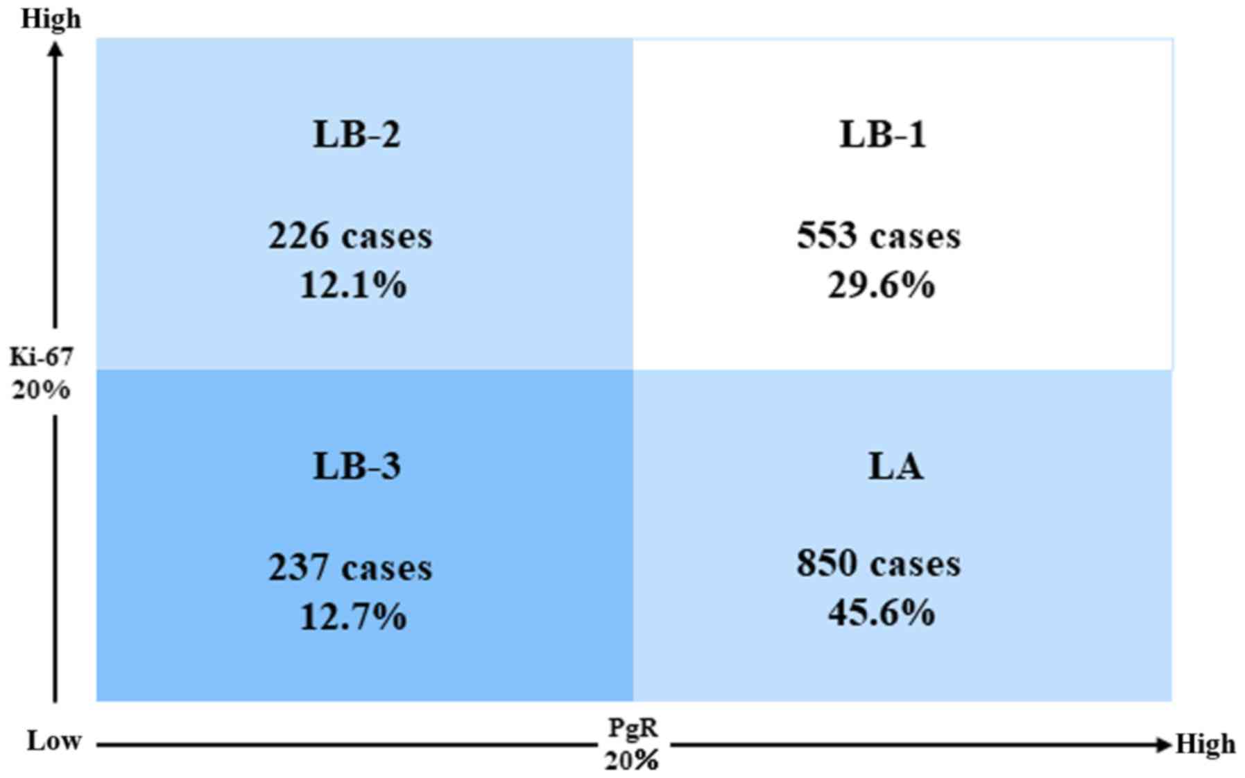
I. The cases were classified as follows (Fig. 1); LA (high PgR/low Ki-67; 850 cases),
LB-1 (high PgR/high Ki-67; 553 cases), LB-2 (low PgR/high Ki-67;
226 cases), and LB-3 (low PgR/low Ki-67; 237 cases). Out of all
these cases, 1,510 were treated with endocrine therapy alone. The
median observation period was 78.1 months. Moreover, 41 of the
cases underwent a 21-gene expression assay and the RS (≤25 and
>25) was compared with our above mentioned classification.
 | Table I.Patient characteristics (n=1866). |
Table I.
Patient characteristics (n=1866).
| Characteristics | N (%) |
|---|
| Age (years) | 58.5+/-0.31
(median+/-SEM) |
| Menopausal
status |
|
| Pre | 687 (36.8) |
| Post | 1,179 (63.2) |
| Tumor size (cm) |
|
|
<2 | 1,400 (75.0) |
| ≥2 | 462 (24.8) |
|
Unknown | 4 cases |
| Nuclear grade |
|
| 1 | 1,142 (62.6) |
| 2 | 527 (28.9) |
| 3 | 154 (8.5) |
|
Unknown | 43 cases |
| ER-positive |
|
| PgR
≥20% | 1,404 (75.2) |
| PgR
<20% | 462 (24.8) |
| p53
overexpression |
|
|
Without | 1,724 (93.2) |
| With | 127 (6.8) |
|
Unknown | 15 cases |
| Ki-67 |
|
|
<20% | 1,087 (58.3) |
| ≥20% | 779 (41.7) |
| Adjuvant therapy |
|
| None | 137 (7.3) |
|
Endocrine | 1,510 (80.9) |
|
Chemo-endocrine | 219 (11.8) |
Histopathological examination
The factors investigated were the lymph nodal
status, tumor size, nuclear grade, ER/PgR and HER2 status,
overexpression of p53 protein and the Ki-67 index value. ER, PgR,
HER2, p53 and Ki-67 were evaluated using IHC with an autostainer
(Benchmark XT; Ventana Medical Systems, Inc., Tucson, USA) in
accordance with the procedure previously reported (13). The antibody used was ER (clone SP1;
rabbit monoclonal), PgR (clone 1E2; rabbit monoclonal), HER2 (clone
4B5; rabbit monoclonal; all Ventana Medical Systems, Inc.), p53
(clone DO7; mouse monoclonal) and Ki-67 (clone MIB-1; mouse
monoclonal; both Dako; Agilent Technologies, Inc., Santa Clara, CA,
USA). The ER- and PgR-positive cell rates were calculated using
IHC. The ASCO/CAP guidelines recommend a value of ≥1% stained tumor
nuclei as being positive.
The percentage of positive nuclei for Ki-67 was
calculated based on a count of at least 500 tumor cells in the hot
spot. The p53 overexpression was determined in the cases with
positive cells ≥50% (8). Positive for
HER2 is either a HER2 score of 3 + (strong and diffuse staining) or
FISH amplified in equivocal cases (score 2+). The other staining
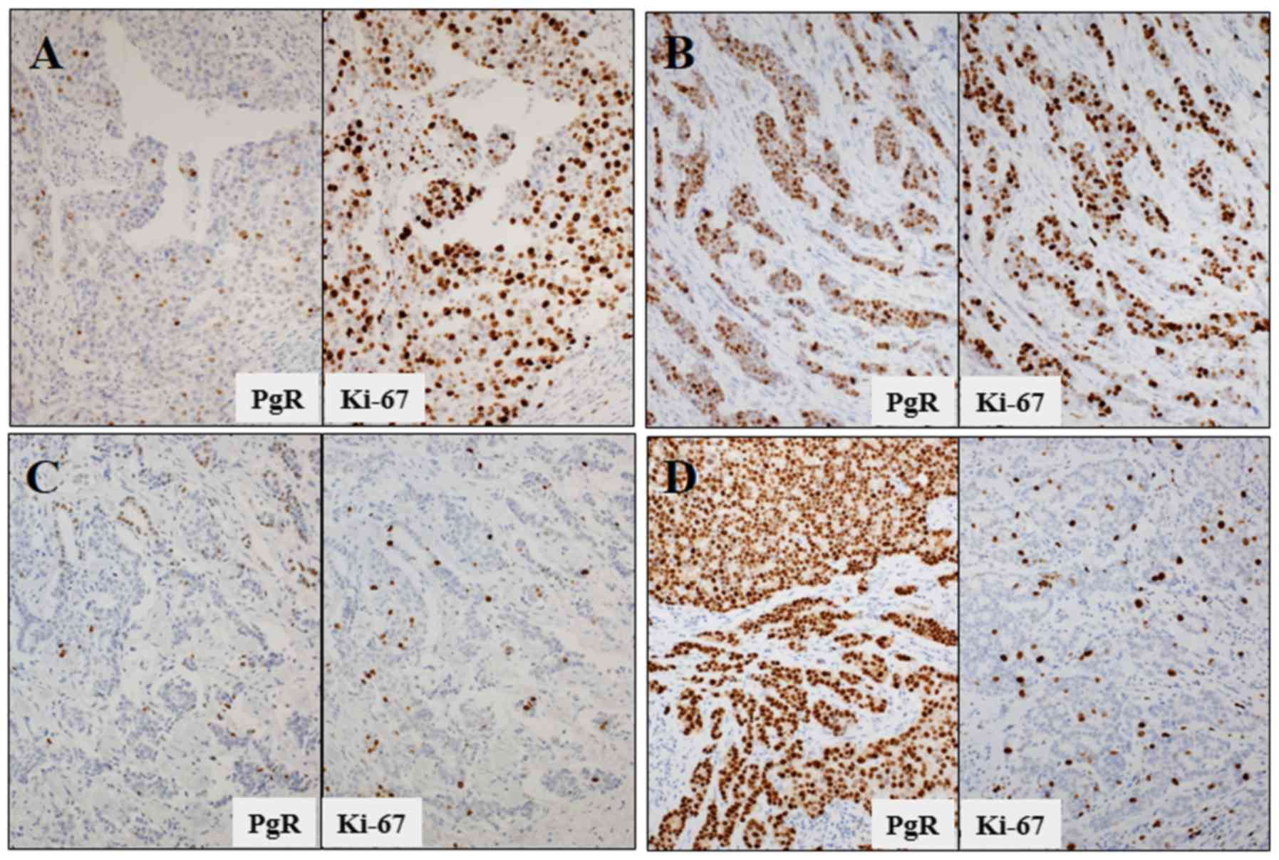
pattern of HER2 was determined as HER2-negative. Fig. 2 shows the subclassified cases of
ER-positive and HER2-negative breast cancer according to the PgR
and the Ki-67 status.
Statistical analysis
Statistical analysis was performed using SPSS
version 21 (IBM Corp., Armonk, NY, USA). Comparisons between groups
(Tables II and III) were analyzed using the Chi-square
test and Fisher's exact test. Age was determined using the
Student's t-test. Cumulative DFS was calculated using the
Kaplan-Meier method and evaluated using the log-rank test.
| Table II.The biological classification and
clinicopathological characteristics in ER-positive and
HER2-negative breast cancer. |
Table II.
The biological classification and
clinicopathological characteristics in ER-positive and
HER2-negative breast cancer.
|
| Cancer subtypes |
|
|
|---|
|
|
|
|
|
|---|
| Characteristics | LA | LB-1 | LB-2 | LB-3 | Total | P-valuea |
|---|
| Age (years,
median+/-SEM) | 58.4+/-0.46 | 55.5+/-0.57 | 60.5+/-0.89 | 63.8+/-0.71 | 58.5 | <0.0001 |
| Menopausal
status |
| Pre | 324 | 283 | 54 | 26 | 687 | <0.001 |
|
Post | 526 (61.9) | 270 (48.8) | 172 (76.1) | 172 (89.0) | 1,179 |
|
| Tumor size
(cm) |
|
<2 | 700 | 390 | 120 | 190 | 1,400 | <0.0001 |
| ≥2 | 150 (17.6) | 161 (29.2) | 104 (46.4) | 47 (19.8) | 462 |
|
| Nuclear grade |
| 1 | 649 | 238 | 72 | 182 | 1,142 | <0.0001 |
| 2 | 171 | 235 | 78 | 43 | 527 |
|
| 3 | 11 (1.3) | 72 (13.2) | 66 (30.6) | 5 (2.2) | 154 |
|
| p53
overexpression |
|
Without | 834 | 497 | 165 | 228 | 1,724 | <0.0001 |
|
With | 15 (1.8) | 54 (9.8) | 52 (24.0) | 6 (2.6) | 127 |
|
| Adjuvant
therapy |
|
None | 77 | 20 | 21 | 19 | 137 | <0.0001 |
|
Endocrine | 743 (87.4) | 428 (77.4) | 133 (58.8) | 206 (86.9) | 1,510 |
|
|
Chemo-endocrine | 30 (3.5) | 105 (19.0) | 72 (31.9) | 12 (5.1) | 219 |
|
| Table III.Adjuvant therapy and tumor
characteristics in the node-negative cases. |
Table III.
Adjuvant therapy and tumor
characteristics in the node-negative cases.
|
| Adjuvant
therapy |
|
|
|---|
|
|
|
|
|
|---|
| Variables | None | Endocrine |
Chemo-endocrine | Total | P-value |
|---|
| Grade |
| 1 | 93 (8.1) | 1,005 (87.8) | 46 (4.0) | 1,144 |
<0.0001a |
| 2 | 28 (5.3) | 416 (78.9) | 83 (15.7) | 527 |
|
| 3 | 13 (8.4) | 56 (36.1) | 86 (55.5) | 155 |
|
|
Total | 134 | 1477 | 215 | 1,826 |
|
| Tumor size
(cm) |
|
<2 | 110 (7.8) | 1,174 (83.6) | 121 (8.6) | 1,405 | 0.04 |
| ≥2 | 30 (6.5) | 335 (72.2) | 99 (21.3) | 464 |
|
|
Total | 140 | 1,509 | 220 | 1,869 |
|
| p53
overexpression |
|
Without | 120 (7.0) | 1,442 (83.6) | 163 (9.4) | 1,725 |
<0.0001a |
|
With | 17 (13.2) | 58 (45.0) | 54 (41.9) | 129 |
|
|
Total | 137 | 1,500 | 217 | 1,854 |
|
Results
Patient characteristics and biological
classification
Table II shows the
relationship between the characteristics of the study cohort and
the biological classification. The mean age was 58.5 years (range,
25–94). T1 (<2 cm) tumors were often seen in the LA group and
rare in the LB-2 group. Nuclear grade 3 and p53 overexpression
significantly correlated with LB-2. Endocrine therapy alone was
performed in 87.4% (LA), 77.4% (LB-1), 58.8% (LB-2) and 86.9%
(LB-3), respectively.
DFS based on the PgR/Ki-67 status in
node-negative cases
The Kaplan-Meier curves show the outcomes for DFS
according to the PgR/Ki-67 status in the node-negative cases
(Fig. 3). There were significant
differences in DFS between the LA group (5-year DFS, 98%; 10-year
DFS, 95.9%), the LB-2 group (5 years, 89.9%; 10 years, 83.6%;
P<0.0001) and the LB-1 group (5 years, 94.9%; 10 years, 89.5%;
P<0.0001), but there was no difference in the LB-3 group (5
years, 98.6%; 10 years, 94.7%; P=0.88). In the cases with endocrine
therapy alone (Fig. 4), LA and LB-3
had a similar DFS rate (P=0.25). LB-2 had a significantly worse DFS
in all cases and in the cases with endocrine therapy alone.
Chemotherapy in combination with endocrine therapy was administered
to cases with a higher nuclear grade, a larger tumor and p53
overexpression (Table III). In the
LB-2 group (Table IV), no difference
in DFS was observed between the cases treated with endocrine
therapy and the cases treated with chemo-endocrine therapy.
However, in the other groups, the cases treated with endocrine
therapy had a significantly more favorable DFS than those treated
with chemo-endocrine therapy.
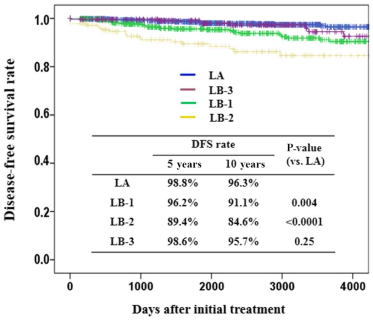
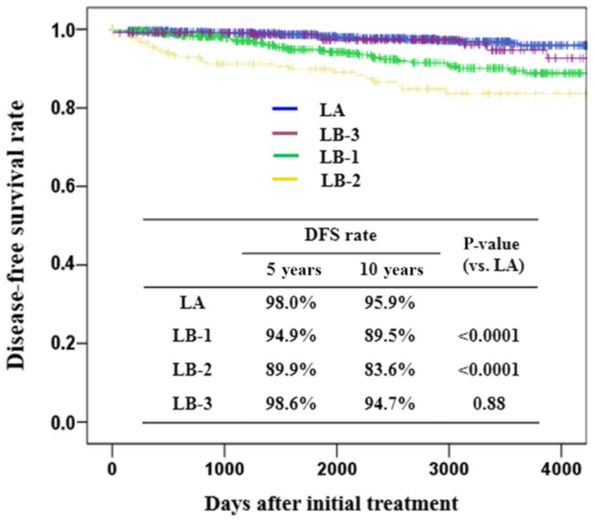 | Figure 3.DFS according to PgR/Ki-67 status in
the node-negative cases. There were significant differences in DFS
between the LA group (5-year DFS, 98%; 10-year DFS, 95.9%), the
LB-2 group (5 years, 89.9%; 10 years, 83.6%; P<0.0001) and the
LB-1 (5 years, 94.9%; 10 years, 89.5%; P<0.0001), but there was
no difference with the LB-3 group (5 years, 98.6%; 10 years, 94.7%;
P=0.88). DFS, disease-free survival; LB-1, high PgR/high Ki-67;
LB-2, low PgR/high Ki-67; LB-3, low PgR/low Ki-67. |
| Table IV.Comparison of DFS between endocrine
therapy alone and chemo-endocrine therapy in terms of PgR/Ki-67
status in the node-negative cases. |
Table IV.
Comparison of DFS between endocrine
therapy alone and chemo-endocrine therapy in terms of PgR/Ki-67
status in the node-negative cases.
|
| DFS (rate) |
|---|
|
|
|
|---|
|
| Endocrine
therapy | Chemo-endocrine
therapy |
|
|---|
|
|
|
|
|
|---|
| Grade | No. of cases | 5 years (%) | 10 years (%) | No. of cases | 5 years (%) | 10 years (%) |
P-valuea |
|---|
| LA | 745 | 98.9 | 97.1 | 31 | 83.2 | 83.2 | <0.0001 |
| LB-1 | 428 | 96.7 | 92.3 | 105 | 88.1 | 81.5 | <0.0001 |
| LB-2 | 131 | 91.7 | 85.0 | 72 | 91.5 | 81.4 | 0.24 |
| LB-3 | 205 | 98.9 | 95.7 | 12 | 80.9 | 80.0 | 0.07 |
RS using a 21-gene expression assay
and biological classification
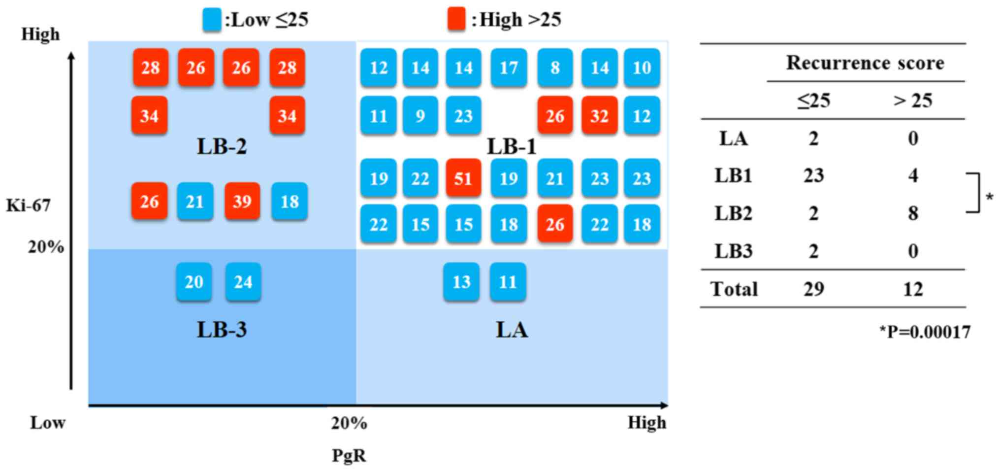
A relationship was found between the RS derived by
using a 21-gene expression assay and biological classification
using Ki-67 and PgR expressions (Fig.
5). There were 29 cases with RS ≤25 and 12 cases with RS
>25. Moreover, most of the cases with LA (2/2), LB-3 (2/2) and
LB-1 (23/27) had a RS of ≤25, and most of the LB-2 (8/10) cases had
a RS of >25. There was a significant difference in RS between
the LB-1 and LB-2 groups (P=0.00017).
Discussion
The purpose of this study was to investigate the
clinical efficacy of a biological classification using the PgR and
the Ki-67 status (cutoff points: 20%) so that it could be used as
an effective prognostic and predictive classification for
determining a suitable postoperative treatment for primary breast
cancer with luminal/HER2-negative and negative nodes. Moreover, the
above mentioned classification was also compared with the RS
derived from the Oncotype DX assay. In this study, it was found
that only the LB-2 cases (low PgR/high Ki-67) benefited from
chemotherapy.
Luminal A type tumors tend to have a better
prognosis, a higher survival rate and a lower recurrence rate among
all the breast cancer subtypes and luminal B type tumors tend to
have a more unfavorable prognosis (14–17). In
this study, all of the Luminal type tumors (1,866 cases) were
categorized into the following 4 groups; LA and three LB groups.
Luminal A occupied the majority and correlated with a smaller
tumor, lower nuclear grade and lower p53 overexpression. On the
other hand, nuclear grade 3, larger tumor and p53 overexpression
significantly correlated with LB-2. In addition, LB-3 correlated
with postmenopausal status, smaller tumor and a lower nuclear
grade. These findings indicate that there are biological
differences among LB tumors.
In terms of prognosis after initial surgery,
significant differences in DFS were observed between the LA group,
the LB-2 group and the LB-1 group, but there was no difference in
the LB-3 group. In the cases with endocrine therapy alone, LA
showed a similar DFS as LB-3. LB-2 had a significantly worse DFS in
all of the cases and in the cases with endocrine therapy alone.
These findings suggest that the tumors with low Ki-67 values (LA
and LB-3) had a favorable DFS irrespective of the PgR status.
However, the tumors with a high Ki-67 value but a low PgR
expression (LB-2) tended to have a worse DFS rate.
Oncotype DX was originally used in ER-positive
tumors and is supported by the ASCO (18) and the NCCN Guidelines (19). The RS result predicts the possibility
of obtaining a beneficial effect from chemotherapy and a higher
score increases the potential benefit of chemotherapy (20,21). In
this study, RS correlated with the biological classification. RS
was lower in the LA, LB-1 and LB-3 groups but higher in the LB-2
group. These data suggest that the LB-2 group has a poorer survival
rate but benefits from chemotherapy. There was no difference in DFS
between the cases with endocrine therapy alone and the cases
treated with chemo-endocrine therapy. On the other hand, the LA,
LB-1 and LB-3 groups showed a favorable survival rate in the cases
treated with endocrine therapy alone. The cases with a high Ki-67
value and low PgR benefited from chemotherapy and this was also
predicted in the RS data.
A multivariate analysis revealed that the Ki-67
index was a significant factor for DFS and that the Ki-67 index
value was a significant prognostic factor only in luminal type
tumors (3,22). Moreover, according to the data from a
large cohort clinical study (23),
the Ki-67 index value is frequently evaluated in routine clinical
work. The Ki-67 expression is an independent prognostic factor for
DFS and OS in addition to common histopathological variables and
the Ki-67 index independently raises the possibility of predicting
the treatment response and prognosis in a group of breast cancer
patients who underwent neoadjuvant treatment (24). The Ki-67 index was also found to be an
independent predictive factor for pCR (OR 3.5; 95% CI, 1.4, 10.1).
The mean Ki-67 value in patients with pCR was 50.6±23.4%, and the
average Ki-67 value in non-pCR patients was 26.7±22.9%. In a
previous study it was found (25)
that ER-positive and PgR-negative/HER2-negative tumors were
associated with poorer survival than cases with ER-positive and
PgR-positive tumors and had a comparatively poorer survival rate to
that of the triple negative breast cancers. Moreover, Prat et
al (5) found that the IHC
expression of PgR increases the prognostic value within the current
IHC-based luminal A definition by improving the identification of
favorable breast cancers and the percentage of PgR-positive cells.
Moreover, they found that the optimal PgR expression cutoff point
to predict outcome was 20%. However, the ER-positive cell rates did
not correlate with DFS even after matching for the standard
clinicopathologic parameters. A retrospective analysis of three
adjuvant clinical trials found that low ER and low PgR expression,
and potentially low PgR expression within ER-positive patients were
efficacious factors for determining the validity of adding
chemotherapy to endocrine therapy (26). These data indicate that the Ki-67
index value and PgR status are important predictors for prognosis
and chemotherapy.
In conclusion, the biology, prognosis and suitable
treatment for Luminal tumors were evaluated based on the PgR and
Ki-67 index value. The patients with a Ki-67 value <20% (LA and
LB-3) had a favorable DFS even in the endocrine therapy alone
group, whereas those with a Ki-67 value ≥20% (LB-1 and LB-2) had a
poorer DFS. Moreover, LB-2 (PgR<20% and Ki-67≥20%) significantly
correlated with a higher degree of malignancy but benefited from
chemotherapy. The LA and LB-3 cases with low Ki-67 values were
considered to be a part of the Luminal A group. These findings
suggest that the PgR and Ki-67 status are useful in predicting
prognosis and determining the most effective treatment strategy for
patients with ER-positive and HER2-negative breast cancer.
Acknowledgements
The authors would like to thank the staff of the
Department of Pathology at Kumamoto Shinto General Hospital
(Kumamoto, Japan) for their technical assistance and for the
collection of cancer tissue samples. The major content of this
study was presented at 2017 San Antonio Breast Cancer Symposium
(http://cancerres.aacrjournals.org/content/78/4_Supplement/P2-09-32).
Funding
No funding was received.
Availability of data and materials
The datasets used and/or analyzed during the current
study are available from the corresponding author on reasonable
request.
Authors' contributions
RN and YT conceived and designed the experiments.
RN, TO, YN, YO, MN and MF performed the experiments and NA, RN, TO,
YN, YO, MN and MF analyzed the data. NA and YT contributed
reagents/materials/analysis tools. NA and RN wrote the
manuscript.
Ethics approval and consent to
participate
The study was approved by the Ethics Committee of
Kumamoto Shinto General Hospital (no. 30-J01-001). All patients
gave a written informed consent to participate in the study.
Patient consent for publication
Written informed consent was provided for the
publication of any data/associated images.
Competing interests
The authors have declared that they have no
competing interests.
References
|
1
|
Goldhirsch A, Winer EP, Coates AS, Gelber
RD, Piccart-Gebhart M, Thürlimann B and Senn HJ; Panel members, .
Personalizing the treatment of women with early breast cancer:
Highlights of the st gallen international expert consensus on the
primary therapy of early breast cancer 2013. Ann Oncol.
24:2206–2223. 2013. View Article : Google Scholar : PubMed/NCBI
|
|
2
|
Arima N, Nishimura R, Osako T, Nishiyama
Y, Fujisue M, Okumura Y, Nakano M, Tashima R and Toyozumi Y: A
Comparison of the hot Spot and the average cancer cell counting
methods and the optimal cutoff point of the Ki-67 index for luminal
type breast cancer. Oncology. 90:43–50. 2016. View Article : Google Scholar : PubMed/NCBI
|
|
3
|
Tashima R, Nishimura R, Osako T, Nishiyama
Y, Okumura Y, Nakano M, Fujisue M, Toyozumi Y and Arima N:
Evaluation of an optimal cut-off point for the Ki-67 index as a
prognostic factor in primary breast cancer: A retrospective study.
PLoS One. 10:e01195652015. View Article : Google Scholar : PubMed/NCBI
|
|
4
|
Bustreo S, Osella-Abate S, Cassoni P,
Donadio M, Airoldi M, Pedani F, Papotti M, Sapino A and Castellano
I: Optimal Ki67 cut-off for luminal breast cancer prognostic
evaluation: A large case series study with a long-term follow-up.
Breast Cancer Res Treat. 157:363–371. 2016. View Article : Google Scholar : PubMed/NCBI
|
|
5
|
Prat A, Cheang MC, Martín M, Parker JS,
Carrasco E, Caballero R, Tyldesley S, Gelmon K, Bemard PS, Nielsen
TO and Perou CM: Prognostic significance of progesterone
receptor-positive tumor cells within immunohistochemically defined
luminal A breast cancer. J Clin Oncol. 31:203–209. 2013. View Article : Google Scholar : PubMed/NCBI
|
|
6
|
Tashima R, Nishimura Y, Arima N, Fujisue
M, Nakano M, Okumura Y, Osako T and Toyozumi Y: P260 Evaluation of
PgR expression as a prognostic factor in luminal HER2-negative
breast cancer. The Breast. 24 Suppl 1:S1162015. View Article : Google Scholar
|
|
7
|
O'Brien KM, Cole SR, Tse CK, Perou CM,
Carey LA, Foulkes WD, Dressler LG, Geradts J and Millikan RC:
Intrinsic breast tumor subtypes, race, and long-term survival in
the Carolina breast cancer study. Clin Cancer Res. 16:6100–6110.
2010. View Article : Google Scholar : PubMed/NCBI
|
|
8
|
Sorlie T, Tibshirani R, Parker J, Hastie
T, Marron JS, Nobel A, Deng S, Johnsen H, Pesich R, Geisler S, et
al: Repeated observation of breast tumor subtypes in independent
gene expression data sets. Proc Natl Acad Sci USA. 100:8418–8423.
2003. View Article : Google Scholar : PubMed/NCBI
|
|
9
|
Brenton JD, Carey LA, Ahmed AA and Caldas
C: Molecular classification and molecular forecasting of breast
cancer: Ready for clinical application? J Clin Oncol. 23:7350–7360.
2005. View Article : Google Scholar : PubMed/NCBI
|
|
10
|
Paik S, Shak S, Tang G, Kim C, Baker J,
Cronin M, Baehner FL, Walker MG, Watson D, Park T, et al: A
multigene assay to predict recurrence of tamoxifen-treated,
node-negative breast cancer. N Engl J Med. 351:2817–2826. 2004.
View Article : Google Scholar : PubMed/NCBI
|
|
11
|
Gianni L, Zambetti M, Clark K, Baker J,
Cronin M, Wu J, Mariani G, Rodriguez J, Carcangiu M, Watson D, et
al: Gene expression profiles in paraffin-embedded core biopsy
tissue predict response to chemotherapy in women with locally
advanced breast cancer. J Clin Oncol. 23:7265–7277. 2005.
View Article : Google Scholar : PubMed/NCBI
|
|
12
|
Barcenas CH, Raghavendra A, Sinha AK, Syed
MP, Hsu L, Patangan MG Jr, Chavez-MacGregor M, Shen Y, Hortobagyi
GH, Valero V, et al: Outcomes in patients with early-stage breast
cancer who underwent a 21-gene expression assay. Cancer.
123:2422–2431. 2017. View Article : Google Scholar : PubMed/NCBI
|
|
13
|
Kai K, Nishimura R, Arima N, Miyayama H
and Iwase H: p53 expression status is a significant molecular
marker in predicting the time to endocrine therapy failure in
recurrent breast cancer: A cohort study. Int J Clin Oncol.
11:426–433. 2006. View Article : Google Scholar : PubMed/NCBI
|
|
14
|
Voduc KD, Cheang MC, Tyldesley S, Gelmon
K, Nielsen TO and Kennecke H: Breast cancer subtypes and the risk
of local and regional relapse. J Clin Oncol. 28:1684–1691. 2010.
View Article : Google Scholar : PubMed/NCBI
|
|
15
|
Arvold ND, Taghian AG, Niemierko A, Abi
Raad RF, Sreedhara M, Nguyen PL, Bellon JR, Wong JS, Smith BL and
Harris JR: Age, breast cancer subtype approximation, and local
recurrence after breast-conserving therapy. J Clin Oncol.
29:3885–3891. 2011. View Article : Google Scholar : PubMed/NCBI
|
|
16
|
Metzger-Filho O, Sun Z, Viale G, Price KN,
Crivellari D, Snyder RD, Gelber RD, Castiglione-Gertsch M, Coates
AS, Goldhirsch A and Cardoso F: Patterns of recurrence and outcome
according to breast cancer subtypes in lymph node-negative disease:
Results from international breast cancer study group trials VIII
and IX. J Clin Oncol. 31:3083–3090. 2013. View Article : Google Scholar : PubMed/NCBI
|
|
17
|
McGuire A, Lowery AJ, Kell MR, Kerin MJ
and Sweeney KJ: Locoregional recurrence following breast cancer
surgery in the trastuzumab era: A systematic review by subtype. Ann
Surg Oncol. 24:3124–3132. 2017. View Article : Google Scholar : PubMed/NCBI
|
|
18
|
Harris L, Fritsche H, Mennel R, Norton L,
Ravdin P, Taube S, Somerfield MR, Hayes DF and Bast RC Jr; American
Society of Clinical Oncology, . American Society of Clinical
Oncology 2007 update of recommendations for the use of tumor
markers in breast cancer. J Clin Oncol. 25:5287–5312. 2007.
View Article : Google Scholar : PubMed/NCBI
|
|
19
|
National Comprehensive Cancer Network
(NCCN) guidelines, Breast Cancer Version 2.2011. http://www.nccn.org/professionals/physician_gls/pdf/breast.pdf
|
|
20
|
Paik S, Tang G, Shak S, Kim C, Baker J,
Kim W, Cronin M, Baehner FL, Watson D, Bryant J, et al: Gene
expression and benefit of chemotherapy in women with node-negative,
estrogen receptor-positive breast cancer. J Clin Oncol.
24:3726–3734. 2006. View Article : Google Scholar : PubMed/NCBI
|
|
21
|
Albain KS, Barlow WE, Shak S, Hortobagyi
GN, Livingston RB, Yeh IT, Ravdin P, Bugarini R, Baehner FL,
Davidson NE, et al: Prognostic and predictive value of the 21-gene
recurrence score assay in postmenopausal women with node-positive,
oestrogen-receptor-positive breast cancer on chemotherapy: A
retrospective analysis of a randomised trial. Lancet Oncol.
11:55–65. 2010. View Article : Google Scholar : PubMed/NCBI
|
|
22
|
Nishimura R, Osako T, Okumura Y, Hayashi
M, Toyozumi Y and Arima N: Ki-67 as a prognostic marker according
to breast cancer subtype and a predictor of recurrence time in
primary breast cancer. Exp Ther Med. 1:747–754. 2010. View Article : Google Scholar : PubMed/NCBI
|
|
23
|
Inwald EC, Klinkhammer-Schalke M,
Hofstädter F, Zeman F, Koller M, Gerstenhauer M and Ortmann O:
Ki-67 is a prognostic parameter in breast cancer patients: Results
of a large population-based cohort of a cancer registry. Breast
Cancer Res Treat. 139:539–552. 2013. View Article : Google Scholar : PubMed/NCBI
|
|
24
|
Fasching PA, Heusinger K, Haeberle L,
Niklos M, Hein A, Bayer CM, Rauh C, Schulz-Wendtland R, Bani MR,
Schrauder M, et al: Ki67, chemotherapy response, and prognosis in
breast cancer patients receiving neoadjuvant treatment. BMC Cancer.
11:4862011. View Article : Google Scholar : PubMed/NCBI
|
|
25
|
Bae SY, Kim S, Lee JH, Lee HC, Lee SK, Kil
WH, Kim SW, Lee JE and Nam SJ: Poor prognosis of single hormone
receptor-positive breast cancer: Similar outcome as triple-negative
breast cancer. BMC Cancer. 15:1382015. View Article : Google Scholar : PubMed/NCBI
|
|
26
|
Viale G, Regan MM, Maiorano E,
Mastropasqua MG, Golouh R, Penin T, Brown RW, Kovács A, Pillay K,
Ohlschlegel C, et al: Chemoendocrine compared with endocrine
adjuvant therapies for node-negative breast cancer: Predictive
value of centrally reviewed expression of estrogen and progesterone
receptors-An International Breast Cancer Study Group. J Clin Oncol.
26:1404–1410. 2008. View Article : Google Scholar : PubMed/NCBI
|