Introduction
Renal cell carcinoma (RCC) accounts for 90% of renal
cancer cases, and is the most common carcinoma of the adult kidney
(1). The incident rate of RCC has
risen during the previous decade (2),
making it the 7 and 8th most common cancer in the USA among males
and females, respectively (3).
Although surgery is currently the most effective treatment approach
for localized RCC tumors and commonly used molecular drugs have
improved over the past several decades (4), 30% of patients continue to develop
metastatic disease following surgical resection (5). Development of specific targets in
relevant biological pathways has been the primary advancement in
treatment (6). In spite of these
advances RCC continues to be a devastating cancer with a low number
of effective treatment options. Therefore, increased efforts should
be made to improve early diagnosis and the monitoring of patients,
particularly patients that undergo resection and treatment for
metastatic RCC.
Coiled-coil-helix-coiled-coil-helix
domain-containing protein 2 (CHCHD2), a member of a family of
proteins containing the CHCH domain, was identified to be
co-expressed with other genes of the oxidative phosphorylation
pathway by using a computational expression screening technique
(7). In addition, Seo et al
(8) revealed that CHCHD2 may be
identified by an in vitro functional genetic selection
strategy as a novel cell migration determinant. Liu et al
(9) reported that CHCHD2 is able to
inhibit apoptosis by interacting with B cell lymphoma-extra large
(Bcl-xL) to regulate Bcl-associated X protein (BAX) activation.
However, less is known regarding the expression and biological
function of CHCHD2 in RCC.
In the present study, CHCHD2 staining in RCC cells
lines and tissues was evaluated. Then, the association between
CHCHD2 expression and clinicopathological variables was assessed.
The possible association between CHCHD2, and movement and
angiogenesis of RCC was also investigated. It was identified that
CHCHD2 expression was increased in human RCC cells and tissue.
CHCHD2 expression was significantly increased in tumor grades II–IV
compared with grade I in accordance with the World Health
Organization classification (P<0.001) (10,11);
however, no significant association was identified between CHCHD2
expression and other clinicopathological variables, including age,
tumor size and Tumor-Node-Metastasis (TNM) stage (10,11). The
present study identified that the migratory capacity of RCC was
inhibited following suppression of CHCHD2 expression using a stable
small interfering (si)RNA compared with a scrambled siRNA control
in vitro. The matrix metalloproteinase-2 (MMP-2) protein
level and enzyme activity were inhibited following CHCHD2
knockdown, confirmed using western blotting and gelatin zymography.
In addition, data revealed that tube formation of human umbilical
vascular endothelial cells (HUVECs) was suppressed following CHCHD2
knockdown, and the ELISA assay identified that the secretion of
vascular endothelial growth factor was inhibited following CHCHD2
knockdown. Taken together these data indicate that CHCHD2 may be a
novel target for the treatment of RCC.
Materials and methods
Ethics statement
The Institutional Review Board of Xuzhou Medical
University (Xuzhou, China) approved the present study, conducted in
accordance with the approved guidelines. All patients provided
written informed consent for their colorectal tissue samples to be
used in the present study.
Patients and samples
The suprarenal epithelioma tissue microarray (TMA)
was purchased from Shanghai Outdo Biotech (Shanghai, China)
(http://superchip.bioon.com.cn/). It
included 75 patients (mean age, 59) who underwent radical
nephrectomy between 2006 and 2008. In this TMA, the array dot
diameter was 1.5 mm and each dot represented a tissue spot from one
individual specimen. The patients consisted of 50 (66.7%) men and
25 (33.3%) women. A total of 42 (56%) patients were aged ≤57 years
and 32 (42.7%) ages >57 years. A total of 57 (76%) patients were
classified TNM I–II and 18 (24%) TNM stage III–IV. Details of the
patient characteristics and CHCHD2 expression are provided in
Table I. Additionally, three RCC
tissues and paired non-cancerous tissues were obtained from The
Affiliated Hospital of Xuzhou Medical University. Patients with
ccRCC who underwent radical nephrectomy without prior treatment
were recruited from the Affiliated Hospital of Xuzhou Medical
University between January 2006 and December 2008. The
clinicopathological information, including age at diagnosis, sex,
tumor diameter, depth of invasion, lymph node metastasis and TNM
stage, was obtained from the medical records in the Affiliated
Hospital of Xuzhou Medical University. All the tissue specimens
were obtained for the present research with patients' informed
consent, and the use of human specimens was approved by the Review
Board of the Affiliated Hospital of Xuzhou Medical University.
 | Table I.CHCHD2 staining and
clinicopathological characteristics of 75 patients with renal
cancer. |
Table I.
CHCHD2 staining and
clinicopathological characteristics of 75 patients with renal
cancer.
|
| CHCHD2 staining |
|---|
|
|
|
|---|
| Variables | Weak, n (%) | Strong, n (%) | Total, n | P-valuea |
|---|
| Age, years | 38 (50.7) | 37 (49.3) | 75 |
|
| ≤56 | 19 (52.8) | 17 (47.2) | 36 | 0.725 |
|
>56 | 19 (48.7) | 20 (51.3) | 39 |
|
| Sex |
|
Male | 25 (50.0) | 25 (50.0) | 50 | 0.870 |
|
Female | 13 (52.0) | 12 (48.0) | 25 |
|
| Tumor size, cm |
| ≤7 | 24 (55.8) | 19 (44.2) | 43 | 0.301 |
|
>7 | 14 (43.8) | 18 (56.2) | 32 |
|
| Grade |
| I | 2 (66.7) | 1 (33.3) | 3 | <0.001 |
| II | 29 (80.6) | 7 (19.4) | 36 |
|
|
III | 7 (25.0) | 21 (75.0) | 28 |
|
| IV | 0 (0.0) | 8 (100.0) | 8 |
|
| TNM stage |
| I | 21 (63.6) | 12 (36.4) | 33 | 0.166 |
| II | 11 (45.8) | 13 (54.2) | 24 |
|
|
III | 6 (35.3) | 11 (64.7) | 17 |
|
| IV | 0 (0.00) | 1 (100.0) | 1 |
|
Cell lines and transfection
All cell lines were obtained from the Shanghai
Institute of Biochemistry and Cell Biology, Chinese Academy of
Sciences (Shanghai, China). HK-2 and ACHN were grown in Dulbecco's
modified Eagle's medium (DMEM; Invitrogen; Thermo Fisher
Scientific, Inc., Waltham, MA, USA) supplemented with 15% fetal
bovine serum (FBS; Invitrogen; Thermo Fisher Scientific, Inc.),
while 786-O and Ketr-3 cells were cultured in RPMI-1640
(Invitrogen; Thermo Fisher Scientific, Inc.) supplemented with 10%
FBS. All cells were maintained as monolayer cultures at 37°C in a
humidified incubator with an atmosphere of 5% CO2.
The CHCHD2 siRNA and scrambled siRNA were purchased
from GenePharma (Shanghai, China) (Stealth RNAi targeting CHCHD2
5′-GGGCACACATTGGGTCACGCCATTA-3′). Transfection of 1.25 µl 20 mM
siRNAs into 40% confluent 786-O and ACHN cells was performed using
siLentFect Lipid reagent (Bio-Rad Laboratories, Inc., Hercules, CA,
USA) according to the manufacturer's protocol. Cell migration
assays were performed 48 h after transfection.
Immunohistochemistry of TMA
Immunohistochemistry was performed using a standard
avidin biotinylated-HRP complex (ABC) kit (Zhongshan Belling
Biotechnology Co., Ltd., Beijing, China). TMA sections were
deparaffinized in xylene and then rehydrated with graded ethanol
and distilled water. According to the manufacturer's protocol,
diaminobenzidine (DAB; Zhongshan Jinqiao Biotechnology Co., Ltd.,
Beijing, China) was used to produce a brown precipitate. TMA slides
were incubated with homemade rabbit anti-CHCHD2 antibody (1:200
dilution; a gift from Dr Yanping Zhang, University of North
Carolina at Chapel Hill, Chapel Hill, NC, USA) overnight at 4°C,
and incubated with a goat anti-rabbit biotinylated secondary
antibody (ready to use; catalog no. PV-9001, Jinqiao Biotechnology
Co., Ltd.) at room temperature for 30 min. The sections were
incubated with streptavidin-peroxidase (Zhongshan Belling
Biotechnology Co., Ltd.) at room temperature for an additional 30
min, washed with phosphate-buffered saline (PBS) and stained using
DAB at room temperature for 15 min. Subsequently, sections were
rinsed in distilled water and counterstained with hematoxylin
(Beyotime Institute of Biotechnology, Shanghai, China) at room
temperature for 30 sec. Using a light microscope (Olympus BX-51
light microscope) and a Camedia Master C-3040 digital camera (both
from Olympus Corporation, Tokyo, Japan), the CHCHD2 expression in
TMA was identified as positive when ≥5% of tumor cells exhibited
immunopositivity, while biopsies with <5% tumor cells exhibiting
immunostaining were graded negative. Samples with an
immunoreactivity score (IRS) of 0–3 and 4–12 were classified as
weak and strong expression of CHCHD2, respectively.
Cell migration assays
The cell migration assay was executed using 6.5 mm
Transwell Boyden chambers (pore size, 8 µm; Corning Incoporated,
Corning, NY, USA) as described previously, but with a number of
modifications (12). Cell suspension
(100 µl) without serum at a density of 4×10⁴ cells/well
was seeded in the Transwell chambers. After 12 h for migration
assays, cells in the upper chamber were carefully removed with a
cotton swab. The cells that had migrated to the basal side of the
membrane were fixed in 100% methanol at room temperature for 30 min
and stained with crystal violet (0.04% in water, 100 µl; Beyotime
Institute of Biotechnology) at room temperature for 15–30 min. The
permeating cells were counted under an inverted microscope (Ti-U;
Nikon Corporation, Tokyo, Japan) and images were captured at a
magnification of ×10.
Western blotting and antibodies
Cells were transiently transfected with siRNA for 48
h. The cells were then lysed with 0.5% NP-40 lysis buffer and
protein concentrations were assessed using a bicinchoninic acid
assay. The loading protein (100 µg) was separated by 12% SDS-PAGE
under reducing conditions, and then transferred onto nitrocellulose
membranes. The membranes were blocked at room temperature for 2 h
in Blotto (5% skimmed milk and PBS). The primary antibodies
utilized at 4°C overnight were rabbit anti-CHCHD2 (1:1,000;
supplied by Dr Yanping Zhang), rabbit anti-MMP2 (1:1,000; catalog
no. 4022s; Cell Signaling Technology, Inc., Danvers, MA, USA) and
mouse anti-β-actin (1:5,000; catalog no. BM0626; Wuhan Boster
Biological Technology, Wuhan, China). Each antibody was used
according to the manufacturer's protocol. Fluorescence-conjugated
secondary antibody (IRDye 700/800; Rockland Immunochemicals Inc.,
Limerick, PA) was used at a dilution of 1:10,000 at room
temperature for 2 h. Analysis was performed using the Odyssey
Infrared Imaging system (LI-COR Biosciences, Lincoln, NE, USA). All
experiments were performed at least three times unless otherwise
indicated.
Gelatin zymography analysis
The gelatinolytic activity was analyzed using
gelatin zymography according to the method previously described
with a number of modifications (13).
Prior to gelatin zymography, cells were cultured in serum-free
conditioned culture medium overnight. Next, the culture conditioned
medium was harvested and the proteins in the medium were
concentrated with Amicon Ultra-4–30 k centrifugal filters (EMD
Millipore, Billerica, MA, USA) at 7,500 × g for 20 min at 4°C. A
total of 30 µg protein was loaded in non-redenaturing conditions on
a 10% polyacrylamide gel containing 0.1% gelatin. The SDS was
removed from the gel using 2.5% Triton X-100 for 1 h at room
temperature. The gel was incubated overnight at 37°C in development
buffer (20 mM Tris-HCl, pH 8.0, 150 mM NaCl, 5 mM CaCl2,
and 0.01% NaN3), stained with 0.5% Coomassie blue R250
(Sigma-Aldrich; Merck KGaA, Darmstadt, Germany) at room temperature
for 1 h, and destained with 30% methanol and 10% glacial acetic
acid for 2 h at room temperature.
Tube formation assay
Transfected 786-O and ACHN cells (1×10⁶)
were cultured in a 6-well plate with fresh complete medium for 48
h, then collected and centrifuged at 800 × g at room temperature
for 5 min to remove any cell debris prior to its use as a
conditioned medium. Next, 48-well plates coated with Matrigel were
kept at 37°C for 30 min prior to the endothelial cell tube
formation assay. HUVECs (Shanghai Institute of Biochemistry and
Cell Biology, Chinese Academy of Sciences, Shanghai, China)
(2×10⁴/well) were suspended in 100 µl conditioned medium
and applied to the pre-coated 48-well plate. The number of
capillary-like tubes was quantified after 24 h in three random
microscopic fields (magnification, ×10) with a computer-assisted
microscope (Nikon Corporation).
ELISA for VEGF
In order to determine the concentration of VEGF,
786-O and ACHN cells were transfected with siRNA subsequent to
being plated in 6-well tissue culture plates at a density of
1×10⁶ cells/well. The supernatants were harvested 48 h
after transfection. The VEGF concentration was measured using a
Quantikine ELISA kit (catalog no. DVE00; R&D Systems, Inc.,
Minneapolis, MN, USA) according to the manufacturer's protocol.
Statistical analysis
All the data in the figures and text are presented
as the mean ± standard deviation from >3 independent
experiments. The differences in the TMA were analyzed using SPSS
software (version 16.0; SPSS, Inc., Chicago, IL, USA). The
association between staining of CHCHD2, and the clinicopathological
parameters of the patients with suprarenal epithelioma, including
age, sex, World Health Organization grade and histological type
(10,11), was evaluated by the χ2
test. Differences in treatment groups were assessed by two-way
analysis of variance followed by the Dunnett's test. P<0.05 was
considered to indicate a statistically significant difference.
Results
CHCHD2 expression is increased in
human RCC tissues and cell lines
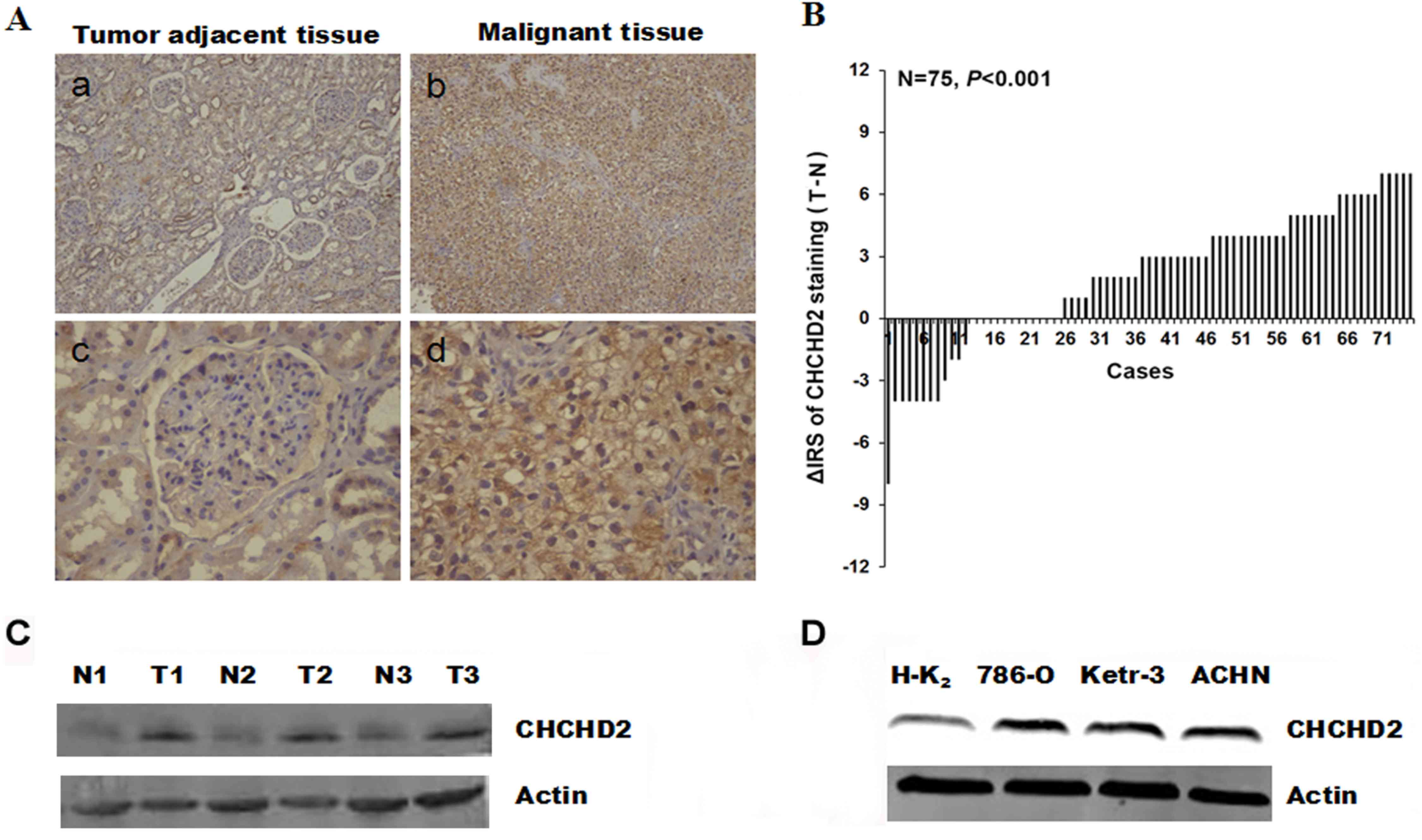
The expression levels of CHCHD2 were evaluated in
human RCC. Immunohistochemical staining was performed in RCC
tissues and paired non-cancerous tissues. The representative images
presented in Fig. 1A revealed that
CHCHD2 protein in cytoplasm was stained brown. Significantly higher
expression of CHCHD2 was observed in the carcinoma tissues
(P<0.001; Fig. 1B). In order to
confirm the aforementioned observation, western blotting was
performed using three RCC tissues together with paired
non-cancerous tissues. Notably, the level of CHCHD2 protein was
markedly increased in malignant tumor tissues compared with
non-cancerous tissues (Fig. 1C).
Additionally, the differences in expression of CHCHD2 between
normal renal cells and carcinoma cells were investigated. Western
blotting was performed to measure the CHCHD2 expression in distinct
renal cell lines. The results identified that CHCHD2 expression
levels were markedly increased in human 786-O, Ketr-3 and ACHN RCC
cells, compared with HK-2 normal renal cells (Fig. 1D). This result was consistent with the
level of CHCHD2 protein expression in RCC tissues.
Association between CHCHD2 expression
levels and clinicopathological parameters
To further investigate the possible function of
CHCHD2 in RCC, the associations between CHCHD2 expression in
primary tumor tissue samples from 75 patients and
clinicopathological parameters were assessed. The mean age of the
patients with suprarenal epithelioma was 58.7 years (50 males and
25 females with a median age of 56 years; range, 29–82 years). The
comparisons of numerous clinicopathological characteristics with
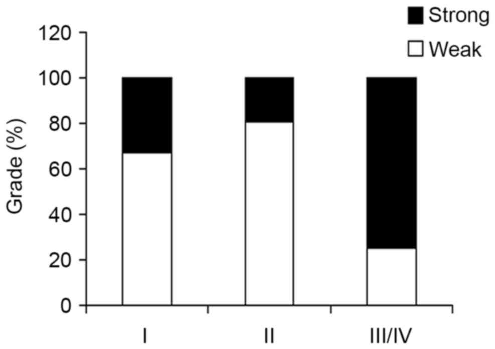
CHCHD2 protein expression are presented in Table I. The results revealed that CHCHD2
staining was significantly increased in tumor grade II–IV compared
with tumor grade I (P<0.001; Fig.
2). Although TNM stage is an important prognostic marker for
patients with RCC, no significant association was observed between
CHCHD2 expression and age (P=0.725), sex (P=0.870), tumor size
(P=0.301) and TNM stage (P=0.166).
Knockdown of CHCHD2 inhibits migration
of RCC cells in vitro
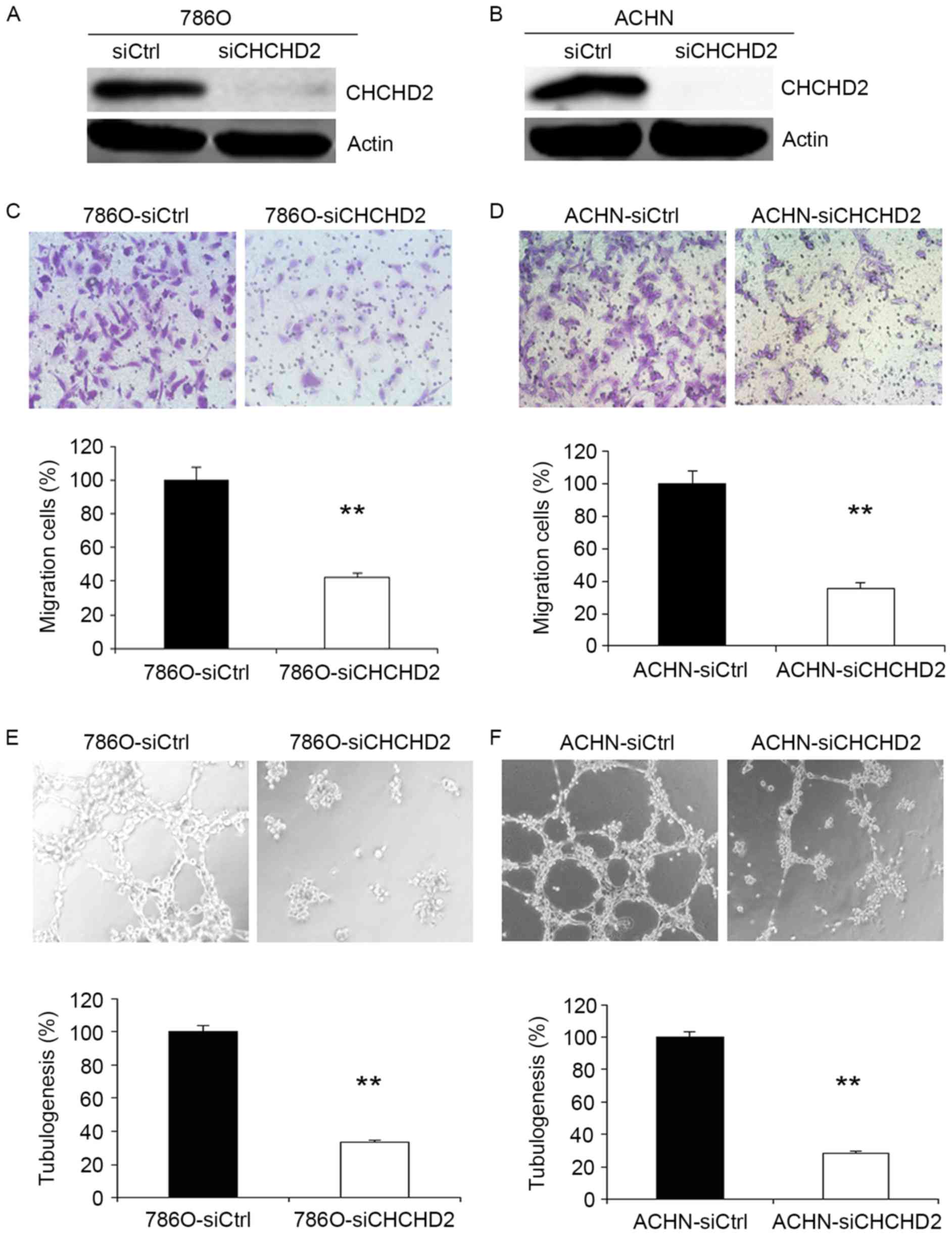
To further investigate the function of CHCHD2 in
tumor cell migration, human 786-O and ACHN cells were transfected
with control siRNA and CHCHD2 siRNA. At 48 h after transfection,
CHCHD2 protein was markedly downregulated in cancer cells (Fig. 3A and B). As presented in Fig. 3C, silencing of CHCHD2 significantly
inhibited the 786-O cells ability to migrate through the Boyden
chambers. In order to examine the effect of CHCHD2 knockdown on
other RCC cells, ACHN cell migratory assays were performed. The
data gathered revealed that the migratory ability of ACHN cells was
also suppressed by silencing of CHCHD2 (Fig. 3D).
Knockdown of CHCHD2 suppresses
angiogenesis in RCC cells
Angiogenesis induces the growth and metastasis of
tumors (14). Therefore, the effect
of angiogenesis following knockdown of CHCHD2 was investigated. The
endothelial cell tube formation assay (Fig. 3E and F) identified that conditioned
medium from CHCHD2 knockdown cancer cells significantly reduced the
average number of complete tubular structures formed by HUVECs
compared with those of control cells.
Knockdown of CHCHD2 decreases MMP-2
expression, activities and VEGF secretion in RCC cells
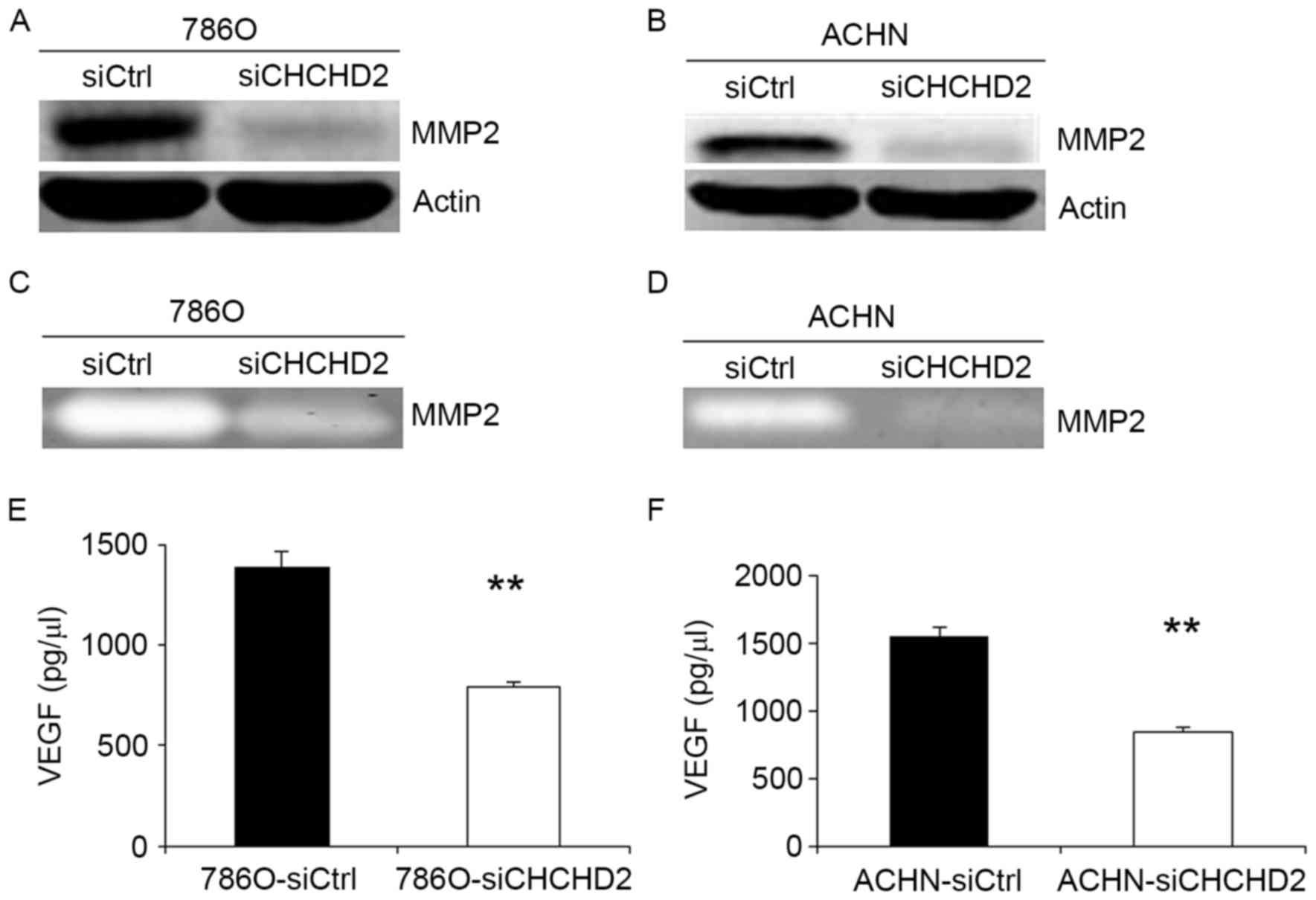
In order to improve understanding of the mechanism
of siCHCHD2-induced cell migration and angiogenesis inhibition in
RCC cells, the MMP expression levels and activities, and VEGF
expression levels in 786-O and ACHN cells were measured. The
invasive ability of cancer cells may be regulated by MMPs (15–17), and
VEGF is considered one of the most prominent pro-angiogenic factors
(18–21). The results of the present study
revealed that MMP-2 levels were markedly suppressed in
786-O-siCHCHD2 and ACHN-siCHCHD2 cells compared with the control
cells (Fig. 4A and B). The activities
of MMP-2 protein were also markedly decreased in 786-O and ACHN
cells transfected with siCHCHD2 using gelatin zymography (Fig. 4C and D). Additionally, it was
identified that VEGF secretion was significantly suppressed in
786-O and ACHN cells transfected with siCHCHD2 compared with the
control cells (Fig. 4E and F).
Discussion
Previous research identified that Cytochrome c
oxidase (COX) is the terminal enzyme of the electron transport
chain, and COX subunit 4 (COX4) is a key regulatory subunit
(22). Aras et al (22) demonstrated that oxygen responsive
element (ORE) is a highly conserved 13-bp sequence in the proximal
promoter of the tissue-specific (predominantly lung) COX4 isoform
2, and further confirmed that CHCHD2, as a transcription factor,
bound this conserved ORE and activated the ORE at 4% oxygen. Seo
et al (8) reported that CHCHD2
protein was able to mutually regulate the balance of cell migration
by directly interacting with hyaluronic acid-binding protein 1, and
CHCHD2 was identified as a novel cell migration determinant using
an in vitro functional genetic selection strategy. In
addition, CHCHD2 is involved in mitochondrial function (23) and has been identified in a screen for
HTT interacting proteins (24,25). Liu
et al (9) demonstrated that
CHCHD2 was able to inhibit apoptosis by interacting with Bcl-xL to
regulate BAX activation. Furthermore, increasing evidence suggests
a potential function of CHCHD2 in CHCHD2-Bcl-xL protein
interactions.
In the present study, CHCHD2 expression was first
examined at the protein level in RCC cells and tissues using
western blotting. The results indicate that the expression of
CHCHD2 protein was more abundant in suprarenal epithelioma cells
and tissues compared with that of the normal renal cells and
tissues. The expression level of CHCHD2 was markedly associated
with tumor grade in data gathered from a TMA. However, little is
known about the association between CHCHD2 expression and patients
with RCC, and the functional role of CHCHD2 in the progress of RCC
remains unresolved. To determine whether CHCHD2 regulates the
development of RCC, RNA interference (RNAi) was used in further
functional studies (26,27). CHCHD2 siRNA or control siRNA were
transfected into 786-O and ACHN cells to observe the effect on the
cells ability to migrate. The results demonstrate that silencing
CHCHD2 with RNAi resulted in significantly reduced cell migration
rates in both cell lines. To investigate the associated mechanism
of this phenomenon in 786O and ACHN cells, the level of various
movement-associated proteins was analyzed. MMPs may allow cancer
cell to migrate and invade by virtue of degrading components of the
basement membrane and the extracellular matrix (28,29).
Therefore, it was hypothesized that siCHCHD2-induced inhibition of
cell migration was also associated with MMPs. The results suggested
an association between CHCHD2 and migration of RCC. In gelatin
zymography, the activities of MMP-2 protein were markedly decreased
in 786O-siCHCHD2 and ACHN-siCHCHD2 cells compared with the control
groups.
Previous research reported that 30% of RCC patients
develop metastatic disease following surgical resection (4), and the growth and metastasis of
neoplasms is dependent on angiogenesis (30,31). The
results of the present study identified that knockdown of CHCHD2
suppressed angiogenesis, and further research revealed that
knockdown of CHCHD2 inhibited VEGF secretion. VEGF is the most
prominent of these pro-angiogenic factors (19–21).
Additionally, several studies reported that increased VEGF
expression was identified to be associated with poorer prognosis in
RCC (32). Therefore, these findings
suggest an association between CHCHD2, and migration and
angiogenesis of RCC.
In summary, an increase of CHCHD2 expression was
observed in the groups presenting with RCC as compared with the
normal renal cells and tissues. This observation implied a
potential function of CHCHD2 in the development and progression of
renal carcinoma. The gathered data demonstrated that migration of
RCC cells was promoted through increasing MMP-2 protein expression
and ability. The knockdown of CHCHD2 suppressed angiogenesis of RCC
cells through decreasing VEGF secretion. Although the underlying
molecular mechanisms of CHCHD2 promoting suprarenal epithelioma
cell migration and angiogenesis require further investigation,
CHCHD2 may be eventually be utilized as a novel molecular marker
and a potential target in the therapy for RCC.
Acknowledgements
Not applicable.
Funding
The present study was supported by a grant from the
National Natural Science Foundation of China (no. 81201637).
Availability of data and materials
The datasets used and/or analyzed during the current
study are available from the corresponding author on reasonable
request.
Authors' contributions
LZ designed the study and applied for approval from
the Research Ethics Board. ZL recruited the patients and collected
the data. QC and DQ analyzed the data and prepared draft figures,
tables and the manuscript draft.
Ethics approval and consent to
participate
The Institutional Review Board of Xuzhou Medical
University approved the present study, conducted in accordance with
the approved guidelines. All patients provided written informed
consent for their colorectal tissue samples to be used in the
present study.
Patient consent for publication
All patients provided written informed consent for
their colorectal tissue samples to be used in the present
study.
Competing interests
The authors declare that they have no competing
interests.
References
|
1
|
Chow WH, Dong LM and Devesa SS:
Epidemiology and risk factors for kidney cancer. Nat Rev Urol.
7:245–257. 2010. View Article : Google Scholar : PubMed/NCBI
|
|
2
|
Volpe A and Patard JJ: Prognostic factors
in renal cell carcinoma. World J Urol. 28:319–327. 2010. View Article : Google Scholar : PubMed/NCBI
|
|
3
|
Rathmell WK and Godley PA: Recent updates
in renal cell carcinoma. Curr Opin Oncol. 22:250–256. 2010.
View Article : Google Scholar : PubMed/NCBI
|
|
4
|
Al-Ali BM, Ress AL, Gerger A and Pichler
M: MicroRNAs in renal cell carcinoma: Implications for
pathogenesis, diagnosis, prognosis and therapy. Anticancer Res.
32:3727–3732. 2012.PubMed/NCBI
|
|
5
|
Zisman A, Pantuck AJ, Wieder J, Chao DH,
Dorey F, Said JW, deKernion JB, Figlin RA and Belldegrun AS: Risk
group assessment and clinical outcome algorithm to predict the
natural history of patients with surgically resected renal cell
carcinoma. J Clin Oncol. 20:4559–4566. 2002. View Article : Google Scholar : PubMed/NCBI
|
|
6
|
Rini BI, Campbell SC and Escudier B: Renal
cell carcinoma. Lancet. 373:1119–1132. 2009. View Article : Google Scholar : PubMed/NCBI
|
|
7
|
Baughman JM, Nilsson R, Gohil VM, Arlow
DH, Gauhar Z and Mootha VK: A computational screen for regulators
of oxidative phosphorylation implicates SLIRP in mitochondrial RNA
homeostasis. PLoS Genet. 5:e10005902009. View Article : Google Scholar : PubMed/NCBI
|
|
8
|
Seo M, Lee WH and Suk K: Identification of
novel cell migration-promoting genes by a functional genetic
screen. FASEB J. 24:464–478. 2010. View Article : Google Scholar : PubMed/NCBI
|
|
9
|
Liu Y, Clegg HV, Leslie PL, Di J, Tollini
LA, He Y, Kim TH, Jin A, Graves LM, Zheng J and Zhang Y: CHCHD2
inhibits apoptosis by interacting with Bcl-x L to regulate Bax
activation. Cell Death Differ. 22:1035–1046. 2015. View Article : Google Scholar : PubMed/NCBI
|
|
10
|
Guinan P, Sobin LH, Algaba F, Badellino F,
Kameyama S, MacLennan G and Novick A: TNM staging of renal cell
carcinoma: Workgroup No 3. Union International Contre le Cancer
(UICC) and the American Joint Committee on Cancer (AJCC). Cancer.
80:992–993. 1997. View Article : Google Scholar : PubMed/NCBI
|
|
11
|
Sobin LH and Fleming ID: TNM
classification of malignant tumors, fifth edition (1997) union
internationale contre le cancer and the American joint committee on
cancer. Cancer. 80:1803–1804. 1997. View Article : Google Scholar : PubMed/NCBI
|
|
12
|
Baumann P, Cremers N, Kroese F, Orend G,
Chiquet-Ehrismann R, Uede T, Yagita H and Sleeman JP: CD24
expression causes the acquisition of multiple cellular properties
associated with tumor growth and metastasis. Cancer Res.
65:10783–10793. 2005. View Article : Google Scholar : PubMed/NCBI
|
|
13
|
Chu YW, Yang PC, Yang SC, Shyu YC, Hendrix
MJ, Wu R and Wu CW: Selection of invasive and metastatic
subpopulations from a human lung adenocarcinoma cell line. Am J
Respir Cell Mol Biol. 17:353–360. 1997. View Article : Google Scholar : PubMed/NCBI
|
|
14
|
Wachsberger P, Burd R and Dicker AP: Tumor
response to ionizing radiation combined with antiangiogenesis or
vascular targeting agents: Exploring mechanisms of interaction.
Clin Cancer Res. 9:1957–1971. 2003.PubMed/NCBI
|
|
15
|
Bai J, Mei PJ, Liu H, Li C, Li W, Wu YP,
Yu ZQ and Zheng JN: BRG1 expression is increased in human glioma
and controls glioma cell proliferation, migration and invasion in
vitro. J Cancer Res Clin Oncol. 138:991–998. 2012. View Article : Google Scholar : PubMed/NCBI
|
|
16
|
Kargiotis O, Chetty C, Gondi CS, Tsung AJ,
Dinh DH, Gujrati M, Lakka SS, Kyritsis AP and Rao JS:
Adenovirus-mediated transfer of siRNA against MMP-2 mRNA results in
impaired invasion and tumor-induced angiogenesis, induces apoptosis
in vitro and inhibits tumor growth in vivo in glioblastoma.
Oncogene. 27:4830–4840. 2008. View Article : Google Scholar : PubMed/NCBI
|
|
17
|
Badiga AV, Chetty C, Kesanakurti D, Are D,
Gujrati M, Klopfenstein JD, Dinh DH and Rao JS: MMP-2 siRNA
inhibits radiation-enhanced invasiveness in glioma cells. PLoS One.
6:e206142011. View Article : Google Scholar : PubMed/NCBI
|
|
18
|
Bergers G and Benjamin LE: Tumorigenesis
and the angiogenic switch. Nat Rev Cancer. 3:401–410. 2003.
View Article : Google Scholar : PubMed/NCBI
|
|
19
|
Dvorak HF: Vascular permeability
factor/vascular endothelial growth factor: A critical cytokine in
tumor angiogenesis and a potential target for diagnosis and
therapy. J Clin Oncol. 20:4368–4380. 2002. View Article : Google Scholar : PubMed/NCBI
|
|
20
|
Ferrara N: Vascular endothelial growth
factor: Basic science and clinical progress. Endocr Rev.
25:581–611. 2004. View Article : Google Scholar : PubMed/NCBI
|
|
21
|
Lucio-Eterovic AK, Piao Y and de Groot JF:
Mediators of glioblastoma resistance and invasion during
antivascular endothelial growth factor therapy. Clin Cancer Res.
15:4589–4599. 2009. View Article : Google Scholar : PubMed/NCBI
|
|
22
|
Aras S, Pak O, Sommer N, Finley R Jr,
Hüttemann M, Weissmann N and Grossman LI: Oxygen-dependent
expression of cytochrome c oxidase subunit 4-2 gene expression is
mediated by transcription factors RBPJ, CXXC5 and CHCHD2. Nucleic
Acids Res. 41:2255–2266. 2013. View Article : Google Scholar : PubMed/NCBI
|
|
23
|
Feyeux M, Bourgois-Rocha F, Redfern A,
Giles P, Lefort N, Aubert S, Bonnefond C, Bugi A, Ruiz M, Deglon N,
et al: Early transcriptional changes linked to naturally occurring
Huntington's disease mutations in neural derivatives of human
embryonic stem cells. Hum Mol Genet. 21:3883–3895. 2012. View Article : Google Scholar : PubMed/NCBI
|
|
24
|
Aras S, Bai M, Lee I, Springett R,
Hüttemann M and Grossman LI: MNRR1 (formerly CHCHD2) is a
bi-organellar regulator of mitochondrial metabolism. Mitochondrion.
43–51. 2015. View Article : Google Scholar : PubMed/NCBI
|
|
25
|
Kaltenbach LS, Romero E, Becklin RR,
Chettier R, Bell R, Phansalkar A, Strand A, Torcassi C, Savage J,
Hurlburt A, et al: Huntingtin interacting proteins are genetic
modifiers of neurodegeneration. PLoS Genet. 3:e822007. View Article : Google Scholar : PubMed/NCBI
|
|
26
|
Hannon GJ: RNA interference. Nature.
418:244–251. 2002. View
Article : Google Scholar : PubMed/NCBI
|
|
27
|
Elbashir SM, Harborth J, Weber K and
Tuschl T: Analysis of gene function in somatic mammalian cells
using small interfering RNAs. Methods. 26:199–213. 2002. View Article : Google Scholar : PubMed/NCBI
|
|
28
|
Rundhaug JE: Matrix metalloproteinases and
angiogenesis. J Cell Mol Med. 9:267–285. 2005. View Article : Google Scholar : PubMed/NCBI
|
|
29
|
Deryugina EI and Quigley JP: Matrix
metalloproteinases and tumor metastasis. Cancer Metastasis Rev.
25:9–34. 2006. View Article : Google Scholar : PubMed/NCBI
|
|
30
|
Folkman J: What is the evidence that
tumors are angiogenesis dependent? J Natl Cancer Inst. 82:4–6.
1990. View Article : Google Scholar : PubMed/NCBI
|
|
31
|
Fidler IJ: Angiogenesis and cancer
metastasis. Cancer J. 6 Suppl 2:S134–S141. 2000.PubMed/NCBI
|
|
32
|
Jacobsen J, Rasmuson T, Grankvist K and
Ljungberg B: Vascular endothelial growth factor as prognostic
factor in renal cell carcinoma. J Urol. 163:343–347. 2000.
View Article : Google Scholar : PubMed/NCBI
|