Introduction
Lung cancer was reported as one of the most common
malignancy types in the United States in 2016 (1). Non-small cell lung cancer (NSCLC) is the
most common tissue subtype of lung cancer, accounting for ~80% of
all lung cancer cases (2).
Approximately 75% of patients were diagnosed at the middle and late
clinical stage in Australia, Canada, Denmark, Norway, Sweden and
the UK, which contributed to the low 5-year survival rate (3). Therefore, it is important to identify
biomarkers for the early diagnosis, predicting the prognosis, and
identifying a targeting therapy of NSCLC.
Bone morphogenetic protein 6 (BMP-6) belongs to the
transforming growth factor-β superfamily, which is not only
involved in the growth and development of normal tissues, but is
also associated with the development of various tumor types,
including colorectal cancer, salivary adenocarcinoma, breast
cancer, liver cancer and prostate cancer (4). An in vitro study demonstrated
that BMP-6 was inactivated in lung cancer cells (5). However, the expression of BMP-6 in the
tumor tissues of NSCLC and its clinical significance have not been
well documented. In the present study, reverse
transcription-quantitative polymerase chain reaction analysis
(RT-qPCR) and immunohistochemistry were performed to measure the
expression of BMP-6 mRNA and protein in NSCLC tumor and tumor
adjacent normal lung tissues, while the Kaplan-Meier plotter
database was used to analyze the prognostic value of BMP-6 mRNA in
patients with NSCLC.
Materials and methods
Specimens
A total of 65 NSCLC tumor tissues and their adjacent
normal lung tissues were collected between January 2016 and January
2017 at The Second Xiangya Hospital (Changsha, China) by surgical
resection for RT-qPCR analysis. The patients included 41 males and
24 females with a mean age of 57.6 years (range, 18–74 years). A
total of 73 paraffin-embedded NSCLC and paired adjacent normal lung
tissues were provided by the Department of Pathology of The Second
Xiangya Hospital and used for immunohistochemistry. Tissues were
obtained from 43 male and 30 female patients with a mean age of
58.2 years (range, 19–75 years). The inclusion criteria were as
follows: Definitive diagnosis of NSCLC, aged between 18 and 75
years, stage I to stage III tumor, and Tumor Node Metastasis
classification (6) data were
available. The pathological data, including tumor type, tumor
differentiation degree, tumor size, lymph node metastasis and
clinical stage, were also collected. The protocol of this study was
reviewed by the Ethics Committee of Human Study of the Second
Xiangya Hospital (Changsha, China). This approved study was
performed in accordance with the ethical standards of the
Declaration of Helsinki (as revised in Brazil 2013). Written
informed consent was obtained from the subjects for use of their
tissue.
RT-qPCR
The total RNA was extracted from tissues using
TRIzol® reagent (Invitrogen; Thermo Fisher Scientific,
Inc., Waltham, MA, USA), according to the manufacturer's protocol.
cDNA was synthesized using RevertAid™ First Strand cDNA
Synthesis kit (Fermentas; Thermo Fisher Scientific, Inc.),
according to the manufacturer's protocol. RT-qPCR was performed
using SYBR® Green Master mix for Real-Time RT-PCR kit
(Takara Bio, Inc., Otsu, Japan), according to the manufacturer's
protocol. The BMP-6 gene was amplified using forward primer,
5′-CCCTTCATGGTGGCTTTCTT-3′ and reverse primer,
5′-GAGCGATTACGACTCTGGTTCTGTTGTC-3′. The BMP-6 was amplified at the
following thermocycling conditions: 94°C, 1 min, followed by 28
cycles of 94°C, 30 sec; 59°C, 40 sec; and 72°C, 2 min. β-actin was
amplified using forward primer, 5′-GCACCACACCTTCTACAATGAG-3′ and
reverse primer, 5′-GATAGCACAGCCTGGATAGCA-3′, as an internal
control. Each sample was amplified 3 times and the raw data were
averaged for mean. Relative gene expression was quantified using
the 2−ΔΔCq in log-10 scale method (7). The normalized log10 value of BMP-6
expression was used for statistical analysis.
Immunohistochemistry
The tissues were fixed in 10% formalin solution for
48 h at room temperature and then paraffin-embedded. Tissues were
sectioned at 5 µm. Immunohistochemical staining was performed using
a Two-step Immunohistochemical Staining kit (OriGene Technologies,
Inc., Beijing, China) according to the manufacturer's protocols.
After dewaxing, rehydration in descending alcohol series (100–70%),
antigen repair at 98°C for 20 min in sodium citrate buffer (10 mM
Sodium citrate, 0.05% Tween 20, pH 6.0), and blocking in 10% donkey
serum (Sigma-Aldrich; Merck KGaA, Darmstadt, German) in tris-buffer
for 2 h at room temperature, sections were incubated with rabbit
anti-human BMP-6 monoclonal antibody (dilution, 1:50; cat. no.
ab101056; Abcam, Cambridge, UK) overnight at 4°C. Following rinsing
with PBS three times for 30 sec each, the sections were incubated
with horseradish peroxidase-conjugated goat-anti rabbit secondary
antibody (dilution, 1:1,000 dilution; cat. no. ab6721; Abcam) for 2
h at room temperature. After the slices were subject to
3,3′-diaminobenzidine treatment for 5 min at room temperature,
hematoxylin staining for 30 sec at room temperature and
dehydration, the slices were then sealed and observed under a light
microscope. The normal prostate tissue slices provided with the kit
were used as a positive control and PBS instead of the primary
antibody was used as the negative control. The staining was scored
by two experienced pathologists in the Department of Pathology of
The Second Xiangya Hospital. The brown granules indicated positive
staining of BMP-6. The whole slice was observed with a
magnification of ×40 to determine the tumor infiltration edge. A
total of 10 randomly selected fields of view were selected under a
high magnification (×400) and 100 tumor cells were counted for each
field. The intensity of staining was scored as 1 for negative, 2
for positive and 3 for strong positive. The percentage of
positively stained cells (the score was 2 or 3) was also
calculated. A tissue was defined as negative staining (−) when the
tissue was scored 1 or <10% cells were scored 2 or 3, weakly
positive staining (+) when 10–30% cells were scored 2 or 3,
positive staining (++) when 31–50% cells were scored 2 or 3 and
strongly positive staining (+++) when >50% cells were scored 2
or 3.
Survival analysis
The Kaplan-Meier plotter online database (http://kmplot.com/analysis/index.php?p=service&cancer=lung#)
was computationally used to analyze the association between BMP-6
mRNA expression and overall survival of patients with NSCLC. The
Kaplan-Meier survival curve, hazard ratio (HR), and log-rank
P-value were obtained. The high expression group was defined when
the BMP-6 mRNA expression in the patient was higher or equal to the
median mRNA expression of all NSCLC samples. The low expression
group was the patients with BMP-6 mRNA expression lower than the
median.
Cell culture
The human lung cancer cell lines including H125,
A549, H23, H460, H520, H1299, PC9 and Human Bronchial Epithelial
Cell (HBE) were purchased from the Shanghai Institute for
Biological Sciences (http://english.sibs.cas.cn/rs/fs/). Cells were
cultured in Dulbecco's modified Eagle medium or RPMI-1640 medium
with 10% fetal calf serum. (Thermo Fisher Scientific Inc.) in 5%
CO2 at 37°C.
Cell proliferation assay
H460, H1299, A549, and H520 cells (1×10⁴)
were seeded in 96-well plates overnight and treated without
(control), or with 5 or 50 ng/ml active BMP-6 recombinant protein
(GeneTex, Inc., Irvine, CA, USA) for 48 h. Cell viability was
determined using Cell Counting Kit-8 (CCK-8; Dojindo Molecular
Technologies, Inc., Rockville, MD, USA), according to the
manufacturer's protocol. The plates were read at microplate reader
at 450 nm and the optical density value was normalized with
the control.
ELISA of BMP-6 concentration in cell
supernatant
The supernatant of BMP-6 in the culture of all
tested cells was measured using a Human BMP-6 ELISA kit (cat. no.
ab99984; Abcam), according to the manufacturer's protocol.
Statistical methods
The data were analysed using SPSS 18.00 (SPSS, Inc.,
Chicago, IL, USA) and were presented as the mean ± standard error
of the mean. The Mann-Whitney U and Kruskal-Wallis test were used
to analyze the data for two groups and multiple groups,
respectively. The Nemenyi test was used for a post-hoc test for
multiple comparisons. While the Pearson χ2 test was used
to analyze the data of immunohistochemistry. P<0.05 was
considered to indicate a statistically significant difference.
Results
Expression of BMP-6 mRNA in NSCLC and
its associations with clinicopathological features
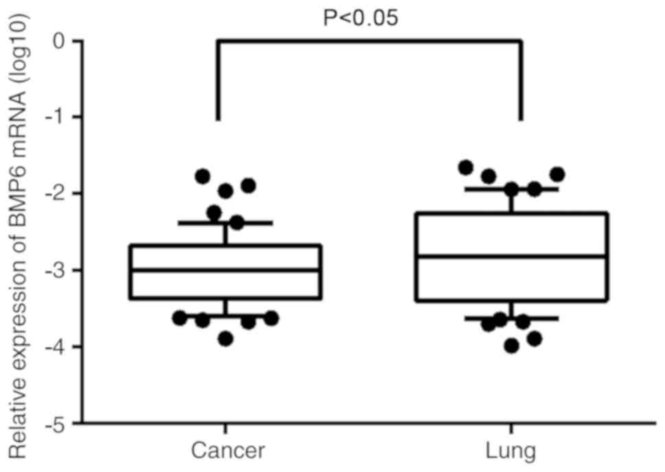
The expression of BMP-6 mRNA in NSCLC and adjacent
tissues was detected by RT-qPCR. The level of BMP-6 mRNA in NSCLC
tissues was significantly reduced, compared with adjacent normal
lung tissues (P<0.05; Fig. 1).
There were no significant differences in BMP-6 mRNA expression
between NSCLC patients with different tumor types, tumor
differentiations, tumor size, clinical stage, and with or without
lymph node metastasis (P>0.05; Fig.
2).
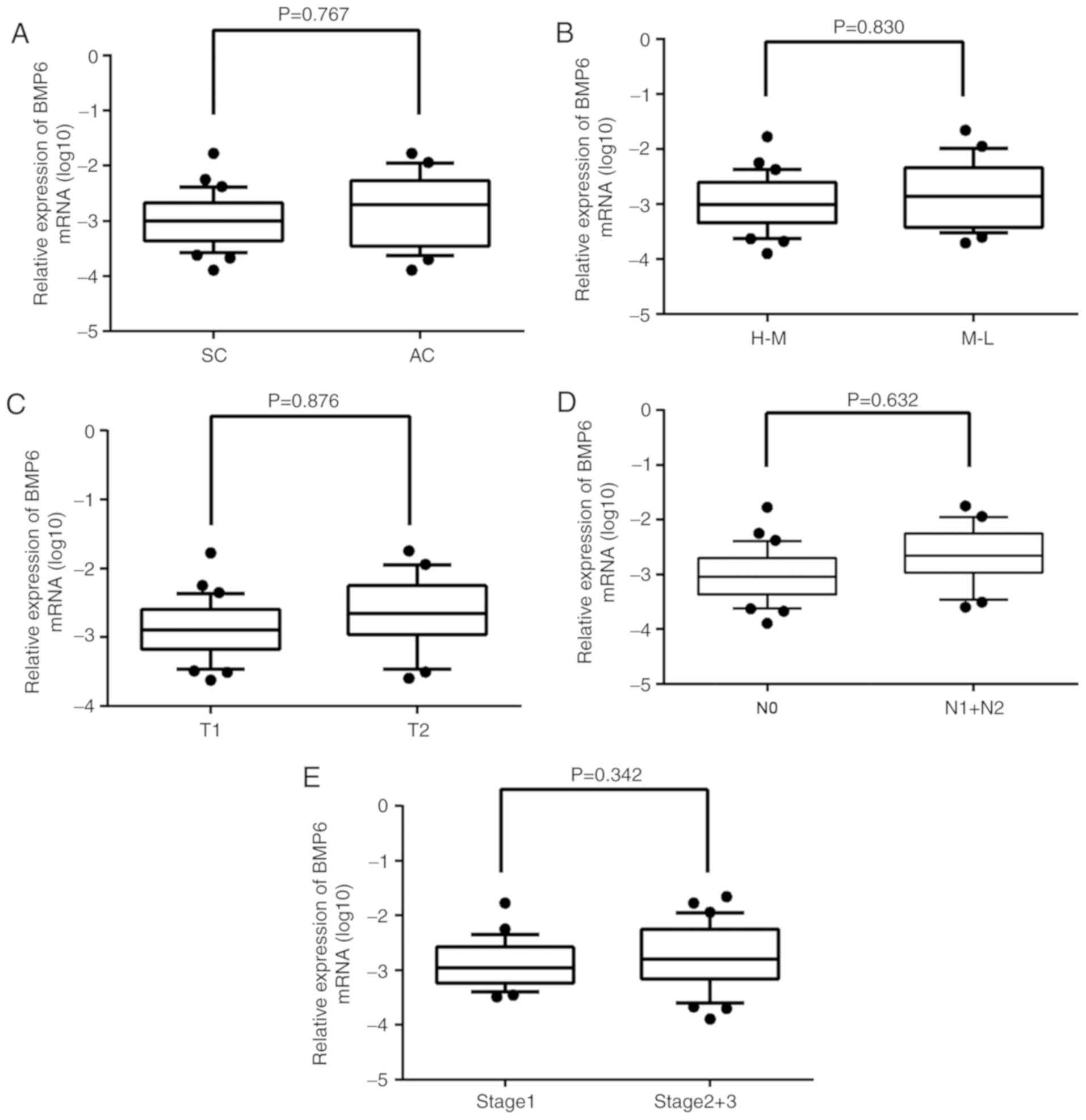 | Figure 2.Comparison of BMP6 mRNA expression
levels in patients with non-small cell lung cancer with different
pathological features. (A) Comparison of BMP6 mRNA expression
between patients with SC and AC. (B) Comparison of BMP6 mRNA
expression between patients with high to middle tumor
differentiation and middle to low differentiation. (C) Comparison
of BMP6 mRNA expression between patients with tumor <2 cm
(longest diameter) (T1) and tumor being 2–5 cm (T2). (D) Comparison
of BMP6 mRNA expression between patients without lymph node
metastasis (N0) and patients with 1–3 (N1) and 4–9 (N2) axillary
(underarm) lymph node metastasis (N1). (E) Comparison of BMP6 mRNA
expression between patients with stage I and stage II/III disease.
BPM6, bone morphogenetic protein 6; SC, squamous lung cancer; AC,
adenocarcinomas; H-M, high to middle differentiation; M-L, middle
to low differentiation; T1, tumor is <2 cm or less (longest
diameter); T2, tumor is 2–5 cm; N0, cancer not spread; N1, cancer
has spread to 1–3 axillary (underarm) lymph nodes; N2, cancer has
spread to 4–9 lymph nodes (n=73). |
Expression of BMP-6 protein in NSCLC
tissues
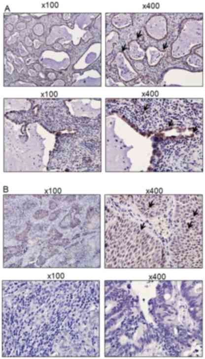
Immunohistochemical staining of BMP-6 expression in
73 NSCLC and adjacent normal lung tissues revealed that BMP-6 was
positively expressed in a number of lung cancer tissues and
negatively expressed in the majority of tumor tissues (Fig. 3). The positive rate of BMP-6
expression in 73 lung cancer tissues was 26.03% (19/73). In normal
lung tissue, BMP-6 expression was positively expressed in the
majority of the samples. The positive rate of BMP-6 expression in
73 adjacent normal lung tissues was 89.04% (65/73). The expression
of BMP-6 was significantly reduced in NSCLC tissues, compared with
adjacent lung tissues (χ2=59.32; P<0.001; Table I).
 | Table I.BMP-6 protein expression in NSCLC and
adjacent tissues. |
Table I.
BMP-6 protein expression in NSCLC and
adjacent tissues.
|
|
| BMP-6 expression |
|
|
|---|
|
|
|
|
|
|
|---|
| Tissue type | Case no. | − | + | ++ | +++ | Pos rate (%) | Neg rate (%) |
|---|
| Adjacent lung
tissues | 73 | 8 | 16 | 35 | 14 | 89.04a | 10.96 |
| NSCLC | 73 | 54 | 19 | 0 | 0 | 26.03 | 73.97 |
| χ2 |
|
| 0.34 | 46.04 | 15.49 | 59.32 |
| P-value |
|
| 0.56 | <0.001 | <0.001 | <0.01 |
Association of BMP-6 mRNA levels with
the prognosis of patients with NSCLC
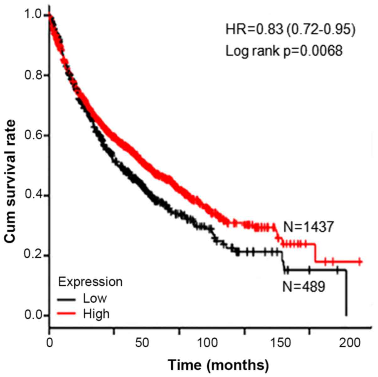
The association of BMP-6 mRNA expression with the
overall survival was analyzed using the Kaplan-Meier plotter online
database. The database contained a total of 1,926 cases of NSCLC
with available data for BMP-6 mRNA expression and overall survival
rate. The online analysis using the Kaplan-Meier plotter database
calculated a HR value of 0.83, and log-rank P-value of 0.0068,
indicating that BMP-6 mRNA level can function as a predictive
factor, and that reduced BMP-6 mRNA expression (lower than the
median) is associated with a poor prognosis of patients. The
Kaplan-Meier survival curve was presented in Fig. 4.
BMP-6 inhibits the proliferation of
human lung cancer cells
The expression of BMP-6 mRNA in cells (Fig. 5A) and the concentration of BMP-6
protein in the supernatant of cultured cells (Fig. 5B) were measured. The mRNA and protein
expression of BMP-6 were significantly decreased in lung cancer
cells, compared with that in HBE cells. To further validate the
role of BMP6 in lung cancer cells, the H460, H1299, A549 and H520
cells were treated with active BMP6 recombinant protein for 48 h,
where it was revealed that 5 or 50 ng/ml BMP-6 protein
significantly inhibited cell proliferation in H460 (Fig. 5C), H1299 (Fig. 5D), A549 (Fig. 5E) and H520 (Fig. 5F) cells, compared with the control
group. These data indicate that BMP-6 is a suppressor in human lung
cancer cells.
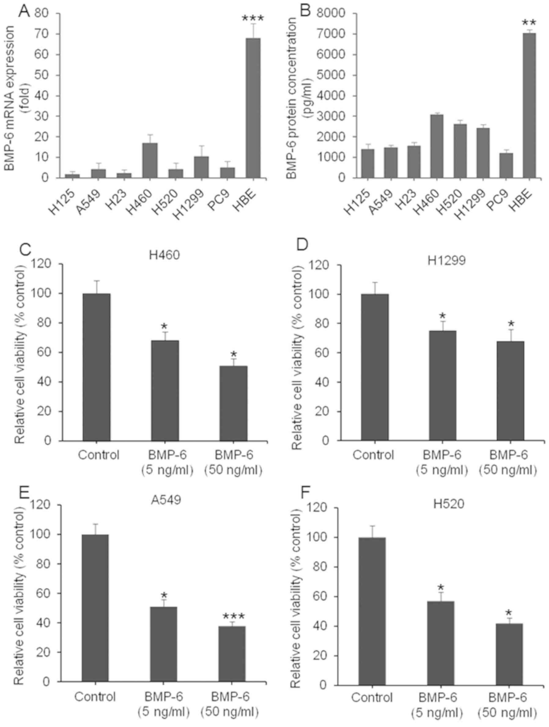 | Figure 5.BMP-6 expression and effect in human
lung cancer cells. (A) The expression of BMP-6 mRNA in H125, A549,
H23, H460, H520, H1299 and PC9 lung cancer, and HBE cells was
measured by reverse transcription-quantitative polymerase chain
reaction. ***P<0.001 vs. all other groups. n=4. (B) The
concentration of BMP-6 protein in the supernatant of cultured cells
was measured by ELISA. **P<0.01 vs. all other groups. n=4. BMP-6
recombinant protein significantly inhibited cell proliferation in
(C) H460, (D) H1299, (E) A549 and (F) H520 cells. BPM6, bone
morphogenetic protein 6; HBE, human bronchial epithelial.
*P<0.05 and ***P<0.001 vs. control group. n=4. |
Discussion
Previous studies reported that BMPs may promote lung
cancer growth or have an anti-cancer effect. For example, BMPs can
regulate the secretion of various cytokines to enhance
proliferation and differentiation of lung cancer cells (8). In contrast, BMPs may also exhibit an
inhibitory role in lung cancer. For instance, an
immunohistochemical study in 35 lung squamous cell carcinoma
samples revealed that the expression of BMP4 in the tumor tissues
was decreased (9). BMP-6 is a
multifunctional growth and differentiation regulatory factor, which
is not only involved in tissue formation and development, and is
also associated with tumor progression (10,11). A
previous study detected a high BMP-6 expression in prostate and
breast cancer, and salivary adenocarcinoma (12). Conversely, other studies demonstrated
that BMP-6 inhibited the growth of breast cancer, plasmacytoma,
renal cell carcinoma, adrenocortical cancer, skin cancer and
myeloma (13–18). BMP-6 has been revealed to be
inactivated in lung cancer cells (5).
In the present study, RT-qPCR revealed that the expression of BMP-6
mRNA in NSCLC tissues was significantly reduced, compared with
adjacent normal lung tissues. The results of immunohistochemical
staining demonstrated that BMP-6 protein expression in NSCLC
tissues was significantly reduced, compared with adjacent normal
lung tissues. The active BMP-6 recombinant protein significantly
inhibited cell proliferation in cultured lung cancer cells.
Collectively, these data indicate that BMP-6 is a suppressor in
human lung cancer cells.
Previous studies reported that the BMP-6 gene was
hypermethylated in lung cancer (19),
breast cancer (20), malignant
lymphoma (21), malignant pleural
mesothelioma (22) and adult T-cell
leukemia (23). Hypermethylation may
inhibit gene transcription and reduce the protein expression, and
can result in the inactivation of tumor suppressor genes (24).
Although the present study did not measure the
methylation of the BNP6 gene, the mRNA and protein expression of
BMP-6 gene was identified to be significantly decreased in the
tumor tissues of NSCLC and seven lung cancer cells, compared with
adjacent normal lung tissues and human bronchial epithelial cells,
respectively. Furthermore, the BMP-6 protein significantly
inhibited cell proliferation in the 4 examined lung cancer cell
lines. The present study indicates that the loss of BMP-6
expression may be a crucial factor associated with tumor growth in
NSCLC.
The Kaplan-Meier plotter database is a prognostic
associated online analytical database. This database contains
10,188 tumor samples (4,142 cases of breast cancer, 1,648 cases of
ovarian cancer, 2,437 cases of lung cancer and 1,065 cases of
gastric cancer) and can analyze the association of 54,675 genes
with the prognosis of these patienst with cancer (25,26). The
present study first used the Kaplan Meier plotter database to
analyze the prognostic value of the BMP-6 gene in patients with
NSCLC. The results demonstrated that low mRNA expression of the
BMP-6 gene was associated with a poor prognosis in patients with
NSCLC. However, data regarding the mRNA level require further
verification at the protein level, although via ELISA, the BMP-6
protein was significantly reduced in 73 NSCLC tissues, compared
with adjacent normal tissues.
In conclusion, BMP-6 expression is reduced in NSCLC
tumor tissues indicating that it serves an inhibitory role in the
development of the disease and is a predictive factor of poor
prognosis in patients with NSCLC.
Acknowledgements
Not applicable.
Funding
Not applicable.
Availability of data and materials
All data generated or analyzed during this study are
included in this published article.
Authors' contributions
WX conducted the experiments. LW collected the
patients' clinical data and performed statistical analysis. FY
designed the study and was a major contributor in writing the
manuscript. All authors read and approved the final manuscript.
Ethics approval and consent to
participate
The protocol of this study was reviewed by the
Ethics Committee of Human Study of the Second Xiangya Hospital.
This approved study was performed in accordance with the ethical
standards of the Declaration of Helsinki (as revised in Brazil
2013). Written informed consent was obtained from the subjects for
use of their tissue.
Patient consent for publication
Not applicable.
Competing interests
All authors declared that they have no competing
interests.
References
|
1
|
Hoffman RM and Sanchez R: Lung cancer
screening. Med Clin North Am. 101:769–785. 2017. View Article : Google Scholar : PubMed/NCBI
|
|
2
|
Zappa C and Mousa SA: Non-small cell lung
cancer: Current treatment and future advances. Transl Lung Cancer
Res. 5:288–300. 2016. View Article : Google Scholar : PubMed/NCBI
|
|
3
|
Blandin Knight S, Crosbie PA, Balata H,
Chudziak J, Hussell T and Dive C: Progress and prospects of early
detection in lung cancer. Open Biol. 7(pii): 1700702017. View Article : Google Scholar : PubMed/NCBI
|
|
4
|
Clement JH, Sänger J and Höffken K:
Expression of bone morphogenetic protein 6 in normal mammary tissue
and breast cancer cell lines and its regulation by epidermal growth
factor. Int J Cancer. 80:250–256. 1999. View Article : Google Scholar : PubMed/NCBI
|
|
5
|
Ro TB, Holt RU, Brenne AT, Hjorth-Hansen
H, Waage A, Hjertner O, Sundan A and Borset M: Bone morphogenetic
protein-5, −6 and −7 inhibit growth and induce apoptosis in human
myeloma cells. Oncogene. 23:3024–3032. 2004. View Article : Google Scholar : PubMed/NCBI
|
|
6
|
Chheang S and Brown K: Lung cancer
staging: Clinical and radiologic perspectives. Semin Intervent
Radiol. 30:99–113. 2013. View Article : Google Scholar : PubMed/NCBI
|
|
7
|
Chen Q, Wang L, Ma Y, Wu X, Jin L and Yu
F: Increased hepcidin expression in non-small cell lung cancer
tissue and serum is associated with clinical stage. Thorac Cancer.
5:14–24. 2014. View Article : Google Scholar : PubMed/NCBI
|
|
8
|
Domvri K, Zarogoulidis P, Darwiche K,
Browning RF, Li Q, Turner JF, Kioumis I, Spyratos D, Porpodis K,
Papaiwannou A, et al: Molecular targeted drugs and biomarkers in
NSCLC, the evolving role of individualized therapy. J Cancer.
4:736–754. 2013. View
Article : Google Scholar : PubMed/NCBI
|
|
9
|
Kraunz KS, Nelson HH, Liu M, Wiencke JK
and Kelsey KT: Interaction between the bone morphogenetic proteins
and Ras/MAP-kinase signalling pathways in lung cancer. British J
Cancer. 93:949–952. 2005. View Article : Google Scholar
|
|
10
|
Vukicevic S and Grgurevic L: BMP-6 and
mesenchymal stem cell differentiation. Cytokine Growth Factor Rev.
20:441–448. 2009. View Article : Google Scholar : PubMed/NCBI
|
|
11
|
Otani H, Otsuka F, Inagaki K, Suzuki J and
Makino H: Roles of bone morphogenetic protein-6 in aldosterone
regulation by adrenocortical cells. Acta Med Okayama. 64:213–218.
2010.PubMed/NCBI
|
|
12
|
Tandon M, Gokul K, Ali SA, Chen Z, Lian J,
Stein GS and Pratap J: Runx2 mediates epigenetic silencing of the
bone morphogenetic protein-3B (BMP-3B/GDF10) in lung cancer cells.
Mol Cancer. 11:272012. View Article : Google Scholar : PubMed/NCBI
|
|
13
|
Seckinger A, Meissner T, Moreaux J,
Goldschmidt H, Fuhler GM, Benner A, Hundemer M, Rème T, Shaughnessy
JD Jr, Barlogie B, et al: Bone morphogenic protein 6: A member of a
novel class of prognostic factors expressed by normal and malignant
plasma cells inhibiting proliferation and angiogenesis. Oncogene.
28:3866–3879. 2009. View Article : Google Scholar : PubMed/NCBI
|
|
14
|
Wach S, Schirmacher P, Protschka M and
Blessing M: Overexpression of bone morphogenetic protein-6 (BMP-6)
in murine epidermis suppresses skin tumor formation by induction of
apoptosis and downregulation of fos/jun family members. Oncogene.
20:7761–7769. 2001. View Article : Google Scholar : PubMed/NCBI
|
|
15
|
Takahashi M, Otsuka F, Miyoshi T, Otani H,
Goto J, Yamashita M, Ogura T, Makino H and Doihara H: Bone
morphogenetic protein 6 (BMP-6) and BMP7 inhibit estrogen-induced
proliferation of breast cancer cells by suppressing p38
mitogen-activated protein kinase activation. J Endocrinol.
199:445–455. 2008. View Article : Google Scholar : PubMed/NCBI
|
|
16
|
Kim IY, Lee DH, Lee DK, Kim BC, Kim HT,
Leach FS, Linehan WM, Morton RA and Kim SJ: Decreased expression of
bone morphogenetic protein (BMP) receptor type II correlates with
insensitivity to BMP-6 in human renal cell carcinoma cells. Clin
Cancer Res. 9:6046–6051. 2003.PubMed/NCBI
|
|
17
|
Johnsen IK, Kappler R, Auernhammer CJ and
Beuschlein F: Bone morphogenetic proteins 2 and 5 are
down-regulated in adrenocortical carcinoma and modulate adrenal
cell proliferation and steroidogenesis. Cancer Res. 69:5784–5792.
2009. View Article : Google Scholar : PubMed/NCBI
|
|
18
|
Kraunz KS, Nelson HH, Liu M, Wiencke JK
and Kelsey KT: Interaction between the bone morphogenetic proteins
and Ras/MAP-kinase signaling pathways in lung cancer. Br J Cancer.
93:949–952. 2005. View Article : Google Scholar : PubMed/NCBI
|
|
19
|
Radpour R, Kohler C, Haghighi MM, Fan AX,
Holzgreve W and Zhong XY: Methylation profiles of 22 candidate
genes in breast cancer using high-throughput MALDI-TOF mass array.
Oncogene. 28:2969–2978. 2009. View Article : Google Scholar : PubMed/NCBI
|
|
20
|
Daibata M, Nemoto Y, Bandobashi K, Kotani
N, Kuroda M, Tsuchiya M, Okuda H, Takakuwa T, Imai S, Shuin T and
Taguchi H: Promoter hypermethylation of the bone morphogenetic
protein-6 gene in malignant lymphoma. Clin Cancer Res.
13:3528–3535. 2007. View Article : Google Scholar : PubMed/NCBI
|
|
21
|
Kimura K, Toyooka S, Tsukuda K, Yamamoto
H, Suehisa H, Soh J, Otani H, Kubo T, Aoe K, Fujimoto N, et al: The
aberrant promoter methylation of BMP3b and BMP-6 in malignant
pleural mesotheliomas. Oncol Rep. 20:1265–1268. 2008.PubMed/NCBI
|
|
22
|
Taniguchi A, Nemoto Y, Yokoyama A, Kotani
N, Imai S, Shuin T and Daibata M: Promoter methylation of the bone
morphogenetic protein-6 gene in association with adult T-cell
leukemia. Int J Cancer. 123:1824–1831. 2008. View Article : Google Scholar : PubMed/NCBI
|
|
23
|
Yang S, Zhong C, Frenkel B, Reddi AH and
Roy-Burman P: Diverse biological effect and Smad signaling of bone
morphogenetic protein 7 in prostate tumor cells. Cancer Res.
65:5769–5777. 2005. View Article : Google Scholar : PubMed/NCBI
|
|
24
|
Esteller M: Epigenetic gene silencing in
cancer: The DNA hypermethylome. Hum Mol Genet 16 Spec No.
1:R50–R59. 2007. View Article : Google Scholar
|
|
25
|
Li S, Sheng B, Zhao M, Shen Q, Zhu H and
Zhu X: The prognostic values of signal transducers activators of
transcription family in ovarian cancer. Biosci Rep. 37(pii):
BSR201706502017. View Article : Google Scholar : PubMed/NCBI
|
|
26
|
Ma YM and Zhao S: Prognostic values of
aldehyde dehydrogenase 1 isoenzymes in ovarian cancer. Onco Targets
Ther. 9:1981–1988. 2016. View Article : Google Scholar : PubMed/NCBI
|