Introduction
Surgical resection is the most important treatment
for patients with gastric cancer. The prognosis of patients with
early gastric cancer after gastrectomy is favorable, with a 5-year
survival rate of ~90% (1,2). The prognosis of patients with advanced
gastric cancer remains poor. Even if patients are asymptomatic, the
rate of lymph node metastasis and risk of early recurrence is high
(3).
Prognostic clinicopathologic factors, including
depth of invasion, lymph node status and distant metastasis are
important to assess the malignant potential of gastric cancer.
Recently, gene expression profiling was developed as a method for
defining the phenotypes of malignant tumors at the molecular level.
Therefore, indexes based on gene expression may indicate the
benefit of systemic therapy and serve as predictive, prognostic and
therapeutic biomarkers in gastric cancer (4).
The homeobox (HOX) gene family consists of 39
members with a shared and highly conserved 61 amino acid
homeodomain motif. These genes are associated with embryologic
regulation, and their roles in neoplastic transformation and tumor
progression are now being recognized (5). HOXB9 is a direct transcriptional target
of WNT/TCF-4, whose expression is normally restricted to
embryogenesis (5,6).
The relationship between HOXB9 expression and tumor
malignancy was identified previously. Nguyen et al (7) reported that WNT/TCF signaling through
lymphoid enhancer-binding factor 1 (LEF1) and HOXB9 mediated lung
adenocarcinoma metastasis. It was reported that elevated HOXB9
expression correlated with high tumor grade in breast cancer
patients, and that HOXB9 expression was an independent prognostic
factor for disease-free survival (8,9). Fang
et al (10) clarified that
higher levels of HOXB9 expression were significantly associated
with advanced clinical stage in patients with glioma. HOXB9
overexpression also stimulated the proliferation, migration and
sphere formation of glioma cells in vitro, and induced the
tumorigenicity of glioma cells in vivo.
Induction of HOXB9 in breast cancer cells may
increase the concentrations of several angiogenic factors,
including vascular endothelial growth factor (VEGF), basic
fibroblast growth factor (bFGF) and interleukin (IL)-8. In
addition, HOXB9 promotes the formation of large, well-vascularized
breast cancer tumors in vivo (8). HOXB9 also induces angiogenesis and
tumor proliferation of colon cancer cells in vitro resulting
in high tumorigenicity in vivo and poor overall survival
(11). Bevacizumab, an anti-VEGF
antibody, remarkably suppresses tumor proliferation by inhibiting
angiogenesis in HOXB9-overexpressing xenografts, improving overall
survival and providing prolonged progression-free survival in
HOXB9-overexpressing patients (11).
These data suggest that HOXB9 has a significant association with
tumor progression and therefore may be a prognostic factor in
clinical outcomes.
Breast and colon cancer often spread through the
blood stream to lung, liver, brain or bones. Regional lymph nodes
are the most common site of tumor spread, and lymph node metastasis
is a major prognostic factor in gastric cancer (12). Thus, understanding the mechanism of
lymphatic metastasis may contribute to the identification of a new
therapeutic target for the treatment of gastric cancer. VEGF and
its receptors (VEGFRs) have crucial roles in physiological and
pathological vasculogenesis. Among VEGFs, VEGF-C and VEGF-D, which
bind only to VEGFR-3, are known to regulate lymphangiogenesis
(13). These ligands and receptors
are often used as lymphangiogenic markers.
In the present study it was hypothesized that, HOXB9
promotes tumor lymphangiogenesis and induces tumor progression,
invasion and metastasis in gastric cancer. The aim of this present
study was to evaluate the correlation between HOXB9 expression,
prognosis and clinicopathologic factors in patients with gastric
cancer, and to assess the role of HOXB9 on tumor cell
lymphangiogenesis in vitro.
Materials and methods
Patients
A panel of 58 patients, 42 male and 16 female, with
gastric cancer were recruited between June 2008 and February 2012
and were selected from the database from the Department of Surgery
of Keio University Hospital (Tokyo, Japan). Their age ranged
between 32 and 88, and the median was 69 years old. The clinical
staging was described based on the Union for International Cancer
Control TMN Classification of Malignant Tumors (7th edition)
(14). The study was approved by the
Ethics Committee of Keio University School of Medicine (Tokyo,
Japan). Written informed consent was obtained from each
patient.
Immunohistochemical staining of
HOXB9
Specimens were cut into 4 µm sections, mounted onto
glass slides, dewaxed in xylene and dehydrated in alcohol. For
antigen retrieval, these sections were autoclaved in citric acid
solution (Ph 6.0) at 121°C for 10 min. Endogenous peroxidase
activity was blocked with a peroxidase blocking reagent (0.03%
hydrogen peroxide containing sodium azide; Dako; Agilent
Technologies, Inc., Santa Clara, CA, USA) for 10 min at 4°C.
Samples were incubated with a rabbit polyclonal anti-human
HOXB9-specific antibody (1:50; cat. no., sc-133671; Santa Cruz
Biotechnology, Inc., Dallas, TX, USA) at 4°C for 12 h, followed by
incubation using a horseradish peroxidase-labeled secondary
antibody (cat. no., K4003; Dako; Agilent Technologies, Inc.) at
room temperature for 30 min. The slides were stained with
3,3′-diaminobenzidine (Dako; Agilent Technologies, Inc.) at room
temperature for 2 min, according to manufacturer's protocol. Slides
were rinsed in PBS. Sections were counterstained with hematoxylin,
dehydrated and mounted. HOXB9 expression was evaluated
independently by two authors, both blinded to the
clinicopathological parameters. A light microscope was used at a
magnification of ×200. The percentage of positive cells and
staining intensity were evaluated (Table
I).
 | Table I.Immunoreactive scores of HOXB9. |
Table I.
Immunoreactive scores of HOXB9.
| Variables | Scores |
|---|
| Percentage of
positive cells |
|
|
<33% | 0 |
|
33-66% | 1 |
|
>66% | 2 |
| Intensity of
reaction |
|
| No
reaction | 0 |
| Weak
reaction | 1 |
| Intense
reaction | 2 |
Reverse transcription-quantitative
polymerase chain reaction (RT-qPCR)
The expression levels of HOXB9 mRNA were determined
in four human gastric cancer cell lines (TMK-1, MKN-45, MKN-74 and
KATO-III) using RT-qPCR. RNA was extracted from the cells using the
RNeasy kit (Qiagen, Inc., Valencia, CA, USA), according to
manufacturer's protocol. The concentration of total RNA was
determined using a NanoDrop ND-1000 Spectrophotometer (Thermo
Fisher Scientific, Inc., Waltham, MA, USA). Total RNA was reverse
transcribed into cDNA using Transcriptor High Fidelity cDNA
Synthesis kit (Roche Diagnostics, Rotkreuz, Switzerland) according
manufacturer's protocol. The quality and quantity of the cDNA
samples were assessed by standard electrophoresis and NanoDrop
ND-1000 spectrophotometer. The following primer pairs were used for
the qPCR: HOXB9: Sense, 5′-CCGGCTACGGGGACAATAA-3′ and antisense,
5′-GGTGTAGGGACAGCGCTTTTT-3′; GAPDH, sense,
5′-ATCATCCCTGCCTCTACTGG-3′ and antisense,
5′-TTTCTAGACGGCAGGTCAGGT-3′; and β-actin: Sense,
5′-CTCTTCCAGCCTTCCTTCCT-3′ and antisense,
5′-AGCACTGTGTTGGCGTACAG-3′. The following thermocycling conditions
were used: Initial denaturation at 95°C for 2 min; 25 cycles of
95°C for 30 sec; 56°C for 30 sec and 72°C for 60 sec; and a final
extension at 72°C for 10 min. Each experiment was performed using
triplicates. qPCR was subsequently performed using a Light Cycler
480 Real-Time PCR System and SYBR Green 1 Master Mix (Roche
Diagnostics, Basel, Switzerland) which contained Taq DNA
polymerase. The following thermocycling conditions were used for
PCR: Pre-denaturation at 95°C for 5 min, followed by 45 cycles of
denaturation at 95°C for 10 sec, annealing at 57°C for 10 sec and
extension at 72°C for 10 sec. Subsequently, melting curve analysis
was performed. The gene expressions were quantified using
2−ΔΔCq method (15).
Transfection of HOXB9 gene
TMK-1 cells with low HOXB9 expression were infected
with HOXB9-expressing lentiviral vector using pLenti6.3/V5-DEST
Gateway Vector kit (Invitrogen; Thermo Fisher Scientific, Inc.),
according to the manufacturer's protocols. The time interval
between transfection and subsequent experimentation was >1
week.
Gel electrophoresis of PCR
products
KOD FX Neo (Toyobo Life Science, Osaka, Japan) was
used for PCR. The following primer pairs were used: VEGF-C, sense,
5′-AAAGAACCTGCCCCAGAAAT-3′ and antisense,
5′-TGGTGGTGGAACTTCTTTCC-3′; VEGF-D, sense,
5′-TCCAGGAACCAGCTCTCTGT-3′ and antisense,
5′-TTTTTGGGGTGCTGGATTAG-3′; and VEGFR-3, sense,
5′-GAGACAAGGACAGCGAGGAC-3′ and antisense,
5′-CTGTGTCGTTGGCATGTACC-3′. The following thermocycling conditions
were used: Initial denaturation at 94°C for 2 min, followed by 30
cycles at 98°C for 10 sec and at 68°C 30 sec. Bands were analyzed
by electrophoresis in 2% gel and visualized under UV light. The
expression levels of lymphangiogenic factors in HOXB9 transfected
cells were compared with that in LacZ transfected cells
(control).
RT-qPCR for lymphangiogenic
markers
RT-qPCR was performed to quantify the difference of
expression of lymphangiogenic markers between transfected cells.
cDNA purified from the cultured cell lines (TMK-1, TMK-1/LacZ, and
TMK-1/HOXB9) were amplified. The thermocycling conditions and
fluorophore were the same as described above. The following primer
pairs were used: VEGF-D, sense, 5′-GAACACCAGCACCTCGTACA-3′ and
antisense; and 5′-TTCTTCAGGGATCTGGATGG-3′; VEGFR-3, sense,
5′-CTGCAAGAACGTGCATCTGT-3′ and antisense,
5′-TTCTTGTGGCAGTGCTTGTC-3′. The aforementioned primers were used
for VEGF-C. The gene expressions were quantified using
2−ΔΔCq method (15).
Statistical analysis
The relevant data were presented as mean ± standard
deviation (n=3). HOXB9 mRNA expression levels in each cell line
were compared using the Kruskal-Wallis test. Dunn's multiple
comparisons test was used for pairwise post-hoc comparisons. The
proportions were compared using Fisher's exact test. Overall
survival curves were created by Kaplan-Meier estimates and compared
by log-rank tests. Statistical analyses were performed using SPSS
(version 19; IBM Corp., Armonk, NY, USA). P<0.05 was considered
to indicate a statistically significant difference.
Results
Patient characteristics
Patient characteristics are shown in Table II. The majority of the 58 patients
were male (72.4%) with a median age of 69 years. The median
follow-up period was 59.2 months. The majority of the patients
(75.9%) exhibited advanced gastric cancer that had invaded beyond
the submucosal layer, and 51.7% were positive for lymph node
metastasis.
| Table II.Patient characteristics (n=58). |
Table II.
Patient characteristics (n=58).
| Parameters | Number (%) |
|---|
| Gender |
|
| Male | 42 (72.4) |
|
Female | 16 (27.6) |
| Median
age in years (range) | 69 (32–88) |
| Pathological depth of
tumor invasion (14) |
|
| T1 | 14 (24.1) |
| T2 | 9 (15.6) |
| T3 | 21 (36.2) |
| T4 | 14 (24.1) |
| Pathological nodal
stage |
|
| N0 | 28 (48.3) |
| N1 | 8 (13.8) |
| N2 | 8 (13.8) |
| N3 | 14 (24.1) |
| Pathological
stage |
|
| I | 16 (27.6) |
| II | 21 (36.2) |
| II | 19 (32.8) |
| IV | 2 (3.4) |
HOXB9 expression in patients with
gastric cancer
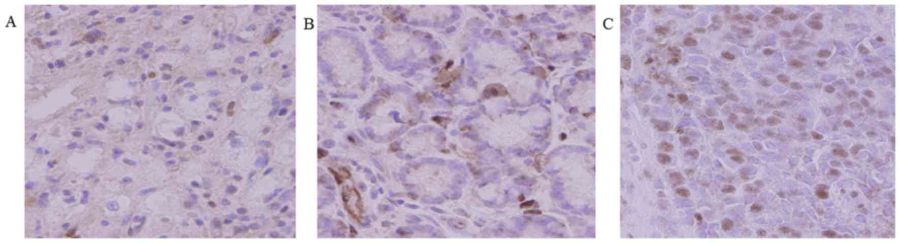
The expression of HOXB9 was evaluated in 58 tumor
specimens. Immunohistochemical staining for HOXB9 (Fig. 1) was performed. Staining intenstity
was graded as none, weak and intense. According to the
classification criteria (Table I), a
total of 30 tumors (51.7%) were positive for HOXB9 expression.
HOXB9 staining was observed in the nucleus of gastric cancer cells.
Association between HOXB9 expression and clinicopathologic
variables is shown in Table
III.
| Table III.Relation between HOXB9 expression and
clinicopathologic factors in 58 gastric cancer patients. |
Table III.
Relation between HOXB9 expression and
clinicopathologic factors in 58 gastric cancer patients.
|
|
| HOXB9 staining |
|
|---|
|
|
|
|
|
|---|
| Characteristics | No. of patients | Negative (n=28) | Positive (n=30) | P-value |
|---|
| Gender |
|
|
| 0.77 |
| Male | 42 | 21 | 21 |
|
|
Female | 16 | 7 | 9 |
|
| Age |
|
|
| 0.19 |
|
<70 | 29 | 17 | 12 |
|
| ≥70 | 29 | 11 | 18 |
|
| T feature |
|
|
| 0.015 |
| T1-2 | 23 | 16 | 7 |
|
|
T3-4 | 35 | 12 | 23 |
|
| LN metastasis |
|
|
| 0.008 |
|
Negative | 28 | 19 | 9 |
|
|
Positive | 30 | 9 | 21 |
|
| Lymphatic
invasion |
|
|
| 0.001 |
|
Negative | 17 | 14 | 3 |
|
|
Positive | 41 | 14 | 27 |
|
| Vascular
invasion |
|
|
| 0.023 |
|
Negative | 18 | 13 | 5 |
|
|
Positive | 40 | 15 | 25 |
|
In addition, overall survival was decreased in
patients with positive HOXB9 expression (P=0.64; Fig. 2). However, this did not reach a
statistically significant difference. The depth of tumor invasion
(P=0.015), the number of node metastases (P=0.008), lymphatic
invasion (P=0.001) and vascular invasion (P=0.023) were
significantly associated with HOXB9 expression (Table III).
Association between HOXB9 and
lymphangiogenic factors
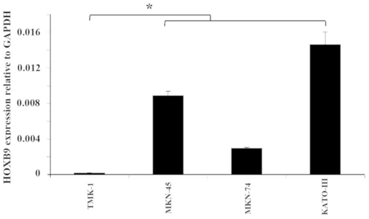
TMK-1 cells expressed the lowest amount of HOXB9
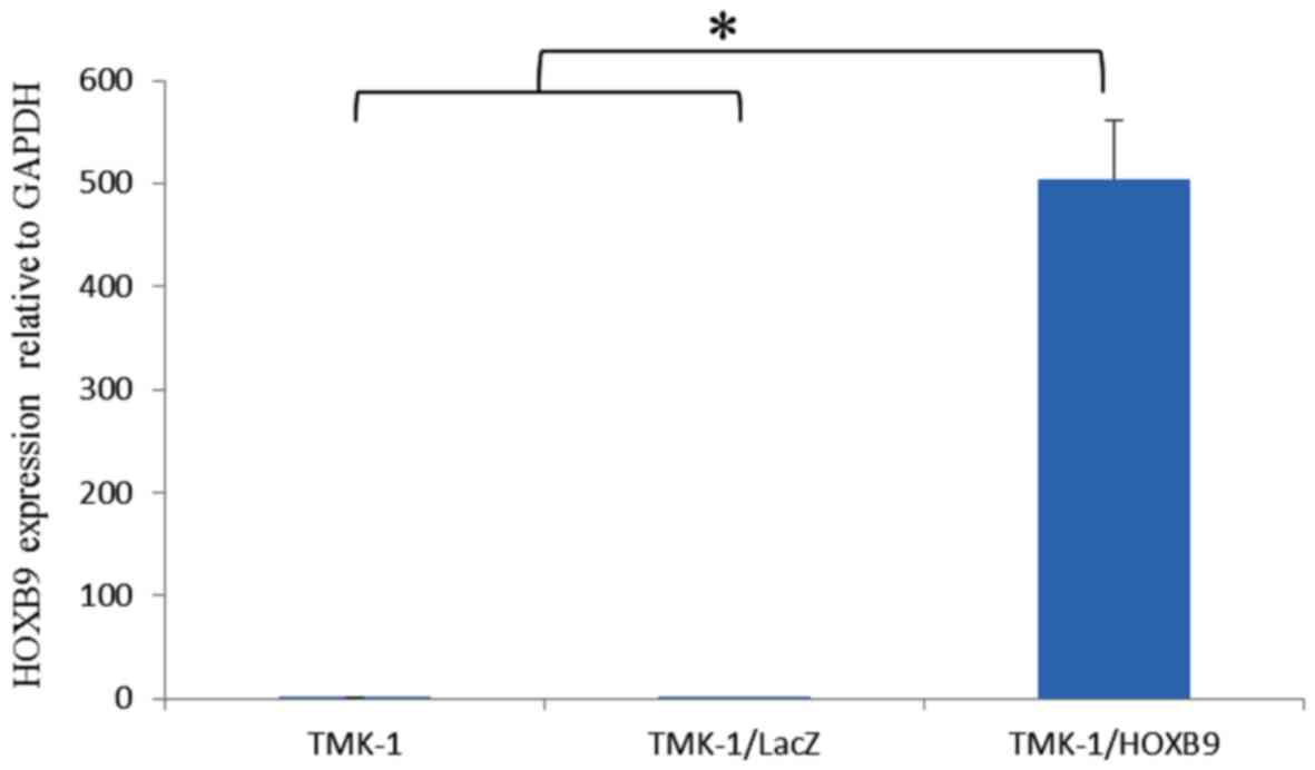
mRNA expression levels among the four cell lines tested (Fig. 3). Using a viral vector, HOXB9 mRNA
expression level was increased by 3,377-fold in TMK-1/HOXB9 cells
compared with that in TMK-1/LacZ cells; this difference reached
statistical significance (P=0.027; Fig.
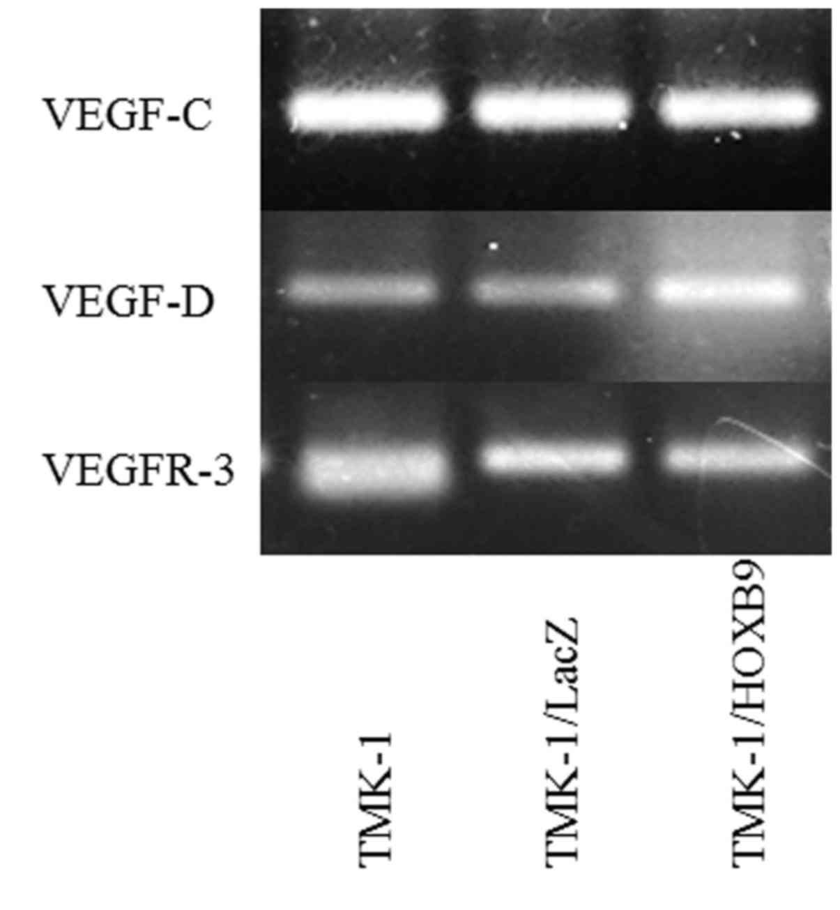
4). Gel electrophoresis of PCR products showed that the
expression levels of VEGF-D mRNA were increased in TMK-1/HOXB9
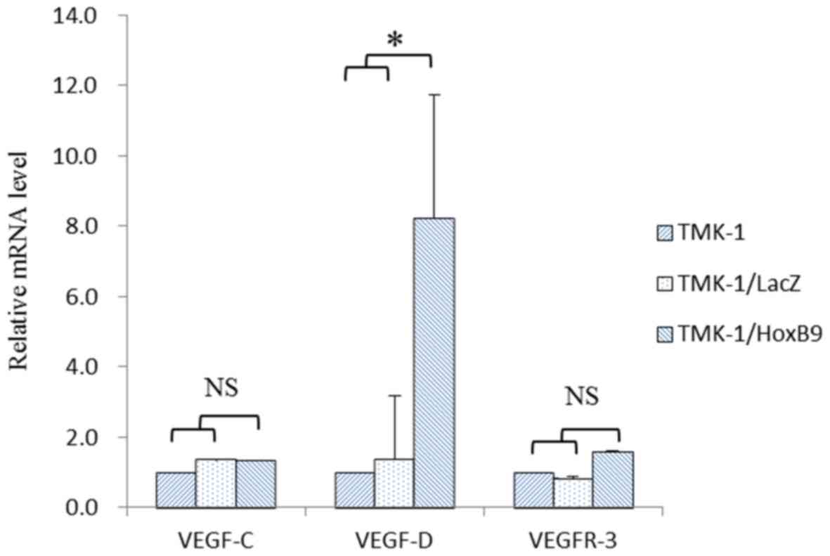
cells compared with TMK-1/LacZ cells (Fig. 5). The relative mRNA level of VEGF-D
was increased by 6.06-fold in TMK-1/HOXB9 compared with that in
TMK-1/LacZ cells (P=0.066; Fig. 6).
VEGF-C and VEGFR-3 levels were unchanged (Figs. 5 and 6).
Discussion
HOXB9 is considered to be a transcription factor.
Transcription factors are proteins, which bind to DNA sequences to
control the expression of genes (16). They may have a dynamic behavior
depending on intracellular/extracellular signals. In the present
study, the expression of HOXB9 and clinicopathological factors were
investigated in patients with gastric cancer at various
pathological stages, in order to compare the association between
HOXB9 expression and gastric cancer progression.
Immunohistochemistry revealed that 51.7% of resected gastric cancer
tissues were positive for HOXB9 expression. In a previous study, it
was showed that HOXB9 was expressed in 49% of resected breast
cancer tissues and in 67% of resected colorectal cancer tissues
(9,11). Although the methods used to evaluate
HOXB9 expression were different in each study, the positive rates
were comparable. In addition, it was indicated that HOXB9
expression was localized to the nucleus of gastric cancer cells,
which is in accordance to a previous study on breast cancer
(9).
In the present study, HOXB9-positive patients with
gastric cancer tended to exhibit a poorer prognosis compared with
those in the HOXB9-negative group. Although this difference did not
reach statistical significance, it is possible that additional data
may clarify the potential association between HOXB9 expression and
survival of patients with gastric cancer. HOXB9 expression was the
only independent prognostic factor for disease-free survival in
breast cancer patients (hazard ratio=15.532; P=0.009) (9), and increased HOXB9 expression in
colorectal cancer was significantly associated with poorer overall
survival (11). However, Sha et
al (17) reported that decreased
expression of HOXB9 was associated with a poor overall survival in
Chinese patients with gastric cancer. The reason for this
discrepancy in the association between survival and HOXB9
expression are unclear, but may be related to differences in the
subjects and methodology.
In our previous study, HOXB9 positivity was
significantly associated with tumor virulence in breast cancer
patients (tumor size, nuclear grade and lymph node metastasis)
(9). In the present study, it was
found that HOXB9 expression was significantly associated with the
depth of invasion, lymph node metastasis, lymphatic invasion and
vascular invasion in patients with gastric cancer, suggesting a
role of this transcription factor in gastric cancer.
Lymph node metastasis and lymphatic invasion are
related to lymphogenic metastasis, a specific phenomenon of gastric
cancer progression (12). A previous
study reported that HOXB9 induces the expression of several
angiogenic factors (epidermal growth factor (EGF), bFGF, IL-8 and
angiopoietin-like 2 (ANGPTL-2)), as well as ErbB (amphiregulin,
epiregulin and neuregulins) and transforming growth factor-β
(TGF-β) in patients with breast cancer (8). These factors activate their respective
pathways, leading to increased cell motility and the acquisition of
mesenchymal phenotypes (8).
Additionally, a study on colon cancer reported that HOXB9 induced
angiogenesis and tumor proliferation in vitro, consistent
with high tumorigenicity in vivo (11). The results of the present study
demonstrated that there was no association between HOXB9 expression
and the angiogenic factors VEGF-A, bFGF, IL8, ANGPTL2, TGF-β1, and
TGF-β2 in patients with gastric cancer (data not shown). However,
the lymphangiogenic factor VEGF-D, but not VEGF-C and VEGFR-3, was
elevated in TMK-1 cells transfected with the HOXB9 gene. To the
best of our knowledge this is the first study suggesting that HOXB9
promotes lymphangiogenesis. Therefore, HOXB9 may be associated with
lymphogenic metastasis.
Overall, HOXB9 expression positively associated with
gastric cancer progression and may be associated with poor
prognosis. These results suggest the potential clinical utility of
HOXB9 as a prognostic factor or therapeutic target in gastric
cancer.
Acknowledgements
The authors would like to thank Dr. Y. Hoshino and
Dr. H. Seki (Department of Surgery, Keio University School of
Medicine) for the help with designing these experiments. The
authors would also like to thank Mrs. Y. Yoshimura, Mr. H. Okazaki,
Mr. K. Miyao and Mrs. S. Fukuhara (Department of Surgery, Keio
University School of Medicine) for their technical assistance with
all aspects of the present study.
Funding
The present work was supported by a JSPS KAKENHI
Grant-in-Aid for Young Scientists (B) (grant no. 26861103) and a
Grant-in-Aid for Scientific Research (C) (grant nos. 22591469 and
25462037).
Availability of data and materials
The datasets used and/or analyzed during the present
study are available from the corresponding author on reasonable
request.
Authors' contributions
FK, NW, TH, and YK conceived the study. RN, TT, HK,
and HT were the major contributors in analyzing and interpreting
the patient data. FK and KF performed the experiments. FK and NW
evaluated immunostained sections. FK wrote the manuscript. YK
supervised the study, and performed the critical review. All
authors read and approved the final manuscript.
Ethics approval and consent to
participate
The study was approved by the Ethics Committee of
Keio University School of Medicine (approval no. 20080189). Written
informed consent was obtained from each patient.
Patient consent for publication
The patients consented to publication.
Competing interests
The authors declare that they have no competing
interests.
References
|
1
|
Sano T, Sasako M, Kinoshita T and Maruyama
K: Recurrence of early gastric cancer. Follow-up of 1475 patients
and review of the Japanese literature. Cancer. 72:3174–3178. 1993.
View Article : Google Scholar : PubMed/NCBI
|
|
2
|
Japanese Gastric Cancer Association
Registration Committee, ; Maruyama K, Kaminishi M, Hayashi K, Isobe
Y, Honda I, Katai H, Arai K, Kodera Y and Nashimoto A: Gastric
cancer treated in 1991 in Japan: Data analysis of nationwide
registry. Gastric Cancer. 9:51–66. 2006. View Article : Google Scholar : PubMed/NCBI
|
|
3
|
Gotoda T, Yanagisawa A, Sasako M, Ono H,
Nakanishi Y, Shimoda T and Kato Y: Incidence of lymph node
metastasis from early gastric cancer: Estimation with a large
number of cases at two large centers. Gastric Cancer. 3:219–225.
2000. View Article : Google Scholar : PubMed/NCBI
|
|
4
|
Bang YJ, Van Cutsem E, Feyereislova A,
Chung HC, Shen L, Sawaki A, Lordick F, Ohtsu A, Omuro Y, Satoh T,
et al: Trastuzumab in combination with chemotherapy versus
chemotherapy alone for treatment of HER2-positive advanced gastric
or gastro-oesophageal junction cancer (ToGA): A phase 3,
open-label, randomised controlled trial. Lancet. 376:687–697. 2010.
View Article : Google Scholar : PubMed/NCBI
|
|
5
|
Abate-Shen C: Deregulated homeobox gene
expression in cancer: Cause or consequence? Nat Rev Cancer.
2:777–785. 2002. View
Article : Google Scholar : PubMed/NCBI
|
|
6
|
Hatzis P, van der Flier LG, van Driel MA,
Guryev V, Nielsen F, Denissov S, Nijman IJ, Koster J, Santo EE,
Welboren W, et al: Genome-wide pattern of TCF7L2/TCF4 chromatin
occupancy in colorectal cancer cells. Mol Cell Biol. 28:2732–2744.
2008. View Article : Google Scholar : PubMed/NCBI
|
|
7
|
Nguyen DX, Chiang AC, Zhang XH, Kim JY,
Kris MG, Ladanyi M, Gerald WL and Massagué J: WNT/TCF signaling
through LEF1 and HOXB9 mediates lung adenocarcinoma metastasis.
Cell. 138:51–62. 2009. View Article : Google Scholar : PubMed/NCBI
|
|
8
|
Hayashida T, Takahashi F, Chiba N,
Brachtel E, Takahashi M, Godin-Heymann N, Gross KW, Vivanco Md,
Wijendran V, Shioda T, et al: HOXB9, a gene overexpressed in breast
cancer, promotes tumorigenicity and lung metastasis. Proc Natl Acad
Sci USA. 107:1100–1105. 2010. View Article : Google Scholar : PubMed/NCBI
|
|
9
|
Seki H, Hayashida T, Jinno H, Hirose S,
Sakata M, Takahashi M, Maheswaran S, Mukai M and Kitagawa Y: HOXB9
expression promoting tumor cell proliferation and angiogenesis is
associated with clinical outcomes in breast cancer patients. Ann
Surg Oncol. 19:1831–1840. 2012. View Article : Google Scholar : PubMed/NCBI
|
|
10
|
Fang L, Xu Y and Zou L: Overexpressed
homeobox B9 regulates oncogenic activities by transforming growth
factor-β1 in gliomas. Biochem Biophys Res Commun. 446:272–279.
2014. View Article : Google Scholar : PubMed/NCBI
|
|
11
|
Hoshino Y, Hayashida T, Hirata A,
Takahashi H, Chiba N, Ohmura M, Wakui M, Jinno H, Hasegawa H,
Maheswaran S, et al: Bevacizumab terminates homeobox B9-induced
tumor proliferation by silencing microenvironmental communication.
Mol Cancer. 13:1022014. View Article : Google Scholar : PubMed/NCBI
|
|
12
|
Pak KH, Jo A, Choi HJ, Choi Y, Kim H and
Cheong JH: The different role of intratumoral and peritumoral
lymphangiogenesis in gastric cancer progression and prognosis. BMC
Cancer. 15:4982015. View Article : Google Scholar : PubMed/NCBI
|
|
13
|
Takahashi S: Vascular endothelial growth
factor (VEGF), VEGF receptors and their inhibitors for
antiangiogenic tumor therapy. Biol Pharm Bull. 34:1785–1788. 2011.
View Article : Google Scholar : PubMed/NCBI
|
|
14
|
Sobin LH, Gospodarowicz MK and Wittekind
C: TNM classification of malignant tumours. (7th). Wiley-Blackwell.
(Hoboken, Oxford). 2009.
|
|
15
|
Livak KJ and Schmittgen TD: Analysis of
relative gene expression data using real-time quantitative PCR and
the 2(-Delta Delta C(T)) method. Methods. 25:402–408. 2001.
View Article : Google Scholar : PubMed/NCBI
|
|
16
|
Bouhlel MA, Lambert M and David-Cordonnier
MH: Targeting transcription factor binding to DNA by competing with
DNA binders as an approach for controlling gene expression. Curr
Top Med Chem. 15:1323–1358. 2015. View Article : Google Scholar : PubMed/NCBI
|
|
17
|
Sha S, Gu Y, Xu B, Hu H, Yang Y, Kong X
and Wu K: Decreased expression of HOXB9 is related to poor overall
survival in patients with gastric carcinoma. Dig Liver Dis.
45:422–429. 2013. View Article : Google Scholar : PubMed/NCBI
|