|
1
|
Neagu M, Caruntu C, Constantin C, Boda D,
Zurac S, Spandidos DA and Tsatsakis AM: Chemically induced skin
carcinogenesis: Updates in experimental models (Review). Oncol Rep.
35:2516–2528. 2016. View Article : Google Scholar : PubMed/NCBI
|
|
2
|
Nedelcu RI, Ion DA, Holeab CA, Cioplea MD,
Brînzea A and Zurac SA: Dendritic cells in melanoma -
immunohistochemical study and research trends. Rom J Morphol
Embryol. 56:997–1002. 2015.PubMed/NCBI
|
|
3
|
West HC and Bennett CL: Redefining the
role of langerhans cells as immune regulators within the skin.
Front Immunol. 8:19412018. View Article : Google Scholar : PubMed/NCBI
|
|
4
|
Serafim A, Petre DG, Moraru L, Cioflan HE,
Vasile E, Mastalier-Manolescu B, Petrutescu M and Stancu IC:
Gelatin-PVP hydrogels with potential skin grafts applications. Key
Eng Mater. 638:38–46. 2015. View Article : Google Scholar
|
|
5
|
Deckers J, Hammad H and Hoste E:
Langerhans cells: Sensing the environment in health and disease.
Front Immunol. 9:932018. View Article : Google Scholar : PubMed/NCBI
|
|
6
|
Nichita L, Zurac S, Popp C, Micu G,
Bastian A, Stăniceanu F and Streinu-Cercel A: Dendritic cells -
immunodeficiency virus (HIV): Early interactions. Rom J Intern Med.
49:251–255. 2011.PubMed/NCBI
|
|
7
|
Malissen B, Tamoutounour S and Henri S:
The origins and functions of dendritic cells and macrophages in the
skin. Nat Rev Immunol. 14:417–428. 2014. View Article : Google Scholar : PubMed/NCBI
|
|
8
|
Boda D: Cellomics as integrative omics for
cancer. Curr Proteomics. 10:237–245. 2013. View Article : Google Scholar
|
|
9
|
Petre M, Zurac S, Andrei R, Tebeica T,
Birceanu A, Chirculescu R, Popp C, Evsei A, Staniceanu F and
Bastian A: Langerhans cells distributions may discriminate early
stage mycosis fungoides and inflammatory dermatoses. Virchows Arch.
463:101–352. 2772013.PubMed/NCBI
|
|
10
|
Tebeică T, Andrei R, Zurac S and
Stăniceanu F: Practical aspects regarding the histopathological
diagnosis of early mycosis fungoides. Rom J Intern Med. 54:3–10.
2016.PubMed/NCBI
|
|
11
|
Zurac S, Neagu M, Constantin C, Cioplea M,
Nedelcu R, Bastian A, Popp C, Nichita L, Andrei R, Tebeica T, et
al: Variations in the expression of TIMP1, TIMP2 and TIMP3 in
cutaneous melanoma with regression and their possible function as
prognostic predictors. Oncol Lett. 11:3354–3360. 2016. View Article : Google Scholar : PubMed/NCBI
|
|
12
|
Neagu M, Constantin C, Dumitrascu GR, Lupu
AR, Caruntu C, Boda D and Zurac S: Inflammation markers in
cutaneous melanoma - edgy biomarkers for prognosis. Discoveries
(Craiova). 3:e382015. View Article : Google Scholar
|
|
13
|
Zaba LC, Fuentes-Duculan J, Eungdamrong
NJ, Abello MV, Novitskaya I, Pierson KC, Gonzalez J, Krueger JG and
Lowe NA: Psoriasis is characterized by accumulation of
immunostimulatory and Th1/Th17 cell-polarizing myeloid dendritic
cells. J Invest Dermatol. 129:79–88. 2008. View Article : Google Scholar : PubMed/NCBI
|
|
14
|
Lowes MA, Chamian F, Abello MV,
Fuentes-Duculan J, Lin SL, Nussbaum R, Novitskaya I, Carbonaro H,
Cardinale I, Kikuchi T, et al: Increase in TNF-alpha and inducible
nitric oxide synthase-expressing dendritic cells in psoriasis and
reduction with efalizumab (anti-CD11a). Proc Natl Acad Sci USA.
102:19057–19062. 2005. View Article : Google Scholar : PubMed/NCBI
|
|
15
|
Căruntu C, Boda D, Căruntu A, Rotaru M,
Baderca F and Zurac S: In vivo imaging techniques for psoriatic
lesions. Rom J Morphol Embryol. 55 (Suppl 3):1191–1196.
2014.PubMed/NCBI
|
|
16
|
Farkas L, Beiske K, Lund-Johansen F,
Brandtzaeg P and Jahnsen FL: Plasmacytoid dendritic cells (natural
interferon-alpha/beta-producing cells) accumulate in cutaneous
lupus erythematosus lesions. Am J Pathol. 159:237–243. 2001.
View Article : Google Scholar : PubMed/NCBI
|
|
17
|
Blanco P, Palucka AK, Gill M, Pascual V
and Banchereau J: Induction of dendritic cell differentiation by
IFN-α in systemic lupus erythematosus. Science. 294:1540–1543.
2001. View Article : Google Scholar : PubMed/NCBI
|
|
18
|
Vermi W, Lonardi S, Morassi M, Rossini C,
Tardanico R, Venturini M, Sala R, Tincani A, Poliani PL,
Calzavara-Pinton PG, et al: Cutaneous distribution of plasmacytoid
dendritic cells in lupus erythematosus. Selective tropism at the
site of epithelial apoptotic damage. Immunobiology. 214:877–886.
2009. View Article : Google Scholar : PubMed/NCBI
|
|
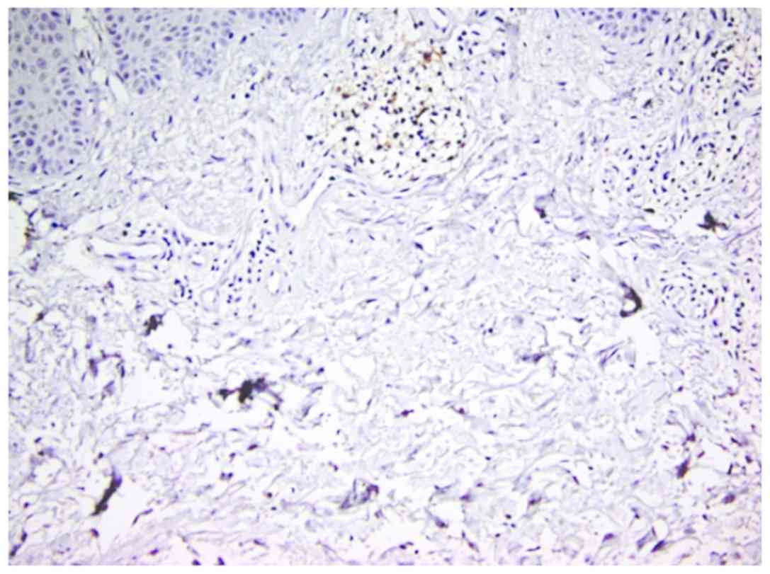
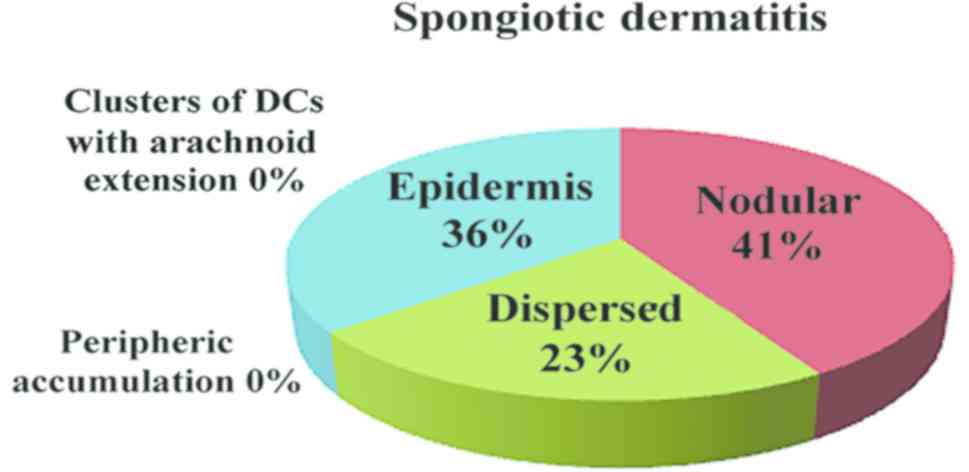
19
|
Phung TI, Wright TS, Pourciau KY and
Smoller BR: Spongiotic dermatitis. Pediatr Dermatol. Jun
21–2017.(Epub ahead of print). doi:
10.1007/978-3-319-44824-4_1.
|
|
20
|
Colmenero I, Torrelo A and Reyes-Múgica M:
Skin. In: Essentials of Surgical Pediatric Pathology. Cohen MC and
Scheimberg I: Cambridge University Press; Cambridge: pp. 1–2. 2014,
PubMed/NCBI
|
|
21
|
Abreu Velez AM, Loebel AM and Howard MS:
Spongiotic dermatitis with a mixed inflammatory infiltrate of
lymphocytes, antigen presenting cells, immunoglobulins and
complement. Dermatol Online. 2:52–57. 2011.
|
|
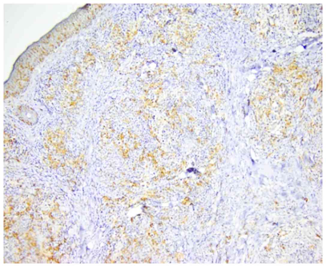
22
|
Deguchi M, Aiba S, Ohtani H, Nagura H and
Tagami H: Comparison of the distribution and numbers of
antigen-presenting cells among T-lymphocyte-mediated dermatoses:
CD1a+, factor XIIIa+, and CD68+
cells in eczematous dermatitis, psoriasis, lichen planus and
graft-versus-host disease. Arch Dermatol Res. 294:297–302. 2002.
View Article : Google Scholar : PubMed/NCBI
|
|
23
|
Lin TC, Wu PY, Lin TY, Yeh SP, Chen SC and
Lee TL: Langerhans cell hyperplasia in the tumor stage of mycosis
fungoides: A mimic of Langerhans cell histiocytosis. Dermatol Sin.
29:101–105. 2011. View Article : Google Scholar
|
|
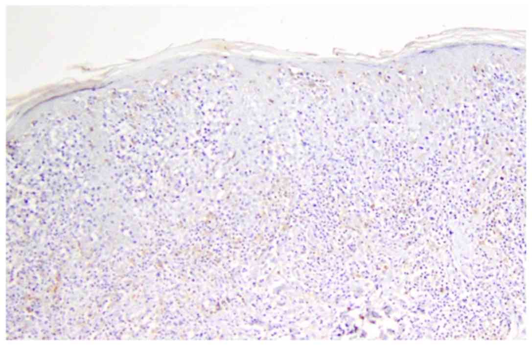
24
|
Goteri G, Filosa A, Mannello B,
Stramazzotti D, Rupoli S, Leoni P and Fabris G: Density of
neoplastic lymphoid infiltrate, CD8+ T cells, and
CD1a+ dendritic cells in mycosis fungoides. J Clin
Pathol. 56:453–458. 2003. View Article : Google Scholar : PubMed/NCBI
|
|
25
|
Berger CL, Hanlon D, Kanada D, Dhodapkar
M, Lombillo V, Wang N, Christensen I, Howe G, Crouch J, El-Fishawy
P, et al: The growth of cutaneous T-cell lymphoma is stimulated by
immature dendritic cells. Blood. 99:2929–2939. 2002.PubMed/NCBI
|
|
26
|
Ion A, Popa IM, Laura ML, Lisievici C,
Lupu M, Voiculescu V, Caruntu C and Boda D: Proteomic approaches to
biomarker discovery in cutaneous T-cell lymphoma. Dis Markers.
2016:96024722016. View Article : Google Scholar : PubMed/NCBI
|
|
27
|
Ni X and Duvic M: Dendritic cells and
cutaneous T-cell lymphomas. G Ital Dermatol Venereol. 146:103–113.
2011.PubMed/NCBI
|