Introduction
Pancreatic cancer is a fast-growing and highly
aggressive malignant digestive system tumor (1), usually occurring in males. The peak age
of its onset is 40–70 years, with the age of onset getting younger
(2). The prognosis of pancreatic
cancer is extremely poor, and the average survival time is
generally less than half a year, with a 5-year survival rate of
only 5% (3). Tumor resection is
still the only way to hopefully cure it. However, since most
patients have been diagnosed with lymph node metastasis and distant
metastasis, failing to meet surgical conditions, the surgical
resection rate is very low, only 5–30% (4,5).
According to reports in the literature, pancreatic cancer is
correlated with congenital and acquired factors such as genetic
factors, smoking, obesity, drinking, diabetes mellitus and chronic
pancreatitis. Those having risk factors described above, you should
take regular physical examinations to prevent pancreatic cancer
(6).
It has been reported in the literature that miRNAs
are involved in cell proliferation, apoptosis, differentiation and
embryonic development (7). In recent
years, the relationship between miRNAs and tumors have become a
research hotspot (8). The miR-425
family, containing miR-425-3p and miR-425-5p (9), is abnormally expressed in tissues such
as the gastrointestinal tract, immune system, and heart, liver and
lung, closely related to the development of various diseases and
participating in their biological processes (10). Related studies have found that
miR-425-5p can promote the proliferation, invasion and migration of
pancreatic cancer cells (11).
Having 35% homology with IL-12, IL-23 is a novel cytokine that
plays an important role in autoimmune diseases, infections and
tumors (12). In the study of
Langowski et al (13), IL-23
was found to play a role in promoting tumor growth in mice
deficient in IL-23p19 (one of two subunits of IL-23).
miR-425-5p and IL-23 have rarely been studied in
clinical aspects of pancreatic cancer. Therefore, the aim of this
study was to investigate their expression levels and clinical
significance in pancreatic cancer, which plays a certain indicative
role in the diagnosis and treatment and in the prediction of
prognosis.
Materials and methods
General information
A retrospective analysis of 76 cases of pancreatic
cancer tissue specimens and normal fresh tissue specimens at 50 mm
adjacent to the corresponding cancer surgically resected in the
first diagnosis from October 2015 to October 2016 in Traditional
Chinese Medical Hospital of Huangdao District (Qingdao, China) and
Weifang Medical University Hospital (Weifang, China) was performed.
The specimens were preserved in liquid nitrogen immediately after
excision and then transferred to a refrigerator at −80°C. Adjacent
tissues were confirmed as no obvious infiltration of cancer cells
or inflammatory cells. The average age of patients was 48.08±16.52
years, with 39 males and 37 females. All subjects were diagnosed as
pancreatic cancer by pancreatic biopsy or pathology, without
chemotherapy or radiotherapy before operation. They were excluded
during pregnancy and lactation. Those with primary and malignant
tumor diseases in other parts and tissues, with heart, liver and
kidney dysfunction or with pancreas-related operation within six
months prior to admission were excluded.
This study was approved by the Ethics Committee of
Traditional Chinese Medical Hospital of Huangdao District and
Weifang Medical University Hospital. All subjects and their family
members signed the informed consent form with complete clinical
information.
Detection of miR-425-5p
TRIzol lysate (1 ml) (Shanghai Pufei Biotechnology
Co., Ltd., Shanghai, China) was added after 100 mg of tissue ground
and pulverized, to extract total RNA from the tissue. After that,
1.5% agarose gel electrophoresis was used to analyze RNA integrity,
and the micronucleic acid analyzer (Beijing Meilinhengtong
Instrument Co., Ltd., Beijing, China) to detect the extracted RNA
purity and concentration. The A260/A280 value was considered to
meet experimental requirements between 1.9 and 2.0. After RNA
extraction, RT-qPCR reaction was performed using a Countess II FL
Automated Cell Counter Real-Time PCR instrument (Applied
Biosystems; Thermo Fisher Scientific, Inc., Waltham, MA, USA). The
reverse transcription reaction system was: 1.0 µl of oligo(dT)
primer, 1.0 µl of dNTP mixture, 1.0 µl of 2.5 U/ µl polymerase, 2
µg of total RNA, and ribonuclease distilled water added to 10 µl;
water bath at 42°C for 90 min, reaction at 95°C for 5 min, and then
immediately ice bath for 5 min. After the reverse transcription
reaction, PCR amplification was performed. The PCR amplification
system was: 2 µl of cDNA template, 25 µl of SYBR-Green Mix, 0.5 µl
of each of upstream primer and downstream primer, and double
distilled water added to 50 µl, pre-denaturation at 95°C for 3 min,
denaturation at 95°C for 30 sec, annealing at 55°C for 30 sec,
extension at 72°C for 60 sec, for a total of 30 cycles, and after
completion of the cycle, extension at 72°C for 5 min. U6 was used
as an reaction internal reference. All samples were repeated for 3
wells. The 2−∆Cq method (14) was used to detect the expression level
of miR-425-5p. The primer sequence was designed and synthesized by
Hepeng (Shanghai) Biotechnology Co., Ltd., Shanghai, China, as
shown in Table I.
 | Table I.miR-425-5p primer and internal
reference sequence. |
Table I.
miR-425-5p primer and internal
reference sequence.
| Gene | Upstream primer | Downstream
primer |
|---|
| miR-425-5p |
5′-GGGGAGTTAGGATTAGGTC-3′ |
5′-TGCGTGTCGTGGAGTC-3′ |
| U6 |
5′-CTCGCTTCGGCAGCACA-3′ |
5′-AACGCTTCACGAATTTGCGT-3′ |
Detection of IL-23
In total 76 cases of cancer tissues and adjacent
normal tissues surgically resected from pancreatic cancer patients
were taken and weighed after the fat and blood clots were removed.
After washed 3 times with D-Hank's cleaning solution containing
double-antibody (Shanghai Chunshi Biotechnology Co., Ltd.,
Shanghai, China), the tissue was cut into small pieces and placed
in an ELISA plate. RPMI-1640 complete medium containing
double-antibody (Shanghai Bosheng Biotechnology Co., Ltd.,
Shanghai, China) was added to the plate, and incubated at 37°C for
24 h. Then, the medium was collected, centrifuged in a centrifuge
tube at 12,600 × g for 5 min at 4°C, with the supernatant taken.
ELISA was used to detect IL-23 levels in cancer tissues and
adjacent tissues. The kit and the sample to be tested were taken
from the refrigerator 30 min in advance to balance the room
temperature, with a sample well, a standard well and a blank well
set. The Antus PHOmo automatic microplate reader (Jinan Yuteng
Biotechnology Co., Ltd., Jinan, China) was used to measure the OD
value of each well at the wavelength of 450 nm, to calculate IL-23
concentration.
Statistical analysis
SPSS 20.0 (Shanghai Kabei Information Technology
Co., Ltd., Shanghai, China) statistical software was used for
analysis, Student's t-test for measurement data which was expressed
as means ± SD, and Pearson for correlation analysis. P<0.05 was
considered to indicate a statistically significant difference.
Results
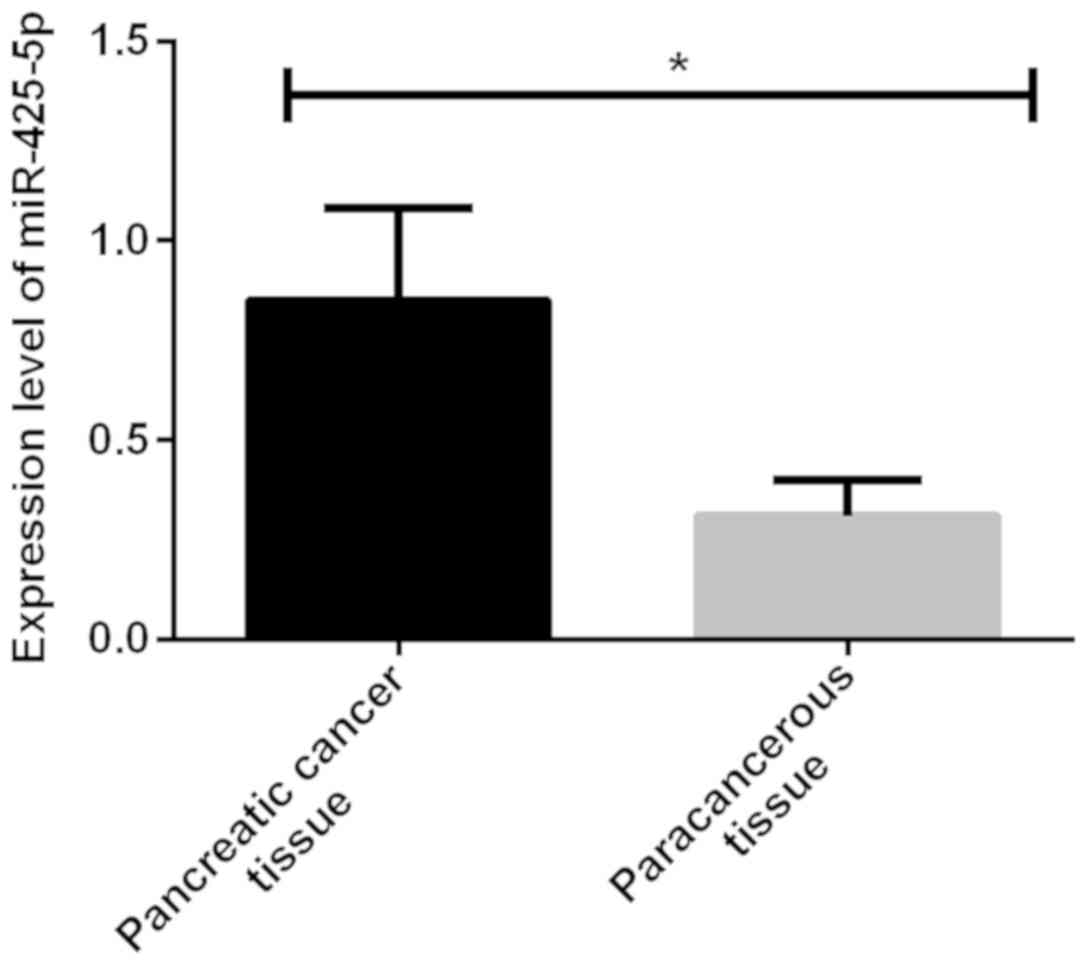
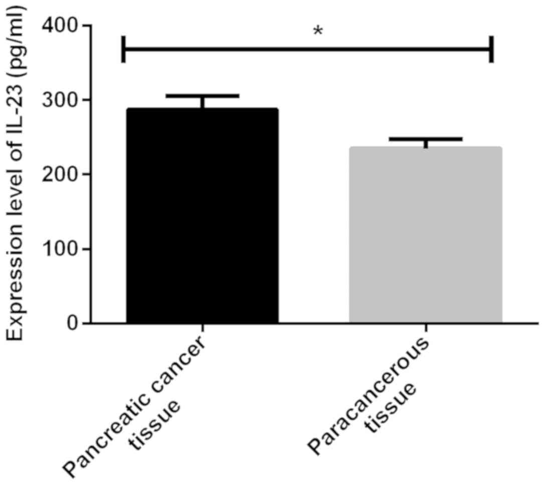
Comparison of miR-425-5p and IL-23
levels between pancreatic cancer tissues and adjacent tissues
The expression levels of miR-425-5p and IL-23 were
significantly higher in pancreatic cancer tissues than those in
adjacent tissues, with statistically significant differences
(P<0.001) (Figs. 1 and 2; and Table
II.
| Table II.Comparison of miR-425-5p and IL-23
levels between pancreatic cancer tissues and adjacent tissues. |
Table II.
Comparison of miR-425-5p and IL-23
levels between pancreatic cancer tissues and adjacent tissues.
| Group | n | miR-425-5p | IL-23 (pg/ml) |
|---|
| Pancreatic cancer
tissues | 76 | 0.85±0.23 | 286.69±18.32 |
| Adjacent tissues | 76 | 0.31±0.09 | 234.36±12.68 |
| t value |
| 19.06 | 20.48 |
| P-value |
| <0.001 | <0.001 |
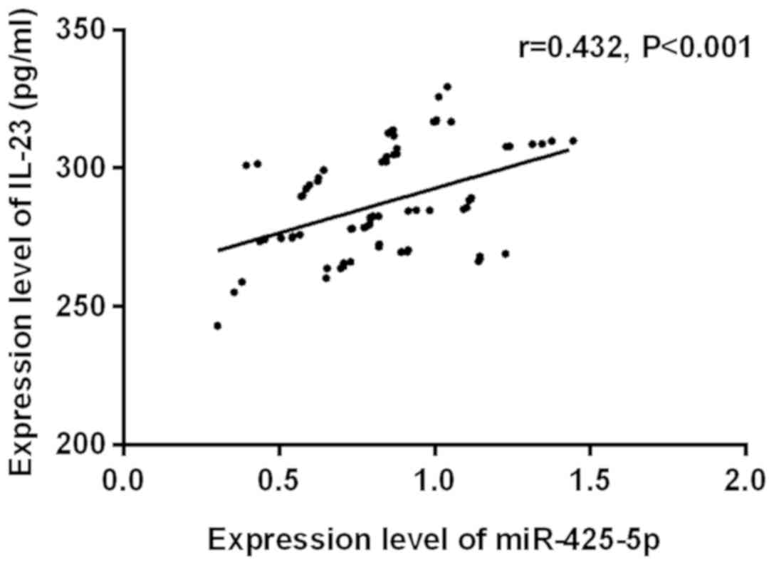
Correlation analysis of miR-425-5p
with IL-23 in pancreatic cancer
The expression level of miR-425-5p was positively
correlated with that of IL-23 in pancreatic cancer tissues
(r=0.432, P<0.001) (Fig. 3).
Correlation of miR-425-5p and IL-23
with clinical pathology of pancreatic cancer
The expression level of miR-425-5p in tumor tissues
of pancreatic cancer patients was not significantly correlated with
age, sex, tumor size and distant metastasis (P>0.05), but
correlated with lymph node metastasis, clinical stage and
differentiation degree (P<0.001). Expression of IL-23 in tumor
tissues of pancreatic cancer patients was not significantly
correlated with age, sex, tumor size, lymph node metastasis,
distant metastasis or differentiation degree (P>0.05), but
correlated with clinical stage (P<0.001) (Table III).
| Table III.Correlation of miR-425-5p and IL-23
with clinical pathology of pancreatic cancer. |
Table III.
Correlation of miR-425-5p and IL-23
with clinical pathology of pancreatic cancer.
| Characteristics | n | miR-425-5p | t value | P-value | IL-23 (pg/ml) | t value | P-value |
|---|
| Age (years) |
|
| 1.803 | 0.076 |
| 1.188 | 0.239 |
| ≥48 | 41 | 0.92±0.25 |
|
| 289.53±23.64 |
|
|
|
<48 | 35 | 0.82±0.23 |
|
| 296.45±27.16 |
|
|
| Sex |
|
| 1.606 | 0.113 |
| 0.896 | 0.373 |
| Male | 39 | 0.89±0.24 |
|
| 283.52±24.16 |
|
|
|
Female | 37 | 0.81±0.19 |
|
| 289.05±29.53 |
|
|
| Tumor size |
|
| 1.570 | 0.121 |
| 1.104 | 0.273 |
| ≥5
cm | 34 | 0.96±0.28 |
|
| 292.58±26.88 |
|
|
| <5
cm | 42 | 0.87±0.22 |
|
| 285.47±28.73 |
|
|
| Lymph node
metastasis |
|
| 4.257 | <0.001 |
| 1.542 | 0.127 |
| Yes | 37 | 0.98±0.31 |
|
| 286.28±24.83 |
|
|
| No | 39 | 0.75±0.13 |
|
| 277.35±25.62 |
|
|
| Distant
metastasis |
|
| 1.407 | 0.164 |
| 1.156 | 0.251 |
| Yes | 24 | 0.91±0.27 |
|
| 293.57±26.38 |
|
|
| No | 52 | 0.83±0.21 |
|
| 286.34±24.85 |
|
|
| Clinical stage |
|
| 4.774 | <0.001 |
| 8.315 | <0.001 |
| I+II | 36 | 0.72±0.15 |
|
| 267.82±20.36 |
|
|
|
III+IV | 40 | 0.97±0.28 |
|
| 315.76±28.69 |
|
|
| Differentiation
degree |
|
| 2.096 | 0.040 |
| 0.564 | 0.575 |
| Low and
medium | 42 | 0.92±0.29 |
|
| 286.15±21.05 |
|
|
|
High | 34 | 0.79±0.24 |
|
| 283.43±20.73 |
|
|
Discussion
According to statistics, the mortality of pancreatic
cancer ranks fourth among cancer patients in the United States and
sixth in China. With the title of ‘king of cancer’, it has clinical
features of short course of disease, rapid progress and low
survival rate (15). Studies have
shown that the miR-425-5p family, correlated with the
post-transcriptional regulation of a variety of genes, is
abnormally expressed in various cancer tissue samples such as liver
cancer, laryngeal cancer and gastric cancer (16). According to reports in the
literature, as an important inflammatory factor in the tumor
microenvironment, IL-23 usually promotes tumor angiogenesis and
inhibits CD8+ cell infiltration, which is of great
significance in the development of tumors (17). In order to improve the early
screening rate and treatment level of pancreatic cancer, in this
study, the correlation of miR-425-5p and IL-23 with the clinical
pathology of pancreatic cancer was studied, providing ideas for
improving the prognosis and survival of pancreatic cancer
patients.
The present study showed that the expression levels
of miR-425-5p and IL-23 were significantly higher in pancreatic
cancer tissues than those in adjacent tissues, with statistically
significant differences (P<0.001). In the study of Zhang et
al (16), it was found that
miR-425-5p, significantly higher in gastric cancer tissues than in
adjacent tissues, promoting invasion and metastasis of gastric
cancer cells. Shime et al (18)showed that IL-23 is a pro-inflammatory
factor. Furthermore, it is highly expressed in mouse breast cancer
tissues (19). IL-23 that is highly
expressed in breast cancer tissues may be involved in the
occurrence of breast cancer (20).
The above conclusions are consistent with our findings. The
expression level of miR-425-5p was positively correlated with that
of IL-23 in pancreatic cancer tissues (r=0.432, P<0.001). No
correlation study of the expression level of miR-425-5p with that
of IL-23 in tumors has been reported. However, inflammatory factors
and immune function are usually abnormal in tumor patients, so
IL-23 may affect miR-425-5p expression by regulating T cells in
tumor tissues. The expression level of miR-425-5p in tumor tissues
of pancreatic cancer patients was not significantly correlated with
age, sex, tumor size and distant metastasis (P>0.05), but
correlated with lymph node metastasis, clinical stage and
differentiation degree (P<0.001). The results of Sun et
al (21) are basically
consistent with ours. They found that the high expression of
miR-425-5p in cervical cancer tissues is correlated with tumor
stage and lymph node metastasis. The expression level of IL-23 in
tumor tissues of pancreatic cancer patients was not significantly
correlated with age, sex, tumor size, lymph node metastasis,
distant metastasis and differentiation degree (P>0.05), but
correlated with clinical stage (P<0.001). Petanidis et al
(22) found that IL-23 is correlated
with the TNM staging of colon cancer, but not significantly
correlated with age, sex, tumor size, lymph node metastasis and
differentiation degree. This is basically consistent with our
results. It is possible that lactic acid produced by tumor cells
can cause mononuclear macrophages in tumors to produce IL-23 and
activate cAMP/PKA signaling pathway, thereby promoting tumor growth
(23).
In summary, both miR-425-5p and IL-23 are highly
expressed in pancreatic cancer tissues. The expression level of
miR-425-5p is positively correlated with that of IL-23 (r=0.432,
P<0.001). Expression of miR-425-5p in tumor tissues of
pancreatic cancer patients is correlated with lymph node
metastasis, clinical stage and differentiation degree, and that of
IL-23 in tumor tissues of pancreatic cancer patients is correlated
with clinical stage. It is suggested that miR-425-5p and IL-23 may
be involved in the pathological development of pancreatic
cancer.
Acknowledgements
Not applicable.
Funding
No funding was received.
Availability of data and materials
The datasets used and/or analyzed during the current
study are available from the corresponding author on reasonable
request.
Authors' contributions
YL was involved in drafting the manuscript. YL and
XW performed PCR and ELISA. JW analyzed the general data of
patients. All authors read and approved the final manuscript.
Ethics approval and consent to
participate
This study was approved by the Ethics Committee of
Traditional Chinese Medical Hospital of Huangdao District (Qingdao,
China) and Weifang Medical University Hospital (Weifang, China).
Signed informed consents were obtained from the patients or the
guardians.
Patient consent for publication
Not applicable.
Competing interests
The authors declare that they have no competing
interests.
References
|
1
|
Li D, Xie K, Wolff R and Abbruzzese JL:
Pancreatic cancer. Lancet. 363:1049–1057. 2004. View Article : Google Scholar : PubMed/NCBI
|
|
2
|
Vincent A, Herman J, Schulick R, Hruban RH
and Goggins M: Pancreatic cancer. Lancet. 378:607–620. 2011.
View Article : Google Scholar : PubMed/NCBI
|
|
3
|
Rochefort MM, Ankeny JS, Kadera BE, Donald
GW, Isacoff W, Wainberg ZA, Hines OJ, Donahue TR, Reber HA and
Tomlinson JS: Impact of tumor grade on pancreatic cancer prognosis:
Validation of a novel TNMG staging system. Ann Surg Oncol.
20:4322–4329. 2013. View Article : Google Scholar : PubMed/NCBI
|
|
4
|
Koliopanos A, Avgerinos C, Farfaras A,
Manes C and Dervenis C: Radical resection of pancreatic cancer.
Hepatobiliary Pancreat Dis Int. 7:11–18. 2008.PubMed/NCBI
|
|
5
|
Abe M, Kondo S, Hirano S, Ambo Y, Tanaka
E, Morikawa T, Okushiba S and Katoh H: Long-term survival after
radical resection of advanced pancreatic cancer: A case report with
special reference to CD8+ T-cell infiltration. Int J
Gastrointest Cancer. 33:107–110. 2003. View Article : Google Scholar : PubMed/NCBI
|
|
6
|
Lowenfels AB and Maisonneuve P:
Epidemiology and risk factors for pancreatic cancer. Best Pract Res
Clin Gastroenterol. 20:197–209. 2006. View Article : Google Scholar : PubMed/NCBI
|
|
7
|
Kashiwagi Y, Kato N, Sassa T, Nishitsuka
K, Yamamoto T, Takamura H and Yamashita H: Cotylenin A inhibits
cell proliferation and induces apoptosis and PAX6 mRNA transcripts
in retinoblastoma cell lines. Mol Vis. 16:970–982. 2010.PubMed/NCBI
|
|
8
|
Tchernitsa O, Kasajima A, Schäfer R, Kuban
RJ, Ungethüm U, Györffy B, Neumann U, Simon E, Weichert W, Ebert
MP, et al: Systematic evaluation of the miRNA-ome and its
downstream effects on mRNA expression identifies gastric cancer
progression. J Pathol. 222:310–319. 2010. View Article : Google Scholar : PubMed/NCBI
|
|
9
|
He B, Li T, Guan L, Liu FE, Chen XM, Zhao
J, Lin S, Liu ZZ and Zhang HQ: CTNNA3 is a tumor suppressor in
hepatocellular carcinomas and is inhibited by miR-425. Oncotarget.
7:8078–8089. 2016.PubMed/NCBI
|
|
10
|
Li L, Chen HZ, Chen FF, Li F, Wang M, Wang
L, Li YQ and Gao DS: Global microRNA expression profiling reveals
differential expression of target genes in
6-hydroxydopamine-injured MN9D cells. Neuromolecular Med.
15:593–604. 2013. View Article : Google Scholar : PubMed/NCBI
|
|
11
|
Yu J, Li A, Hong SM, Hruban RH and Goggins
M: MicroRNA alterations of pancreatic intraepithelial neoplasias.
Clin Cancer Res. 18:981–992. 2012. View Article : Google Scholar : PubMed/NCBI
|
|
12
|
Ding C, Xu J and Li J: ABT-874, a fully
human monoclonal anti-IL-12/IL-23 antibody for the potential
treatment of autoimmune diseases. Curr Opin Investig Drugs.
9:515–522. 2008.PubMed/NCBI
|
|
13
|
Langowski JL, Zhang X, Wu L, Mattson JD,
Chen T, Smith K, Basham B, McClanahan T, Kastelein RA and Oft M:
IL-23 promotes tumour incidence and growth. Nature. 442:461–465.
2006. View Article : Google Scholar : PubMed/NCBI
|
|
14
|
Livak KJ and Schmittgen TD: Analysis of
relative gene expression data using realtime quantitative PCR and
the 2(-Delta Delta C(T)) method. Methods. 25:402–408. 2001.
View Article : Google Scholar : PubMed/NCBI
|
|
15
|
Siegel R, Naishadham D and Jemal A: Cancer
statistics, 2013. CA Cancer J Clin. 63:11–30. 2013. View Article : Google Scholar : PubMed/NCBI
|
|
16
|
Zhang Z, Wen M, Guo J, Shi J, Wang Z, Tan
B, Zhang G, Zheng X and Zhang A: Clinical value of miR-425-5p
detection and its association with cell proliferation and apoptosis
of gastric cancer. Pathol Res Pract. 213:929–937. 2017. View Article : Google Scholar : PubMed/NCBI
|
|
17
|
Ciric B, El-behi M, Cabrera R, Zhang GX
and Rostami A: IL-23 drives pathogenic IL-17-producing
CD8+ T cells. J Immunol. 182:5296–5305. 2009. View Article : Google Scholar : PubMed/NCBI
|
|
18
|
Shime H, Yabu M, Akazawa T, Kodama K,
Matsumoto M, Seya T and Inoue N: Tumor-secreted lactic acid
promotes IL-23/IL-17 proinflammatory pathway. J Immunol.
180:7175–7183. 2008. View Article : Google Scholar : PubMed/NCBI
|
|
19
|
Chen G, Liang Y, Guan X, Chen H, Liu Q,
Lin B, Chen C, Huang M, Chen J, Wu W, et al: Circulating low IL-23:
IL-35 cytokine ratio promotes progression associated with poor
prognosisin breast cancer. Am J Transl Res. 8:2255–2264.
2016.PubMed/NCBI
|
|
20
|
Khodadadi A, Razmkhah M, Eskandari AR,
Hosseini A, Habibagahi M, Ghaderi A and Jaberipour M: IL-23/IL-27
ratio in peripheral blood of patients with breast cancer. Iran J
Med Sci. 39:350–356. 2014.PubMed/NCBI
|
|
21
|
Sun L, Jiang R, Li J, Wang B, Ma C, Lv Y
and Mu N: MicoRNA-425-5p is a potential prognostic biomarker for
cervical cancer. Ann Clin Biochem. 54:127–133. 2017. View Article : Google Scholar : PubMed/NCBI
|
|
22
|
Petanidis S, Anestakis D, Argyraki M,
Hadzopoulou-Cladaras M and Salifoglou A: Differential expression of
IL-17, 22 and 23 in the progression of colorectal cancer in
patients with K-ras mutation: Ras signal inhibition and crosstalk
with GM-CSF and IFN-γ. PLoS One. 8:e736162013. View Article : Google Scholar : PubMed/NCBI
|
|
23
|
Qian X, Gu L, Ning H, Zhang Y, Hsueh EC,
Fu M, Hu X, Wei L, Hoft DF and Liu J: Increased Th17 cells in the
tumor microenvironment is mediated by IL-23 via tumor-secreted
prostaglandin E2. J Immunol. 190:5894–5902. 2013. View Article : Google Scholar : PubMed/NCBI
|