Introduction
Cardiac carcinoma is a kind of malignant tumor in
the cardia, which has the poorest prognosis in gastric carcinoma,
seriously harming the quality of life and health of patients
(1,2). According to the epidemiological survey,
the incidence of cardiac carcinoma shows an increasing trend year
by year in China, and it has increased more than five-fold over the
decades (3). It has been proven that
smoking, stimulating beverages, irregular diet, gastrointestinal
inflammation and gastroesophageal reflux are precipitating factors
for cardiac carcinoma. Currently, surgical resection is the most
effective treatment means for cardiac carcinoma, but the poor
prognosis of patients is still an important reason affecting the
therapeutic effect (4,5).
The p16 gene can inhibit the progression of a
variety of tumors and encode the nucleotide protein (p16 protein)
to significantly inhibit the cyclin-dependent kinase 4 (CDK4), thus
playing an important role in the progression of various tumors
(6). Cyclin D1, as a key cell cycle
regulatory protein, can bind to CDK4/6 to form the binary complex
and keep cells in S phase, thus promoting cell proliferation
(7). Dreyer et al (8) showed that both p16 and cyclin D1 can
competitively bind to CDK4, thereby regulating the occurrence and
development of tumors. Feng et al (9) found that when the expression level of
p16 is blocked, it leads to dominant binding between cyclin D1 and
CDK4 and continuous cell proliferation, thus resulting in tumors.
According to the study of Pu et al (10), p16 is a predisposing gene of
cardiac carcinoma, and it has been shown in several animal models
that the p16 expression level in cardiac carcinoma tissues is
significantly reduced. However, the correlation of p16 and cyclin
D1 with incidence and prognosis of cardiac carcinoma remains
unclear.
This study analyzed the p16 and cyclin D1 gene and
protein expression levels in carcinoma tissues of patients with
cardiac carcinoma, and aimed to clarify the correlation of their
expression with the incidence and prognosis of cardiac carcinoma to
provide a theoretical basis for clinical diagnosis, treatment and
prognosis estimation of cardiac carcinoma.
Patients and methods
Research subjects and sampling
Patients with cardiac carcinoma who attended the
Department of Gastroenterology of The Second Affiliated Hospital of
Zhengzhou University (Zhengzhou, China) from March 2012 to March
2013 were collected, of which 36 cases (28 males, 8 females)
definitely diagnosed with primary cardiac carcinoma and treated
with radical operation of cardiac carcinoma were selected. The
patients were aged 42–69 years with an average age of 58.6 years.
According to the tumor-node-metastasis (TNM) staging system issued
by the Union for International Cancer Control (UICC), there were 3
cases in stage I, 5 cases in stage II, 25 cases in stage III and 3
cases in stage IV. Inclusion criteria were: i) patients without a
history of severe cardiovascular, cerebrovascular diseases and
immune system disease, ii) patients without severe hepatic and
renal impairment, iii) patients receiving no chemotherapy or
radiotherapy before operation, iv) patients definitely diagnosed
with cardiac carcinoma by pathologic examinations, and v) patients
who agreed to participate in follow-up visits and finished 5-year
follow-up visits.
Informed consent was signed by patients themselves
or their family members. All clinical and pathological data and
complete treatment schedule were preserved. The plan of this
experiment was reviewed and ethically approved in The Second
Affiliated Hospital of Zhengzhou University.
For sampling, cardiac carcinoma tissue was excised
surgically while para-carcinoma tissue was resected at the cardia
over 3 cm away from the edge of the cancerous tissue. Specimens of
cancerous tissue and para-carcinoma tissue were partially made into
paraffin sections and were definitely diagnosed by the Department
of Pathology. The remaining cancerous and para-carcinoma tissue was
immediately cryopreserved in liquid nitrogen and then transferred
to a refrigerator at −80°C for standby application.
Detection of expression levels of p16
and cyclin D1 mRNA in cardiac carcinoma tissue and para-carcinoma
tissue by quantitative polymerase chain reaction (qPCR)
Cardiac carcinoma tissue and para-carcinoma tissue
of all the patients were weighed and added into TRIzol (Thermo
Fisher Scientific, Inc., Waltham, MA, USA) at a ratio of 100 mg:1
ml. Tissues were smashed to fragments invisible to the naked eye in
an ice bath using an ultrasonic homogenizer, placed on ice for 10
min of incubation and then centrifuged at 4°C and 12,000 × g for 10
min. The supernatant was collected and added with 700 µl of
trichloromethane, which was placed on ice for 10 min of incubation
after repeated shaking for 10 sec, followed by centrifugation at
4°C and 12,000 × g for 15 min. The upper layer aqueous phase was
collected and added with isopropanol of equal volume. After 10 sec
of repeated shaking, the mixture was centrifuged at 4°C and 10,000
× g for 10 min. The mixture was washed with 1 ml of newly-prepared
75% ethanol after the supernatant was discarded, shaken repeatedly
for 10 sec and centrifuged at 4°C and 12,000 × g for 10 min. After
the supernatant was discarded, the cover of the centrifuge tube was
opened to make it dry naturally. RNA was obtained after 30 µl
DEPC-treated water was added. RNA concentration and purity were
determined using an ultraviolet spectrophotometer. In this
experiment, the absorbance (A) 260/A280 ratio of RNA was between
1.8 and 1.9 and the concentration and purity of RNA were both high.
The agarose gel experiment indicated that RNA was not degraded, so
it could be used in follow-up experiments. Reverse transcriptional
reaction system was configured strictly according to instructions
of the reverse transcription kit (Vazyme Biotech Co., Ltd.,
Nanjing, China). Reverse transcription was carried out after 5 min
of reaction at 65°C and 2 min of incubation at 37°C. qPCR system
was configured strictly following instructions of qPCR kit (Vazyme
Biotech Co., Ltd.), which was amplified on qPCR device.
Amplification conditions were: 15 min of predegeneration at 95°C,
10 sec of degeneration at 95°C and 32 sec of annealing at 60°C, 40
cycles in total. The amplification volume of p16 gene and cyclin D1
gene and that of glyceraldehyde-3-phosphate dehydrogenase
(GAPDH) gene in all specimens were compared using
2−ΔΔCq (11). Expression
levels of p16 and cyclin D1 in cardiac carcinoma and para-carcinoma
tissue were presented as p16/GAPDH and cyclin D1/GAPDH and analyzed
with ImageJ professional image analysis software (NIH, Bethesda,
MD, USA).
The primer sequences in this experiment were
produced by Invitrogen (Thermo Fisher Scientific, Inc.) and the
primer sequences are shown in Table
I.
 | Table I.PCR primer sequences. |
Table I.
PCR primer sequences.
| Gene name | Primers |
|---|
| p16 | F: 5′-
ACCAGAGGCAGTAACCATGC-3′ |
|
| R:
5′-CCTGTAGGACCTTCGGTGAC-3′ |
| Cyclin D1 | F:
5′-ACCTGAGGAGCCCCAACAAC-3′ |
|
| R:
5′-GCTTCGATCTGCTCCTGGC-3′ |
| GAPDH | F:
5′-CTCTGGTAAAGTGGATATTGT-3′ |
|
| R:
5′-GGTGGAATCATATTGGAACA-3′ |
Detection of p16 and cyclin D1 protein
expression in cardiac carcinoma tissues and para-carcinoma tissues
via western blotting
After sodium dodecyl sulphate-polyacrylamide gel
(SDS-PAGE), cardiac carcinoma tissues and para-carcinoma tissues
were taken, added with radioimmunoprecipitation assay (RIPA) lysis
solution (1 mg:1 ml) and then added with 1% protease inhibitor and
1% phosphatase inhibitor, followed by homogenization in an ice bath
using an ultrasonic homogenizer until there were no visible tissues
to the naked eye. After standing for 5 min in an ice bath,
centrifugation was performed at 12,000 × g and 4°C for 10 min. The
supernatant was transferred into a new EP tube, and the total
protein concentration in the sample in each group was detected
using the bicinchoninic acid (BCA) protein assay kit (Pierce). The
5X SDS loading buffer and RIPA lysis solution were added to prepare
the protein sample in the protein loading buffer in an equal
concentration, and the protein was boiled at 95°C for 15 min for
denaturation. Loading buffer (15 µl) was added to each well, and
10% SDS-PAGE was performed under a constant pressure of 80 V, after
which the membrane transfer ‘sandwich’ was prepared. The protein
was transferred onto a polyvinylidene fluoride (PVDF) membrane
(IPVH00010; EMD Millipore, Billerica, MA, USA) after being soaked
in methanol at low temperature and 100 V for 90 min. Subsequently,
the protein band was sealed with freshly-prepared 5% skim milk
powder at room temperature. After 1 h, the band was cut according
to the size of the target band, and rabbit anti-human p16, cyclin
D1 and GAPDH monoclonal antibodies (1:1,000; cat. nos. 80772, 2978
and 2118, respectively; all obtained from Cell Signaling
Technology, Inc., Danvers, MA, USA) were incubated at 4°C
overnight. The next day, the membrane was taken, washed with
Tris-buffered saline with Tween-20 (TBST) 3 times (10 min/time),
incubated with the horseradish peroxidase-labeled secondary goat
anti-rabbit polyclonal antibody (1:5,000; cat. no. 7074; Cell
Signaling Technology, Inc.) at room temperature for 1 h, and washed
again with TBST 3 times (10 min/time). The liquid on the surface of
PVDF membrane was sucked dry using the filter paper, and the
enhanced chemiluminescence (ECL) solution was added (liquid A and B
were mixed in an equal volume), followed by incubation in the dark
for 1 min, and image development using the gel imaging system
(Bio-Rad Laboratories, Inc., Hercules, CA, USA). After the image
was scanned and saved, the gray value of band was analyzed. The
p16/GAPDH and cyclin D1/GAPDH represented the expression levels of
p16 and cyclin D1 in cardiac carcinoma tissues and para-carcinoma
tissues.
Collection of clinicopathological data
and survival analysis
Various clinicopathological data, clinical data and
clinical therapeutic schemes of all included patients were
preserved in detail and the postoperative basic treatment for
patients was consistent. Enrolled patients were divided into the
low- and high-expression p16 group, and the low- and
high-expression cyclin D1 group according to differences in the
expression levels of p16 and cyclin D1. Patients were followed up
for 5 years and relevant follow-up data were recorded, the
correlation of the expression levels of p16 and cyclin D1 with
prognosis was analyzed. Kaplan-Meier survival curves were
plotted.
Statistical analysis
Research data are presented as mean ± standard
deviation. Statistical Product and Service Solutions (SPSS) 19.0
software (IBM Corp., Armonk, NY, USA) was used for analytical
processing of data. The Chi-square test was used for analysis of
enumeration data, which were tested by homogeneity test of
variance. The Bonferronic method was used for pairwise comparison
if variance was homogeneous, while the Welch method was conducted
if not. The Students' t-test was applied for comparison between two
groups and Pearson correlation analysis was performed for
correlation analysis between p16 and cyclin D1. The correlation of
clinicopathologic data with expression levels of p16 and cyclin D1
protein was analyzed using Fisher's precise test and survival
analysis was carried out with Kaplan-Meier and log rank test.
P<0.05 was considered to indicate a statistically significant
difference.
Results
Expression levels of p16 and cyclin D1
mRNA in cardiac carcinoma tissues and para-carcinoma tissues
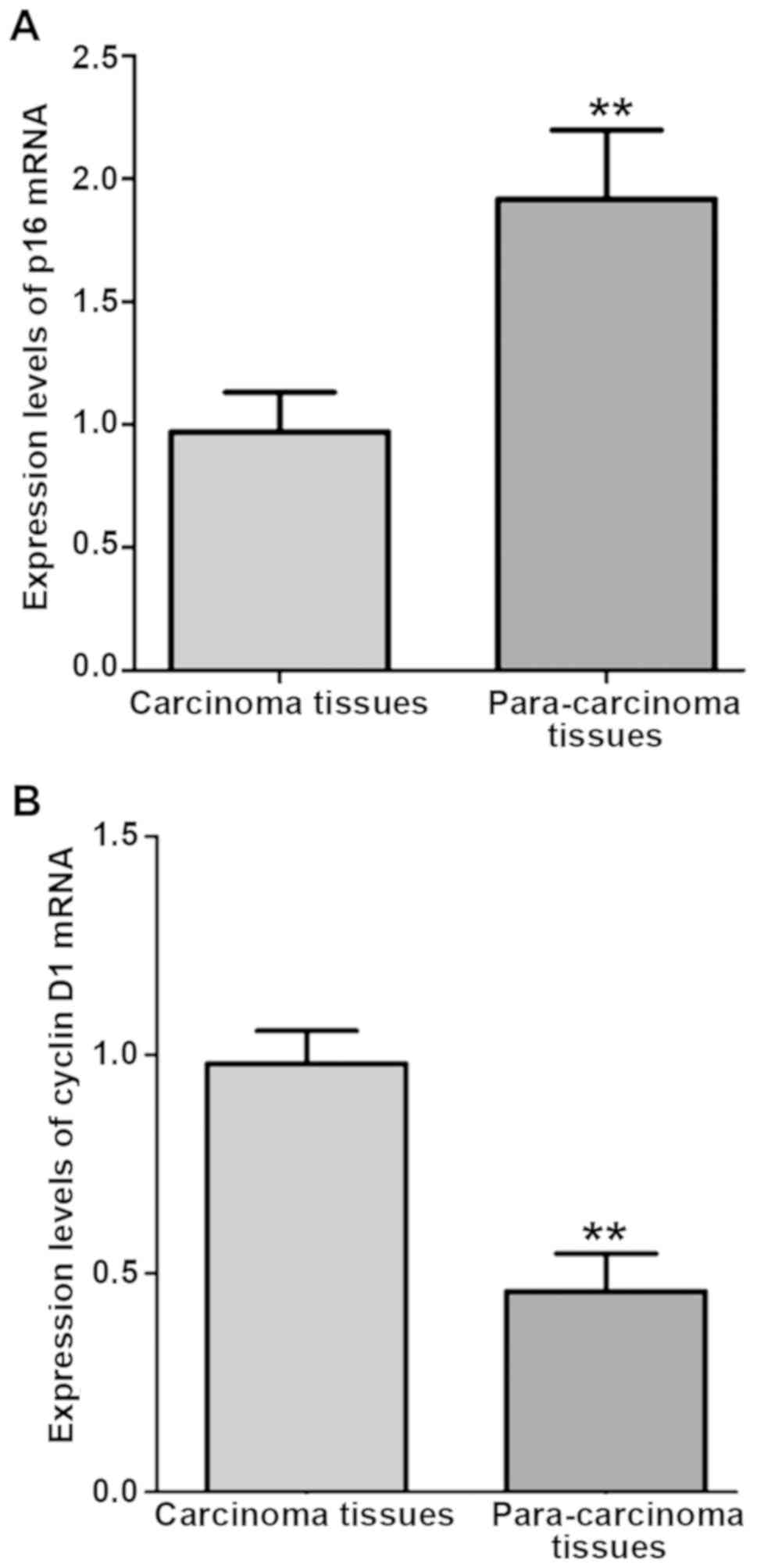
p16 and cyclin D1 mRNA expression in cardiac
carcinoma tissues and para-carcinoma tissues were detected via
qPCR. Results showed that in cardiac carcinoma tissues, the p16
mRNA expression was significantly lower than that in para-carcinoma
tissues (P<0.01), but the cyclin D1 mRNA expression was
significantly higher than that in para-carcinoma tissues
(P<0.01) (Fig. 1).
Expression levels of p16 and cyclin D1
proteins in cardiac carcinoma tissues and para-carcinoma
tissues
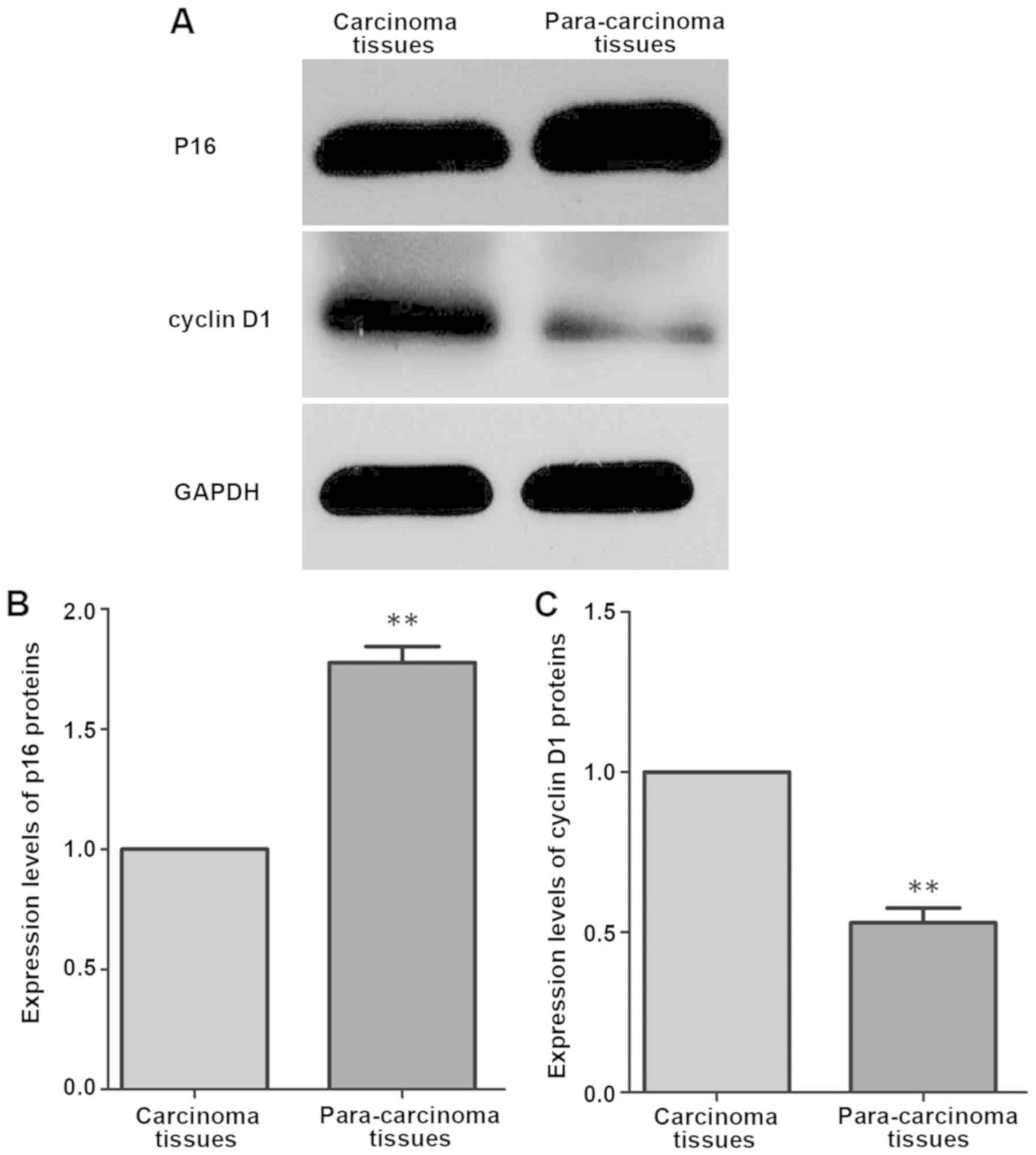
p16 and cyclin D1 protein expression levels in
cardiac carcinoma tissues and para-carcinoma tissues were detected
via western blotting. Results revealed that in cardiac carcinoma
tissues, the p16 protein expression was significantly lower than
that in para-carcinoma tissues (P<0.01), but the cyclin D1
protein expression was significantly higher than that in
para-carcinoma tissues (P<0.01) (Fig.
2).
Correlation analysis between p16 and
cyclin D1 in cardiac carcinoma tissues
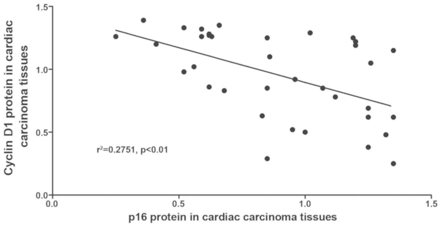
The correlation between p16 and cyclin D1 protein
expression in cardiac carcinoma tissues was analyzed. Results
demonstrated that there was a negative correlation between p16 and
cyclin D1 protein expression in cardiac carcinoma tissues
(r2=0.2751, P<0.01) (Fig.
3).
Correlation of p16 and cyclin D1
protein expression levels with clinicopathological factors of
patients with cardiac carcinoma
The clinicopathological data of patients with
cardiac carcinoma were recorded in detail, and their correlations
with relative expression levels of p16 and cyclin D1 protein were
analyzed. The relative expression levels of p16 and cyclin D1
protein in cardiac carcinoma tissues had no correlation with the
age and sex of patients with cardiac carcinoma (P>0.05), but was
correlated with the tumor size, lymph node metastasis and TNM stage
of cardiac carcinoma (P<0.01) (Table
II).
| Table II.Correlation of relative expression
levels of p16 and cyclin D1 with clinicopathological factors of
patients with cardiac carcinoma. |
Table II.
Correlation of relative expression
levels of p16 and cyclin D1 with clinicopathological factors of
patients with cardiac carcinoma.
| Pathological
detail | n | Relative expression
level of p16 | P-value | Relative expression
level of cyclin D1 | P-value |
|---|
| Carcinoma tissue | 36 | 1.08±0.12 | <0.01 | 0.83±0.15 | <0.01 |
| Para-carcinoma
tissue | 36 | 1.72±0.36 |
| 0.52±0.12 |
|
| Age (years) |
|
|
|
|
|
|
<60 | 15 | 1.28±0.52 | >0.05 | 0.78±0.22 | >0.05 |
| ≥60 | 21 | 1.33±0.45 |
| 0.75±0.26 |
|
| Sex |
|
|
|
|
|
| Male | 28 | 1.22±0.49 | >0.05 | 0.69±0.32 | >0.05 |
|
Female | 8 | 1.26±0.55 |
| 0.72±0.29 |
|
| Tumor size (cm) |
|
|
|
|
|
| ≤3 | 20 | 1.69±0.13 | <0.01 | 0.49±0.11 | <0.01 |
|
>3 | 16 | 1.12±0.18 |
| 0.92±0.08 |
|
| Lymph node
metastasis |
|
|
|
|
|
| No | 8 | 1.76±0.06 | <0.01 | 0.45±0.09 | <0.01 |
| Yes | 28 | 1.05±0.16 |
| 0.86±0.13 |
|
| TNM stage |
|
|
|
| <0.01 |
| I | 3 | 1.42±0.06 | <0.01 | 0.42±0.05 |
|
| II | 5 | 1.25±0.12 |
| 0.56±0.08 |
|
| III | 25 | 1.03±0.18 |
| 0.85±0.13 |
|
| IV | 3 | 0.96±0.05 |
| 0.86±0.07 |
|
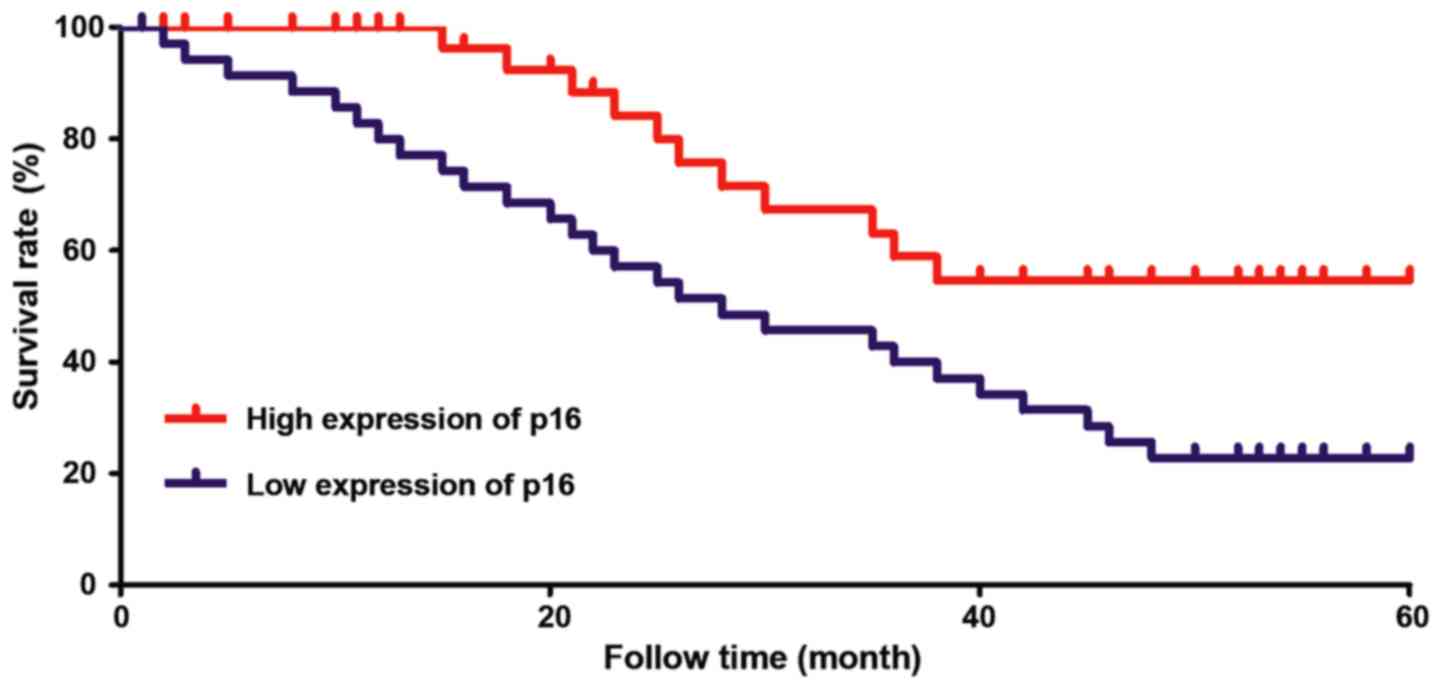
Effects of p16 and cyclin D1
expression on the prognosis of patients with cardiac carcinoma
All the patients enrolled were grouped based on the
relative expression levels of p16 and cyclin D1 proteins in cardiac
carcinoma tissues, and followed up for 5 years. The survival data
of patients were recorded, and the survival curve was drawn.
According to Kaplan-Meier survival analysis, the survival rate of
patients with high expression of p16 was obviously higher (58%)
than that (26%) of patients with low expression of p16 (P<0.01),
while the survival rate of patients with high expression of cyclin
D1 was obviously lower (19%) than that (52%) of patients with low
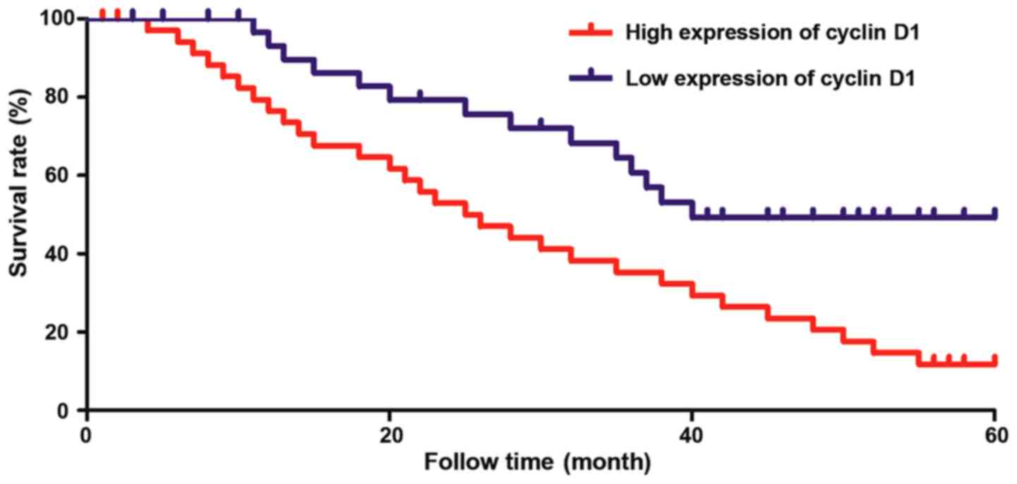
expression of cyclin D1 (P<0.01) (Figs. 4 and 5).
Discussion
With the continuous development of medical
technology, the 5-year survival time of tumor patients has also
increased continuously, and the quality of life and health of
patients have been greatly improved. However, the 5-year survival
rate of patients with cardiac carcinoma is still approximately 30%,
and lymph node metastasis and postoperative recurrence of cardiac
carcinoma are important factors seriously affecting the therapeutic
effect on cardiac carcinoma (12,13).
Research evidence has demonstrated that cardiac carcinoma
frequently occurs in high-incidence regions of gastric carcinoma
and esophageal carcinoma, but neither incidence trend nor
influencing factors of cardiac carcinoma are correlated with
gastric carcinoma and esophageal carcinoma (14). Zhao et al (15) found that the p16/cyclin D1 signaling
pathway plays an important role in glioma, which is involved in the
regulation of the cell cycle. The decrease in p16 expression level
or the increase in cyclin D1 expression level is closely related to
the occurrence and development of glioma. p16 is a kind of cyclin
kinase inhibitor playing an important role in inhibiting
tumorigenesis. In case of deletion, methylation or point mutation
of p16 gene, the p16 protein expression level will decline, and the
number of abnormal cells will increase, leading to the tumor
(16). According to the study of
Zhou and Gu (17), the incidence
rates of various tumors will be significantly increased after
p16-knockout mice are exposed to various carcinogenic conditions.
The expression level of cyclin D1, as a molecule playing an
important role in the regulation of cell cycle, has been found to
be obviously increased in a variety of tumors (18). Moreover, Cao et al (19) found that the expression level of
cyclin D1 in gastric carcinoma tissues is significantly higher than
that in para-carcinoma tissues, and the prognosis of patients with
high expression of cyclin D1 is poorer.
In this study, the p16 gene and protein expression
levels in carcinoma tissues and para-carcinoma tissues of 36
patients with cardiac carcinoma were detected. Results revealed
that the p16 gene and protein expression levels in cardiac
carcinoma tissues were significantly lower than those in
para-carcinoma tissues. The p16 protein expression level in
carcinoma tissues was significantly reduced with the enlargement of
the cardiac carcinoma tumor and the increase of TNM stage. The
above results strongly indicate that the p16 protein expression is
closely related to the occurrence and development of cardiac
carcinoma. Due to the particularity of cardiac carcinoma, lymph
node metastasis is closely associated with the prognosis of
patients. It was found that the p16 expression in carcinoma tissues
of patients with lymph node metastasis of cardiac carcinoma was
significantly lower than that in patients without lymph node
metastasis of cardiac carcinoma. The 5-year follow up showed that
the 5-year survival rate (58%) of patients with high expression of
p16 was significantly higher than that (26%) of patients with low
expression of p16, indicating that p16 is closely related to the
prognosis of patients. Moreover, the correlation between p16 and
cyclin D1 protein expression in cardiac carcinoma tissues was
analyzed in this study, and results showed that p16 was negatively
correlated with cyclin D1 in cardiac carcinoma tissues. Previous
findings have demonstrated (20,21) that
there is a steady balance between p16 and cyclin D1 in normal human
body, both of which jointly regulate the cell cycle. If the p16
expression is inhibited in the body, the cyclin D1 expression will
be increased significantly, thereby leading to a tumor. At the same
time, it was found in this study that the cyclin D1 expression was
closely related to the tumor size, lymph node metastasis and TNM
stage of cardiac carcinoma, and the 5-year survival rate (19%) of
patients with high expression of cyclin D1 was obviously lower than
that (52%) of patients with low expression of cyclin D1.
In conclusion, the expression of p16 and cyclin D1
is closely related to the incidence and prognosis of cardiac
carcinoma, which is expected to be developed into important genes
for predicting the incidence and prognosis of cardiac
carcinoma.
Acknowledgements
Not applicable.
Funding
No funding was received.
Availability of data and materials
The datasets used and/or analyzed during the present
study are available from the corresponding author on reasonable
request.
Authors' contributions
HW performed PCR. HW and JW were responsible for
western blotting. HW and BZ helped with the collection of
clinicopathologic data and survival analysis. All authors read and
approved the final manuscript.
Ethics approval and consent to
participate
The study was approved by the Ethics Committee of
The Second Affiliated Hospital of Zhengzhou University (Zhengzhou,
China) and written informed consents were signed by the patients or
the guardians.
Patient consent for publication
Not applicable.
Competing interests
The authors declare that they have no competing
interests.
References
|
1
|
Wang HF and Lv JQ: The clinical evaluation
of tegafur gimeracil oteracil combined with THP and DDP for
second-line treatment of advanced cardiac carcinoma. Cell Biochem
Biophys. 72:695–699. 2015. View Article : Google Scholar : PubMed/NCBI
|
|
2
|
Chen G, Xu M, Chen J, Hong L, Lin W, Zhao
S, Zhang G, Dan G and Liu S: Clinicopathological features and
increased expression of toll-like receptor 4 of gastric cardia
cancer in a high-risk Chinese population. J Immunol Res.
2018:71328682018. View Article : Google Scholar : PubMed/NCBI
|
|
3
|
Lagergren F, Xie SH, Mattsson F and
Lagergren J: Updated incidence trends in cardia and non-cardia
gastric adenocarcinoma in Sweden. Acta Oncol. 9:1–6. 2018.
|
|
4
|
Padrão AI, Nogueira-Ferreira R, Vitorino
R, Carvalho D, Correia C, Neuparth MJ, Pires MJ, Faustino-Rocha AI,
Santos LL, Oliveira PA, et al: Exercise training protects against
cancer-induced cardiac remodeling in an animal model of urothelial
carcinoma. Arch Biochem Biophys. 645:12–18. 2018. View Article : Google Scholar : PubMed/NCBI
|
|
5
|
Xie CM, Lin XT, Wu D, Tan Y, Cheng CHK and
Zhang J: Cardiac glycoside bufalin blocks cancer cell growth by
inhibition of Aurora A and Aurora B activation via PI3K-Akt
pathway. Oncotarget. 9:13783–13795. 2018. View Article : Google Scholar : PubMed/NCBI
|
|
6
|
Xi S, Payabyab EC, Straughan DM, Reardon
ES, Zhang M, Hong JA, Ripley RT, Hoang CD and Schrump DS: Asbestos
induces epigenetic repression of ras association domain-containing
protein 1, p16 kinase 4a inhibitor, and p14 alternative reading
frame in normal human mesothelial cells. Ann Am Thorac Soc. 15
(Suppl 2):S1232018. View Article : Google Scholar : PubMed/NCBI
|
|
7
|
Liu JQ, Zhang QH and Wang ZL:
Clinicopathological significance of p16, cyclin D1, Rb and MIB-1
levels in skull base chordoma and chondrosarcoma. World J
Otorhinolaryngol Head Neck Surg. 1:50–56. 2015. View Article : Google Scholar : PubMed/NCBI
|
|
8
|
Dreyer JH, Hauck F, Barros MHM and
Niedobitek G: pRb and cyclinD1 complement p16 as
immunohistochemical surrogate markers of HPV infection in head and
neck cancer. Appl Immunohistochem Mol Morphol. 25:366–373. 2017.
View Article : Google Scholar : PubMed/NCBI
|
|
9
|
Feng Z, Chen J, Wei H, Gao P, Shi J, Zhang
J and Zhao F: The risk factor of gallbladder cancer: Hyperplasia of
mucous epithelium caused by gallstones associates with
p16/CyclinD1/CDK4 pathway. Exp Mol Pathol. 91:569–577. 2011.
View Article : Google Scholar : PubMed/NCBI
|
|
10
|
Pu X, Zhu L, Fu Y, Fan Z, Zheng J, Zhang
B, Yang J, Guan W, Wu H, Ye Q, et al: Companied P16 genetic and
protein status together providing useful information on the
clinical outcome of urinary bladder cancer. Medicine (Baltimore).
97:e03532018. View Article : Google Scholar : PubMed/NCBI
|
|
11
|
Livak KJ and Scmittgen TD: Analysis of
relative gene expression data using real-time quantitative PCR and
the 2(-Delta Delta C(T)) method. Methods. 25:402–408. 2001.
View Article : Google Scholar : PubMed/NCBI
|
|
12
|
Ong G, Brezden-Masley C, Dhir V, Deva DP,
Chan KKW, Chow CM, Thavendiranathan D, Haq R, Barfett JJ, Petrella
TM, et al: Myocardial strain imaging by cardiac magnetic resonance
for detection of subclinical myocardial dysfunction in breast
cancer patients receiving trastuzumab and chemotherapy. Int J
Cardiol. 261:228–233. 2018. View Article : Google Scholar : PubMed/NCBI
|
|
13
|
Boisclair Lachance JF, Webber JL, Hong L,
Dinner AR and Rebay I: Cooperative recruitment of Yan via a
high-affinity ETS supersite organizes repression to confer
specificity and robustness to cardiac cell fate specification.
Genes Dev. 32:389–401. 2018. View Article : Google Scholar : PubMed/NCBI
|
|
14
|
Chen Y, Kang Y, Hong L and Yao H:
Hypoglycemia caused by co-secretion of insulin from lung tumor and
cardia cancer: First case report. Sao Paulo Med J. Nov
17–2017.(Epub ahead of print). View Article : Google Scholar
|
|
15
|
Zhao X, Song T, He Z, Tang L and Zhu Y: A
novel role of cyclinD1 and p16 in clinical pathology and prognosis
of childhood medulloblastoma. Med Oncol. 27:985–991. 2010.
View Article : Google Scholar : PubMed/NCBI
|
|
16
|
Prigenzi KCK, Heinke T, Salim RC and
Focchi GRA: Dual p16 and Ki-67 expression in liquid-based cervical
cytological samples compared to Pap cytology findings, biopsies,
and HPV testing in cervical cancer screening: A diagnostic accuracy
study. Acta Cytol. 62:104–114. 2018. View Article : Google Scholar : PubMed/NCBI
|
|
17
|
Zhou N and Gu Q: Prognostic and
clinicopathological value of p16 protein aberrant expression in
colorectal cancer: A PRISMA-compliant Meta-analysis. Genes Nutr.
5:63–75. 2015.
|
|
18
|
Tang Y, Berlind J and Mavila N: Inhibition
of CREB binding protein-beta-catenin signaling down regulates CD133
expression and activates PP2A-PTEN signaling in tumor initiating
liver cancer cells. Cell Commun Signal. 16:92018. View Article : Google Scholar : PubMed/NCBI
|
|
19
|
Cao L, Liu Y, Wang D, Huang L, Li F, Liu
J, Zhang C, Shen Z, Gao Q, Yuan W, et al: MiR-760 suppresses human
colorectal cancer growth by targeting BATF3/AP-1/cyclinD1
signaling. J Exp Clin Cancer Res. 37:832018. View Article : Google Scholar : PubMed/NCBI
|
|
20
|
Fu ZJ, Ma ZY, Wang QR, Lei DP, Wang R, Liu
CX and Pan XL: Overexpression of CyclinD1 and underexpression of
p16 correlate with lymph node metastases in laryngeal squamous cell
carcinoma in Chinese patients. Clin Exp Metastasis. 25:887–892.
2008. View Article : Google Scholar : PubMed/NCBI
|
|
21
|
Liu T, Niu Y, Feng Y, Niu R, Yu Y, Lv A
and Yang Y: Methylation of CpG islands of p16(INK4a) and cyclinD1
overexpression associated with progression of intraductal
proliferative lesions of the breast. Hum Pathol. 39:1637–1646.
2008. View Article : Google Scholar : PubMed/NCBI
|