Introduction
Breast cancer (BCa) is the most common type of
malignancy in females worldwide and is responsible for
cancer-associated mortality (1). BCa
is a highly heterogeneous and malignant disease that can be
classified into a number of subtypes based on the status of
molecular markers, including the expression of estrogen receptor
(ER), progesterone receptor (PR) and human epidermal growth factor
receptor 2 (HER2) (2). BCa is
typically considered a complex malignancy, which is predominantly
due to its varied etiology, including certain environmental factors
and genetic changes (3). Therefore,
the majority of BC cases exhibit a range of different pathological
entities and clinical manifestations, which can lead to
misdiagnosis or delayed diagnosis (4). Despite advancements in diverse
therapeutic strategies, including surgical resection, chemotherapy
and radiotherapy, the overall survival rate of patients with BCa
remains poor (5). Additionally, the
current therapeutic strategies have been reported to possess
certain adverse side effects, including poor specificity or a
limited response to chemotherapy (6). Therefore, the identification of
functional molecules that serve crucial roles during BCa initiation
and development is urgently required to improve understanding of
the underlying mechanisms of BCa and promote the development of
novel treatment strategies.
MicroRNAs (miRNAs) are a group of short, non-coding
RNA molecules that consist of ~23 nucleotides and serve important
regulatory roles in gene expression at the post-transcriptional
level (7). miRNAs can bind to the
3′-untranslated region of a target mRNA, which leads to suppression
of the target mRNA or promotion of mRNA degradation (8). miRNAs have been reported to be involved
in various biological processes, including cell proliferation,
differentiation, invasion, migration and apoptosis, in normal and
tumor cells (9–12). In recent decades, the functional
roles of miRNAs in tumor progression have been identified in
different types of human cancer, which has increased attention
regarding the aberrant expression of miRNAs in tumor samples
(13–15). miRNAs may potentially be used in
cancer-targeted therapy and typically exhibit a high diagnostic and
prognostic significance in patients with cancer (16,17).
Therefore, there is a requirement to identify additional functional
miRNAs in BCa progression, as they may be beneficial in BCa
treatment. miRNA-505 (miR-505) has been reported to act as a tumor
suppressor and inhibit tumor progression in certain types of human
cancer, including osteosarcoma (18), cervical cancer (19), hepatoma (20) and endometrial cancer (21). Matamala et al (22) revealed that miR-505 is downregulated
in breast cancer tissues. However, the clinical and functional role
of miR-505 in breast cancer remains elusive.
The present study investigated the expression levels
of miR-505 in BCa tissues and cells. In addition, the prognostic
significance of miR-505 was evaluated for patients with BCa.
Finally, the effect of miR-505 on the behaviour of BCa cells was
evaluated.
Materials and methods
Patients and tissue collection
A total of 128 patients with a mean age of 58.3±12.9
years (range, 35–80 years), who were pathologically diagnosed with
BCa and underwent surgical resection between July 2008 and June
2012 at Yidu Central Hospital of Weifang (Weifang, China), were
included in the present study. None of the patients had previously
received any antitumor therapy and the electronic medical records
of all patients were complete. The BCa tissue samples and adjacent
normal tissue samples (at least 5 cm from the edge of the tumor
tissue) were collected from the patients during surgery and
immediately frozen with liquid nitrogen for future use. The
demographic and clinicopathologic data are summarized in Table I. The Tumor-Node-Metastasis (TNM)
stage of the patients was determined according to the criteria
published by the American Joint Committee on Cancer classification
(23). Distant metastasis indicated
that the tumor had spread to the whole tissue or other organs,
including the lungs, brain, bone and liver. Each patient provided
written informed consent and their personal information was
anonymized. The experimental procedures of the present study were
approved by the Ethics Committee of Yidu Central Hospital of
Weifang (Weifang, China). The patients were enrolled in a 5-year
follow-up survey following surgery and the survival information was
obatined by telephone communication.
 | Table I.Association between miR-505 expression
and the clinicopathological features of patients with breast
cancer. |
Table I.
Association between miR-505 expression
and the clinicopathological features of patients with breast
cancer.
|
|
| miR-505
expression |
|
|---|
|
|
|
|
|
|---|
| Feature | Total no.
(n=128) | Low (n=66) | High (n=62) | P-value |
|---|
| Age, years |
|
|
| 0.798 |
|
≤50 | 44 | 22 | 22 |
|
|
>50 | 84 | 44 | 40 |
|
| Tumor size, cm |
|
|
| 0.160 |
| ≤2 | 62 | 28 | 34 |
|
|
>2 | 66 | 38 | 28 |
|
| ER status |
|
|
| 0.927 |
|
Negative | 80 | 41 | 39 |
|
|
Positive | 48 | 25 | 23 |
|
| PR status |
|
|
| 0.412 |
|
Negative | 81 | 44 | 37 |
|
|
Positive | 47 | 22 | 25 |
|
| HER2 status |
|
|
| 0.665 |
|
Negative | 87 | 46 | 41 |
|
|
Positive | 41 | 20 | 21 |
|
| Distant
metastasis |
|
|
| 0.013 |
|
Negative | 90 | 40 | 50 |
|
|
Positive | 38 | 26 | 12 |
|
| TNM stage |
|
|
| 0.002 |
|
I–II | 60 | 22 | 38 |
|
|
III–IV | 68 | 44 | 24 |
|
Cell culture and transfection
The four BCa cell lines MCF-7, BT474, T47D and
MDA-MB-231, and the normal mammary epithelial cell line MCF-10A
were obtained from American Type Culture Collection (Manassas, MA,
USA). The cells were cultured in Dulbecco's modified Eagles medium
(DMEM; Gibco; Thermo Fisher Scientific, Inc., Waltham, MA, USA)
supplemented with 10% fetal bovine serum (FBS; Gibco; Thermo Fisher
Scientific, Inc.) and maintained in a 5% CO2. atmosphere
at 37°C.
Two BCa cell lines MCF-7 and MDA-MB-231, which had
low expression of miR-505, were seeded in 24-well plates and
cultured for 24 h at 37°C. Following incubation, the cells were
transfected with 20 nM miR-505 mimic, miR-505 inhibitor, mimic
negative control (NC) or inhibitor NC using
Lipofectamine® 2000 (Invitrogen; Thermo Fisher
Scientific, Inc.), according to the manufacturer's protocol. The
vectors used in transfection were obtained from Shanghai GenePharma
Co., Ltd. (Shanghai, China). The MiR sequences were as follows:
miR-505 mimic, 5′-GGGAGCCAGGAAGUAUUGAUGU-3′; miR-505 inhibitor,
5′-ACAUCAAUACUUCCUGGCUCCC-3′; mimic NC,
5′-UUCUCCGAACGUGUCACGUTT-3′; inhibitor NC,
5′-CAGUACUUUUGUGUAGUACAA-3′. Cells were used for subsequent
experimentation 48 h following transfection.
RNA extraction and reverse
transcription-quantitative polymerase chain reaction (RT-qPCR)
Total RNA was extracted from the tissue samples and
the five cell lines using TRIzol® reagent (Invitrogen;
Thermo Fisher Scientific, Inc.). A NanoDrop 2000 (Thermo Fisher
Scientific, Inc.) was used to evaluate the concentration and
quality of the RNA. Single-stranded complementary DNA was
synthesized from 1 µg RNA using a PrimeScript RT reagent kit
(Takara Bio, Inc., Otsu, Japan) and stored at −20°C.
The expression levels of miR-505 were examined using
qPCR, which was performed with a SYBR-Green I Master mix kit
(Invitrogen; Thermo Fisher Scientific, Inc.) using a 7300 Real-Time
PCR system (Applied Biosystems; Thermo Fisher Scientific, Inc.).
The following thermocycling conditions were used for the qPCR:
initial denaturation at 95°C for 10 min; 40 cycles of 95°C for 30
sec, 58°C for 30 sec, and 72°C for 20 sec. U6 was used as an
internal control. The primers for qPCR were as follows: miR-505
forward, 5′-CTACGTGGGTCACCCCCTC-3′ and reverse,
5′-CCAAAGGAGACCTCGTAG-3′; and U6 forward,
3′-GCTTCGGCAGCACATATACTAAAAT-5′ and reverse,
3′-CGCTTCACGAATTTGCGTGTCAT-5′. The expression data were calculated
using the 2−ΔΔCq method (24) and normalized to the U6 expression
level.
Cell proliferation analysis
The MCF-7 and MDA-MB-231 cells were transfected with
miR-505 mimic, miR-505 inhibitor or NCs as aforementioned.
Following transfection, the stably transfected cells were seeded in
96-well plates at a cell concentration of 4×10⁵
cells/well. Following incubation for 0, 24, 48 or 72 h, 20 µl MTT
(5 g/l; Ameresco) was added to each well and the cells were then
further cultured for 4 h at 37°C. The medium was then removed and
150 µl dimethyl sulfoxide was added to the wells to dissolve the
formazan. Cell proliferation was evaluated by examining the
absorbance at 490 nm using a microplate reader.
Cell migration and invasion
analysis
The BCa cell migration and invasion abilities were
assessed using a Transwell system (Corning, Inc., Corning, NY, USA)
with 8-µm pore size membranes. Membranes pre-coated with Matrigel
(Corning, Inc.) were used for the invasion analysis. The MCF-7 and
MDA-MB-231 cells were harvested at 48 h post-transfection and
seeded in the upper chambers of the Transwell system at a
concentration of 4×10⁵ cells/well. Serum-free DMEM was
added into the upper chambers and the bottom chambers were filled
with DMEM supplemented with 10% FBS. After 48 h, the cells in the
bottom chambers were stained using 0.1% crystal violet
(Sigma-Aldrich; Merck KGaA, Darmstadt, Germany) for 10 min at room
temperature. Cells were subsequently counted using an inverted
light microscope at a magnification of ×200. The experiments were
repeated a minimum of three times.
Statistical analysis
All data are presented as the mean ± standard
deviation. All statistical analysis was performed using SPSS 18.0
software (SPSS, Inc., Chicago, IL, USA) or GraphPad Prism 5.0
software (GraphPad Software, Inc., La Jolla, CA, USA). The data
from two groups or multiple groups were compared using a Student's
t-test or one-way analysis of variance followed by Tukey's post hoc
test. The associations between miR-505 expression and
clinicopathological parameters were analyzed using χ2
test. Survival analysis was performed using the Kaplan-Meier method
and a log-rank test. The prognostic value of miR-505 was evaluated
by Cox regression analysis. All the experiments were repeated at
least three times. P<0.05 was considered to indicate a
statistically significant difference.
Results
Expression of miR-505 in BCa tissues
and cells
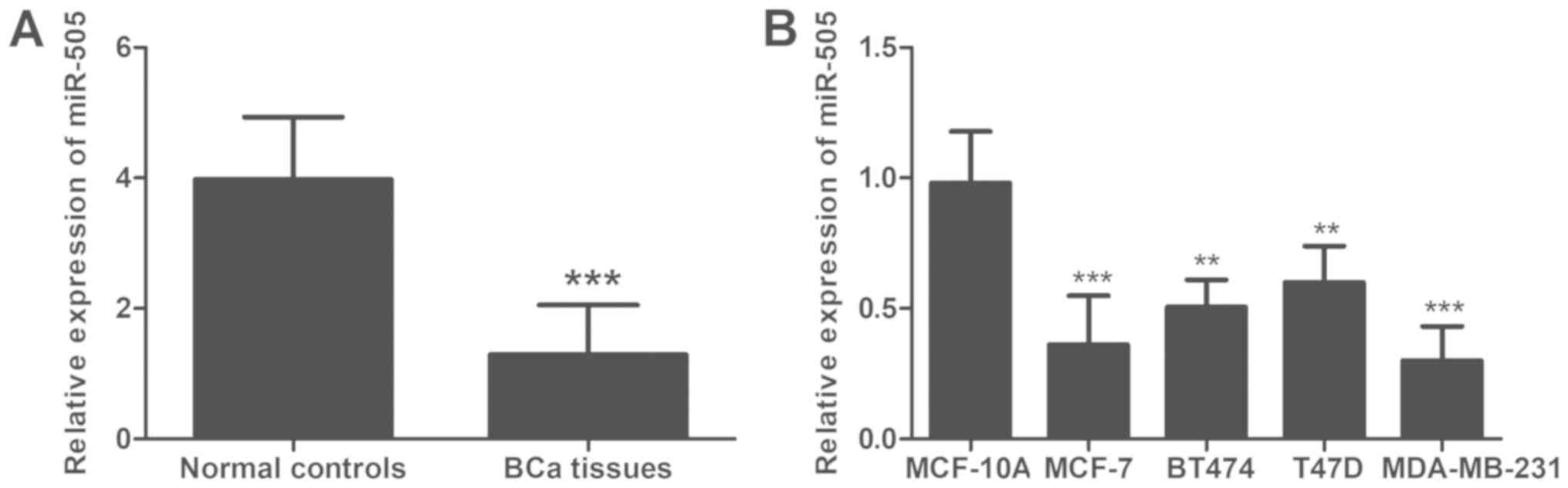
As presented in Fig.
1, RT-qPCR revealed that the expression of miR-505 was
significantly downregulated in BCa tissues compared with adjacent
normal tissues (P<0.001). Similarly, a significantly lower
expression level of miR-505 was identfied in the four BCa cell
lines examined compared with the normal mammary cell line (all
P<0.01).
Associations between miR-505
expression and the clinicopathological features of patients with
BCa
Due to the dysreguled expression of miR-505
identified in BCa, we hypothesized that miR-505 may be involved in
BCa development. Therefore, the associations between miR-505
expression level and the clinicopathological features of patients
with BCa were assessed. The patients with BCa were divided into two
groups based on the median expression value of miR-505 (1.124),
which generated a low miR-505 expression group and a high miR-505
expression group. Notably, the expression of miR-505 was identifed
to be associated with distant metastasis status (P=0.013) and TNM
staging (P=0.002). However, no significant associations were
revealed between miR-505 expression and other clinical parameters,
including age, tumor size, ER status, PR status and HER2 status
(all P>0.05; Table I).
Prognostic value of miR-505 in
patients with BCa
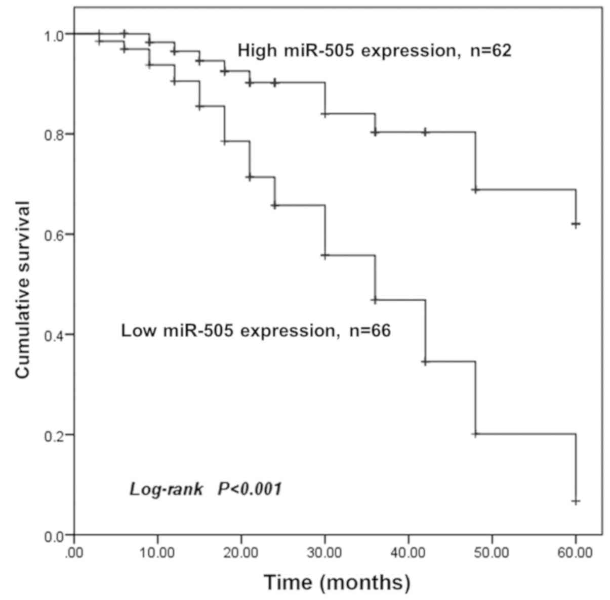
Survival analysis was performed for the patients
with different miR-505 expression levels. As presented in Fig. 2, Kaplan-Meier survival curves
revealed that the overall survival time was significantly shorter
for patients with low miR-505 expression compared with those with
high miR-505 expression (P<0.001). Furthermore, miR-505
expression and other clinicopathological parameters were included
in Cox regression analysis to identify prognostic factors for the
overall survival of patients with BCa. Univariate Cox analysis
revealed that miR-505 expression [hazard ratio (HR), 3.972; 95%
confidence interval (CI), 2.044–7.720; P=0.012], distant metastasis
(HR, 1.974; 95% CI, 0.993–3.927; P=0.022) and TNM stage (HR, 1.678;
95% CI, 0.976–2.886; P=0.031) were significantly associated with
the overall survival rate of patients with BCa. Furthermore,
multivariate analysis identified miR-505 expression (HR, 5.707; 95%
CI, 2.798–11.638; P=0.001) and TNM stage (HR, 2.602; 95% CI,
1.461–4.632; P=0.041) as independent prognostic factors for the
overall survival rate of patients with BCa (Table II).
| Table II.Cox regression analysis for miR-505
expression in patients with breast cancer. |
Table II.
Cox regression analysis for miR-505
expression in patients with breast cancer.
|
| Univariate
analysis | Multivariate
analysis |
|---|
|
|
|
|
|---|
| Variable | HR | 95% CI | P-value | HR | 95% CI | P-value |
|---|
| miR-505
expression | 3.972 | 2.044–7.720 | 0.012 | 5.707 | 2.798–11.638 | 0.001 |
| Age | 1.328 | 0.721–2.446 | 0.364 | 1.032 | 0.544–1.958 | 0.923 |
| Tumor size | 1.398 | 0.807–2.420 | 0.232 | 1.269 | 0.715–2.253 | 0.416 |
| ER status | 1.069 | 0.618–1.849 | 0.811 | 1.023 | 0.566–1.895 | 0.939 |
| PR status | 1.036 | 0.587–1.829 | 0.903 | 1.182 | 0.631–2.215 | 0.601 |
| HER2 status | 1.231 | 0.702–2.159 | 0.469 | 1.232 | 0.669–2.268 | 0.503 |
| Distant
metastasis | 1.974 | 0.993–3.927 | 0.022 | 1.985 | 0.965–4.086 | 0.062 |
| TNM stage | 1.678 | 0.976–2.886 | 0.031 | 2.602 | 1.461–4.632 | 0.041 |
Effects of miR-505 on the
proliferation of BCa cells
In addition to assessing the expression of miR-505
in patients with BCa patients, the functional role of miR-505 in
the progression of BCa was investigated using miR-505 mimic or
miR-505 inhibitor to regulate miR-505 expression in BCa cells.
MCF-7 and MDA-MB-231 cells were selected for transfection
experiments as they exhibited low miR-505 expression levels.
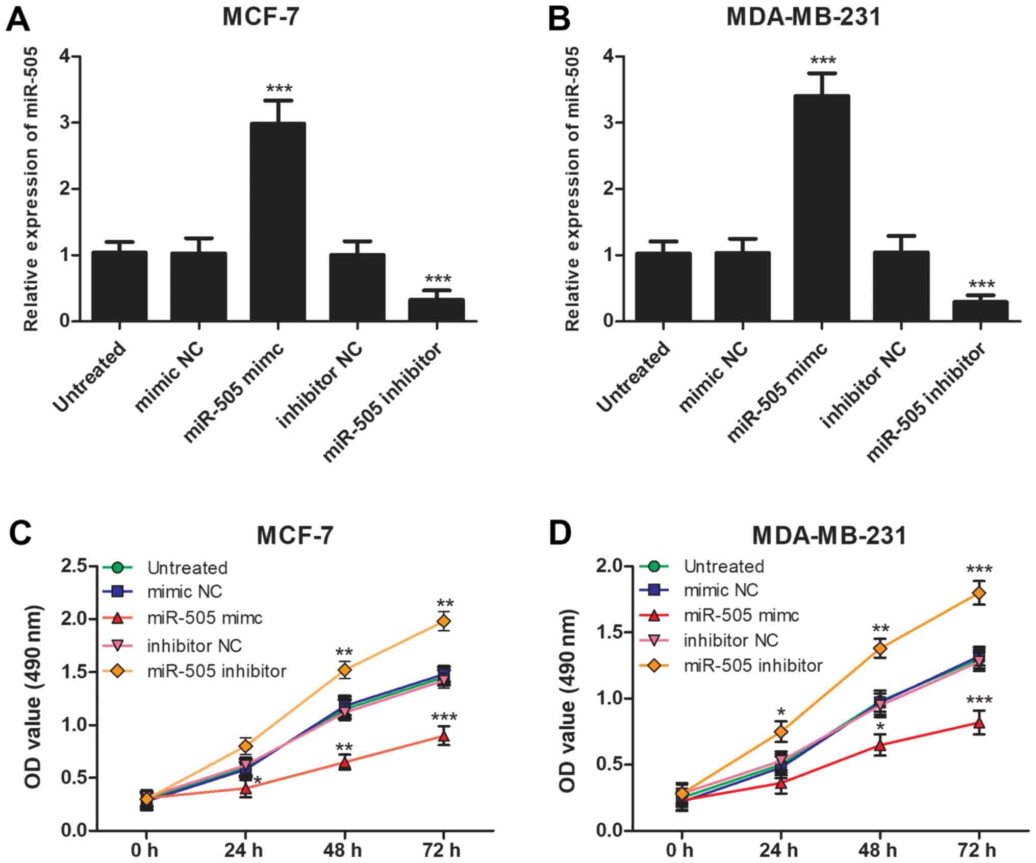
RT-qPCR confirmed that the expression of miR-505 was significantly
higher in cells transfected with miR-505 mimic (P<0.001) and
significantly lower in cells transfected with miR-505 inhibitor
compared with the untreated cells (P<0.001; Fig. 3A and B). Subsequently, MTT assay
demonstrated that an overexpression of miR-505 significantly
inhibited the proliferation and knockdown of miR-505 significantly
increased the proliferation of MCF-7 and MDA-MB-231 cells compared
with the untreated cells (all P<0.05; Fig. 3C and D).
Effects of miR-505 on BCa cell
migration and invasion
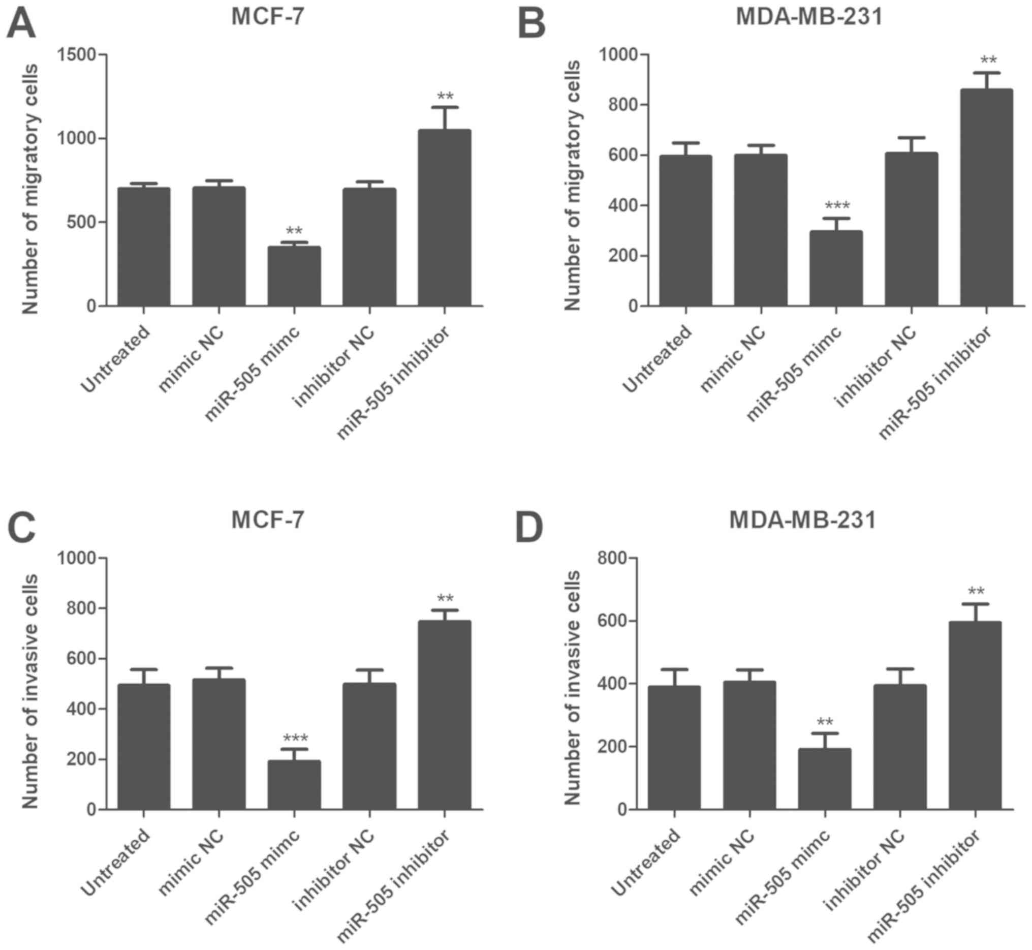
Subsequently, Transwell and Matrigel assays were
performed to assess the influence of miR-505 on BCa cell migration
and invasion, respectively. As presented in Fig. 4A and B, compared with the untreated
cells, the migration abilities of MCF-7 and MDA-MB-231 cells were
significaly suppressed following an upregulation of miR-505
(P<0.01 for MCF-7, P<0.001 for MDA-MB-231) and significantly
increased following downregulation of miR-505 expression (P<0.01
for both). As expected, an overexpression of miR-505 significantly
reduced the number of invasive cells (P<0.001 for MCF-7,
P<0.01 for MDA-MB-231) and a downregulation of miR-505
significantly increased the number of invasive MCF-7 and MDA-MB-231
cells compared with the untreated cells (all P<0.01 for both;
Fig. 4C and D).
Discussion
BCa is one the most frequent types of malignancy
among females worldwide (25).
Despite the development of various therapeutic strategies,
including resection operation, chemotherapy and radiotherapy, the
prognosis of patients diagnosed with BCa remains poor, which is
predominantly due to the limited sensitivity and specificity of
these treatment methods (26,27).
Therefore, this is an urgent requirement for the development of
efficient treatment strategies for patients with BCa.
It has been reported that the initiation and
development of BCa are complex processes that involve a wide range
of genetic changes (28).
Dysregulation of genes typically serves a pivotal role in the
progression of BCa and exhibits significant potential for the
improvement of BCa-targeted therapy (29). For example, increased expression of
prolyl-4-hydroxylase α subunit 2 (P4HA2) in BCa can promote tumor
cell proliferation and aggressive phenotypes, which indicates an
oncogenic role of P4HA2 in tumor progression (30). Zhu et al (31) demonstrated that Sulfatase 2 (SULF2)
could enhance cell proliferation, invasion, mobility and adhesion,
and suppress apoptosis of BCa cells, which suggests that SULF2 may
be a therapeutic target for BCa treatment. Over the past decades, a
number of studies have reported important roles of miRNAs in
numerous types of human cancer (32–34). In
addition, miRNAs have been described as functional molecules during
the progression of various types of malignancy, including BCa
(35). Chai et al (36) indicated that BCa cell proliferation
and cell cycle progression were promoted by miR-498, which was
demonstrated to serve an oncogenic role in BCa progression by
downregulating phosphatatse and tensin homolog expression.
Furthermore, a downregulation of miR-202 expression has been
observed in BCa tissues, and miR-202 was revealed to exert an
inhibitory effect on cell proliferation, migration and invasion in
BCa cells (37). These previous
studies indicate that aberrant expression levels of miRNAs serve
important roles in the tumor progression of BCa.
In the present study, the expression level of
miR-505 was identified to be significantly lower in BCa tissues
compared with adjacent normal tissues. Similarly, the expression
level of miR-505 was significantly lower in BCa cell lines compared
with normal cells. Furthermore, the majority of patients with low
miR-505 expression exhibited distant metastasis and presented with
an advanced TNM stage. Therefore, it can be suggested that miR-505
may be involved in the development of BCa. The present results were
consistent with a previous study, which also identified a decreased
expression of miR-505 in BCa tissues (22). Additionally, aberrant expression
patterns of miR-505 have been detected in other types of human
cancer. In hepatoma cells, the expression of miR-505 has been
demonstrated to be downregulated and miR-505 was identified to
promote cell proliferation and invasion by regulating high-mobility
group box 1 (20). Similarly,
downregulated miR-505 expression has been identified in endometrial
cancer tissues and was involved in tumor progression, with a tumor
suppressor role in this disease (21). Therefore, we hypothesize that miR-505
may be a tumor suppressor in BCa.
Given the dysregulated miR-505 expression in BCa
tissues, the current study evaluated the prognostic value of
miR-505 in BCa. The clinical significance of miRNAs has received
increasing attention due to their high diagnostic and prognostic
potential in different types of human cancer (38,39).
Certain miRNAs have been identified as diagnostic or prognostic
biomarkers in BCa, including miR-204 (40) and miR-301a (41). Ma et al (19) reported that downregulated miR-505
expression predicts poor prognosis in cervical cancer. However, to
the best of our knowledge, the clinical significance of miR-505 has
infrequently been reported in BCa. In the present study, the
Kaplan-Meier method was used to plot survival curves for patients
with BCa, which demonstrated that patients with low miR-505
expression exhibited a shorter overall survival time compared with
those with high miR-505 expression. Additionally, univariate Cox
analysis demonstrated that miR-505 expression, distant metastasis
and TNM stage were associated with overall survival of patients
with BCa. Multivariate Cox analysis further validated that miR-505
expression and TNM stage were independent prognostic factors for
patients with BCa. Although distant metastasis is considered an
aggressive behavior in BCa and may predict a poor prognosis, an
independent association between distant metastasis and overall
survival was not identified during multivariate analysis. This
result may be due to the effects of other parameters in the
equations of multivariate analysis or due to the cohort size used
in the present study, which may be a limitation.
To further investigate the biological function of
miR-505 in BCa progression, the effects of miR-505 on BCa cell
proliferation, migration and invasion were assessed. The expression
of miR-505 was regulated by cell transfection with miR-505 mimic or
miR-505 inhibitor. According to MTT and Transwell assays, it was
identified that upregulation of miR-505 could inhibit the
proliferation, migration and invasion of BCa cells, whereas a
downregulation of miR-505 could promote the proliferation,
migration and invasion of BCa cells. Therefore, it can be suggested
that miR-505 serves an inhibitory role in BCa progression. In
endometrial cancer, miR-505 also acts as a tumor suppressor and
suppresses tumor cell biological behaviors by targeting tumor
growth factor-α (21). In addition,
antitumor effects of miR-505 in cervical carcinoma can be achieved
by downregulating the expression of Frizzled-4 (19). However, to the best of our knowledge,
the molecular mechanisms underlying the role of miR-505 in BCa
remain unknown and require further investigation.
In conclusion, the present results demonstrated that
miR-505 expression is decreased in BCa and serves as an independent
prognostic biomarker. Overexpression of miR-505 was demonstrated to
suppress BCa cell proliferation, migration and invasion, which
indicates miR-505 may potentially be used to improve targeted
therapy for patients with BCa.
Acknowledgements
Not applicable.
Funding
No funding was received.
Availability of data and materials
The analyzed datasets generated during the study are
available from the corresponding author on reasonable request.
Authors' contributions
JW and HL designed the study, performed the clinical
research, analyzed the data and wrote the manuscript. ML performed
the cell experiments. All authors read and approved the final
manuscript.
Ethics approval and patient consent
Each patient provided written informed consent and
their personal information was anonymized. The experimental
procedures of the present study were approved by the Ethics
Committee of Yidu Central Hospital of Weifang (Weifang, China).
Patient consent for publication
Not applicable.
Competing interests
The authors declare that they have no competing
interests.
References
|
1
|
Torre LA, Bray F, Siegel RL, Ferlay J,
Lortet-Tieulent J and Jemal A: Globalcancer statistics, 2012. CA
Cancer J Clin. 65:87–108. 2015. View Article : Google Scholar : PubMed/NCBI
|
|
2
|
Brenton JD, Carey LA, Ahmed AA and Caldas
C: Molecular classification and molecular forecasting of breast
cancer: Ready for clinical application? J Clin Oncol. 23:7350–7360.
2005. View Article : Google Scholar : PubMed/NCBI
|
|
3
|
Bombonati A and Sgroi DC: The molecular
pathology of breast cancer progression. J Pathol. 223:307–317.
2011. View Article : Google Scholar : PubMed/NCBI
|
|
4
|
Reis-Filho JS and Lakhani SR: The
diagnosis and management of pre-invasive breast disease: Genetic
alterations in pre-invasive lesions. Breast Cancer Res. 5:313–319.
2003. View
Article : Google Scholar : PubMed/NCBI
|
|
5
|
Dai K, Qin F, Zhang H, Liu X, Guo C, Zhang
M, Gu F, Fu L and Ma Y: Low expression of BMPRIB indicates poor
prognosis of breast cancer and is insensitive to
taxane-anthracycline chemotherapy. Oncotarget. 7:4770–4784.
2016.PubMed/NCBI
|
|
6
|
Schmitz KH, DiSipio T, Gordon LG and Hayes
SC: Adverse breast cancer treatment effects: The economic case for
making rehabilitative programs standard of care. Support Care
Cancer. 23:1807–1817. 2015. View Article : Google Scholar : PubMed/NCBI
|
|
7
|
Turchinovich A, Weiz L and Burwinkel B:
Extracellular miRNAs: The mystery of their origin and function.
Trends Biochem Sci. 37:460–465. 2012. View Article : Google Scholar : PubMed/NCBI
|
|
8
|
Pillai RS, Bhattacharyya SN, Artus CG,
Zoller T, Cougot N, Basyuk E, Bertrand E and Filipowicz W:
Inhibition of translational initiation by Let-7 MicroRNA in human
cells. Science. 309:1573–1576. 2005. View Article : Google Scholar : PubMed/NCBI
|
|
9
|
Xiang J, Wu Y, Li DS, Wang ZY, Shen Q, Sun
TQ, Guan Q and Wang YJ: MiR-584 suppresses invasion and cell
migration of thyroid carcinoma by regulating the target oncogene
ROCK1. Oncol Res Treat. 38:436–440. 2015. View Article : Google Scholar : PubMed/NCBI
|
|
10
|
Lu Y, Hu J, Sun W, Li S, Deng S and Li M:
MiR-29c inhibits cell growth, invasion, and migration of pancreatic
cancer by targeting ITGB1. Onco Targets Ther. 9:99–109.
2016.PubMed/NCBI
|
|
11
|
Bai J, Zhang Z, Li X and Liu H:
MicroRNA-365 inhibits growth, invasion and metastasis of malignant
melanoma by targeting NRP1 expression. Cancer Biomark. 15:599–608.
2015. View Article : Google Scholar : PubMed/NCBI
|
|
12
|
Zhou C, Lu Y and Li X: MiR-339-3p inhibits
proliferation and metastasis of colorectal cancer. Oncol Lett.
10:2842–2848. 2015. View Article : Google Scholar : PubMed/NCBI
|
|
13
|
Dan B, Luo J, Li K and Chen S: Prognostic
value of miR-375 for survival outcomes in various cancers: A
systematic review and meta-analysis. Oncol Res Treat. 41:47–50.
2018. View Article : Google Scholar : PubMed/NCBI
|
|
14
|
Lin F, Yao L, Xiao J, Liu D and Ni Z:
MiR-206 functions as a tumor suppressor and directly targets K-Ras
in human oral squamous cell carcinoma. Onco Targets Ther.
7:1583–1591. 2014.PubMed/NCBI
|
|
15
|
Zheng K, Liu W, Liu Y, Jiang C and Qian Q:
MicroRNA-133a suppresses colorectal cancer cell invasion by
targeting Fascin1. Oncol Lett. 9:869–874. 2015. View Article : Google Scholar : PubMed/NCBI
|
|
16
|
Yang H, Yu J, Wang L, Ding D, Zhang L, Chu
C, Chen Q, Xu Z, Zou Q and Liu X: miR-320a is an independent
prognostic biomarker for invasive breast cancer. Oncol Lett.
8:1043–1050. 2014. View Article : Google Scholar : PubMed/NCBI
|
|
17
|
Deng Y and Chen Y: Increased expression of
miR-29a and its prognostic significance in patients with
cholangiocarcinoma. Oncol Res Treat. 40:128–132. 2017. View Article : Google Scholar : PubMed/NCBI
|
|
18
|
Liu YJ, Li W, Chang F, Liu JN, Lin JX and
Chen DX: MicroRNA-505 is downregulated in human osteosarcoma and
regulates cell proliferation, migration and invasion. Oncol Rep.
39:491–500. 2018.PubMed/NCBI
|
|
19
|
Ma C, Xu B, Husaiyin S, Wang L,
Wusainahong K, Ma J, Zhu K and Niyazi M: MicroRNA-505 predicts
prognosis and acts as tumor inhibitor in cervical carcinoma with
inverse association with FZD4. Biomed Pharmacother. 92:586–594.
2017. View Article : Google Scholar : PubMed/NCBI
|
|
20
|
Lu L, Qiu C, Li D, Bai G, Liang J and Yang
Q: MicroRNA-505 suppresses proliferation and invasion in hepatoma
cells by directly targeting high-mobility group box 1. Life Sci.
157:12–18. 2016. View Article : Google Scholar : PubMed/NCBI
|
|
21
|
Chen S, Sun KX, Liu BL, Zong ZH and Zhao
Y: MicroRNA-505 functions as a tumor suppressor in endometrial
cancer by targeting TGF-alpha. Mol Cancer. 15:112016. View Article : Google Scholar : PubMed/NCBI
|
|
22
|
Matamala N, Vargas MT, Gonzalez-Campora R,
Minambres R, Arias JI, Menendez P, Andres-Leon E, Gomez-Lopez G,
Yanowsky K, Calvete-Candenas J, et al: Tumor microRNA expression
profiling identifies circulating microRNAs for early breast cancer
detection. Clin Chem. 61:1098–1106. 2015. View Article : Google Scholar : PubMed/NCBI
|
|
23
|
Singletary SE, Allred C, Ashley P, Bassett
LW, Berry D, Bland KI, Borgen PI, Clark GM, Edge SB, Hayes DF, et
al: Staging system for breast cancer: Revisions for the 6th edition
of the AJCC Cancer Staging Manual. Surg Clin North Am. 83:803–819.
2003. View Article : Google Scholar : PubMed/NCBI
|
|
24
|
Livak KJ and Schmittgen TD: Analysis of
relative gene expression data using real-time quantitative PCR and
the 2(-Delta Delta C(T)) method. Methods. 25:402–408. 2001.
View Article : Google Scholar : PubMed/NCBI
|
|
25
|
Akram M, Iqbal M, Daniyal M and Khan AU:
Awareness and current knowledge of breast cancer. Biol Res.
50:332017. View Article : Google Scholar : PubMed/NCBI
|
|
26
|
Shi M and Guo N: MicroRNA expression and
its implications for the diagnosis and therapeutic strategies of
breast cancer. Cancer Treat Rev. 35:328–334. 2009. View Article : Google Scholar : PubMed/NCBI
|
|
27
|
Blank PR, Schwenkglenks M, Moch H and
Szucs TD: Human epidermal growth factor receptor 2 expression in
early breast cancer patients: A swiss cost-effectiveness analysis
of different predictive assay strategies. Breast Cancer Res Treat.
124:497–507. 2010. View Article : Google Scholar : PubMed/NCBI
|
|
28
|
Byler S, Goldgar S, Heerboth S, Leary M,
Housman G, Moulton K and Sarkar S: Genetic and epigenetic aspects
of breast cancer progression and therapy. Anticancer Res.
34:1071–1077. 2014.PubMed/NCBI
|
|
29
|
Lei F, Zhang L, Li X, Lin X, Wu S, Li F
and Liu J: Overexpression of prostate tumor overexpressed 1
correlates with tumor progression and predicts poor prognosis in
breast cancer. BMC Cancer. 14:4572014. View Article : Google Scholar : PubMed/NCBI
|
|
30
|
Xiong G, Deng L, Zhu J, Rychahou PG and Xu
R: Prolyl-4-hydroxylase alpha subunit 2 promotes breast cancer
progression and metastasis by regulating collagen deposition. BMC
Cancer. 14:12014. View Article : Google Scholar : PubMed/NCBI
|
|
31
|
Zhu C, He L, Zhou X, Nie X and Gu Y:
Sulfatase 2 promotes breast cancer progression through regulating
some tumor-related factors. Oncol Rep. 35:1318–1328. 2016.
View Article : Google Scholar : PubMed/NCBI
|
|
32
|
Dragomir M, Mafra ACP, Dias SMG, Vasilescu
C and Calin GA: Using microRNA networks to understand cancer. Int J
Mol Sci. 19:E18712018. View Article : Google Scholar : PubMed/NCBI
|
|
33
|
Huang LL, Huang LW, Wang L, Tong BD, Wei Q
and Ding XS: Potential role of miR-139-5p in cancer diagnosis,
prognosis and therapy. Oncol Lett. 14:1215–1222. 2017. View Article : Google Scholar : PubMed/NCBI
|
|
34
|
Long JP, Dong LF, Chen FF and Fan YF:
miR-146a-5p targets interleukin-1 receptor-associated kinase 1 to
inhibit the growth, migration, and invasion of breast cancer cells.
Oncol Lett. 17:1573–1580. 2019.PubMed/NCBI
|
|
35
|
Sun EH, Zhou Q, Liu KS, Wei W, Wang CM,
Liu XF, Lu C and Ma DY: Screening miRNAs related to different
subtypes of breast cancer with miRNAs microarray. Eur Rev Med
Pharmacol Sci. 18:2783–2788. 2014.PubMed/NCBI
|
|
36
|
Chai C, Wu H, Wang B, Eisenstat DD and
Leng RP: MicroRNA-498 promotes proliferation and migration by
targeting the tumor suppressor PTEN in breast cancer cells.
Carcinogenesis. 39:1185–1196. 2018. View Article : Google Scholar : PubMed/NCBI
|
|
37
|
Gao S, Cao C, Dai Q, Chen J and Tu J:
miR-202 acts as a potential tumor suppressor in breast cancer.
Oncol Lett. 16:1155–1162. 2018.PubMed/NCBI
|
|
38
|
Lu X and Lu J: The significance of
detection of serum miR-423-5p and miR-484 for diagnosis of
colorectal cancer. Clin Lab. 61:187–190. 2015. View Article : Google Scholar : PubMed/NCBI
|
|
39
|
Cong J, Liu R, Wang X, Wang J, Wang H and
Hou J: Low miR-498 expression levels are associated with poor
prognosis in ovarian cancer. Eur Rev Med Pharmacol Sci.
19:4762–4765. 2015.PubMed/NCBI
|
|
40
|
Li W, Jin X, Zhang Q, Zhang G, Deng X and
Ma L: Decreased expression of miR-204 is associated with poor
prognosis in patients with breast cancer. Int J Clin Exp Pathol.
7:3287–3292. 2014.PubMed/NCBI
|
|
41
|
Yu H, Li H, Qian H, Jiao X, Zhu X, Jiang
X, Dai G and Huang J: Upregulation of miR-301a correlates with poor
prognosis in triple-negative breast cancer. Med Oncol. 31:2832014.
View Article : Google Scholar : PubMed/NCBI
|