Introduction
Due to the continued improvement of chemotherapeutic
agents, the remission rate of leukemia has significantly improved
(1–3). However, the recurrence rate remains as
high as 30–40% (4). Extramedullary
infiltration (EMI) of acute leukemia includes a wide variety of
clinically significant phenomena that often pose therapeutic
dilemmas. The acute myeloid leukemia 1 protein/protein ETO
(AML1/ETO; A/E) fusion gene, caused by a t(8;21) translocation,
accounts for 15% of AML cases and is identified in 15–26.7% of
young patients with EMI (5), with a
5-year overall survival (OS) rate of 61% (6). It is hypothesized that being
A/E+ is indicative of a good prognosis and it has been
reported that ≤40% of A/E+ leukemia cases have EMI
(7–9). Our previous study demonstrated that
patients with high expression of amyloid precursor protein (APP)
were more likely to develop EMI (10). The aim of the present study was to
investigate the molecular mechanisms of EMI in detail in
vitro.
C-X-C motif chemokine receptor 4 (CXCR4) serves an
important role in cell migration, with certain studies
hypothesizing that cell migration is predominantly dependent on the
CXCR4/stromal-derived factor-1 (SDF-1) axis (11). CXCR4 and its ligand SDF-1 are
primarily studied for their crucial roles in the homing of stem and
progenitor cells in the bone marrow, chemotaxis, cell arrest,
angiogenesis, metastasis and cell survival (12). Tavor et al (13) reported that AML cells constitutively
secrete and express SDF-1-dependent cell surface elastase, which
regulates their migration and proliferation. Our previous study
revealed that A/E+ patients highly expressed CXCR4.
Furthermore, it was found that APP regulated cell migration via
matrix metalloproteinase (MMP)-2. MMP-2 and MMP-9 serve important
roles in metastasis due to their capacity to degrade the
extracellular matrix (14,15); they are hypothesized to be
particularly important for cell migration, as these proteinases act
on type IV collagen (11).
Experimental evidence has demonstrated that MMP-2 and MMP-9 are not
only involved in the invasion and metastasis of solid tumors, but
are also overexpressed in a variety of acute and chronic leukemia
(13,16), suggesting that they may serve an
important role in breaking through the bone marrow barrier. In the
present study, the different molecular expression levels of MMP-2
and MMP-9 were measured following interference with APP
expression.
MicroRNA (miRNA/miR)-144 is an important
transcriptional regulator in the process of hematopoiesis, and its
abnormal expression is closely associated with the pathogenesis of
hematological malignancies (17,18). At
present, an increasing amount of research is focusing on miR-144
and its role in erythroid formation; however, there are relatively
few studies pertaining to its role in leukemia (19,20). Liu
et al (21) reported that
miR-144 increased the sensitivity of leukemia cells to imatinib and
was closely associated with the c-Myc gene. Liang et al
(22) reported that miR-144 could
inhibit human embryonic trophoblast cell invasion. Based on these
results, we hypothesize that miR-144 may serve an important role in
the migration of Kasumi-1 cells.
The present study investigated the association
between miR-144 and the APP gene, and demonstrated that APP
regulates cell migration through the APP/phosphorylated
extracellular-signal-regulated kinase (p-ERK)/c-Myc/MMP-2 pathway.
This finding may provide novel insights into the development of
therapeutic strategies for the treatment of patients with AML, with
particular focus on A/E+ patients with EMI.
Materials and methods
Patient characteristics
A/E+ AML patients (n=123), diagnosed
according to the World Health Organization 2008 criteria (23), were enrolled in the present study
between February 2002 and June 2013 at the Nanfang Hospital,
Southern Medical University, (Guangzhou, China). All patients
enrolled in the present study provided written informed consent,
and the study was approved by the Ethics Committee of Nanfang
Hospital (Guangzhou, China). Patients were examined using marrow
cytology analysis, karyotype analysis and fluorescence in
situ hybridization (FISH). All patients received follow-up
until December 2013, with a median time of 46 months (6–141
months). As this was a retrospective study, certain data were not
obtained for all individuals included in the present study.
All patients completed 1–2 cycles of induction
chemotherapy, the majority of whom received the ‘3+7’ regimen
consisting of anthracyclines and cytarabine. Following remission,
109 patients received a median of 3 (range, 1–8) intrathecal
injections of 10 mg methotrexate or 50 mg cytarabine plus 5 mg
dexamethasone, which were used alternately. A total of 25 patients
with central nervous system lymphoma received a lumbar intrathecal
injection every other day until cerebrospinal fluid without blast
cells was detected by microscopy (Table
I).
 | Table I.Characteristics of
AML1/ETO+ patients with or without EML. |
Table I.
Characteristics of
AML1/ETO+ patients with or without EML.
|
Characteristics |
EML+ |
EML− | P-value |
|---|
| No. of
patients | 36 | 87 |
|
| Male/female, n | 26/10 | 46/41 | 0.048 |
| Median age (range),
years | 30.5 (2–69) | 25.0 (3–74) | 0.295 |
| Median WBC (range),
×109 cells/l | 21.7
(1.8–79.3) | 13.6
(1.1–85.8) | 0.059 |
| C-KIT mutation, n
(%)a |
|
Positive | 9
(39.1) | 14 (23.7) | 0.163 |
|
Negative | 14 (60.9) | 45 (76.3) |
|
| FLT3-ITD, n
(%)a |
|
Positive | 1 (4.3) | 2 (3.4) | 0.836 |
|
Negative | 22 (95.7) | 57 (96.6) |
|
| Expression of APP
gene, n (%) |
|
High | 11 (91.7) | 20 (41.7) | 0.019 |
|
Low | 1 (8.3) | 28 (58.3) |
|
| Induction therapy,
n (%) |
| DA | 10 (30.3) | 20 (23.3) |
|
| IA | 15 (45.5) | 46 (53.5) | 0.648 |
|
Other | 8
(24.2) | 20 (23.3) |
|
| Consolidation
therapy, n (%) |
| Regimen
1 | 17 (58.6) | 31 (38.8) |
|
| Regimen
2 | 8
(27.6) | 34 (42.5) | 0.179 |
| Regimen
3 | 4
(13.8) | 15 (18.8) |
|
| IT, n (%) |
| ≤2 | 11 (31.0) | 37 (42.5) | 0.415 |
|
>2 | 25 (69.0) | 50 (57.5) |
|
| Outcomes of
therapy, % |
| Rate of
RFS | 34.5 | 68.8 | <0.001 |
| Rate of
OS | 48.3 | 73.8 | 0.003 |
Patients without extramedullary leukemia (EML) were
designated as the control group, and those with EML were designated
as the study group. The complete remission (CR) rate, relapse-free
survival (RFS) time and OS time were compared following two cycles
of chemotherapy. RFS was calculated from time of CR until relapse
and OS was defined as the time from diagnosis until mortality. APP
expression was determined using bone marrow samples (n=65) of the
AML/ETO+ patients prior to treatments via reverse
transcription-quantitative polymerase chain reaction (RT-qPCR). The
prognosis was analyzed and compared between these two groups
(Table II).
| Table II.Association between APP expression
and survival. |
Table II.
Association between APP expression
and survival.
|
| High expression of
APP (n=30), % | Low expression of
APP (n=30), % | P-value |
|---|
| Cumulative CR rate
after the second cycle of chemotherapy | 84.4 | 100.0 | 0.020 |
| Rate of RFS | 40.0 |
80.0 | 0.001 |
| Rate of OS | 60.0 |
83.3 | 0.029 |
Cell culture and detection
Kasumi-1 cells (American Type Culture Collection,
Manassas, VA, USA), which are a human A/E+ cell line
derived from AML, were plated at 3×10⁵ cells/ml in
RPMI-1640 medium (Gibco; Thermo Fisher Scientific, Inc., Waltham,
MA, USA) supplemented with 20% fetal bovine serum (FBS; Gibco;
Thermo Fisher Scientific, Inc.), 4 mM glutamine, 100 U/ml
penicillin and 100 µg/ml streptomycin at 37°C in a humidified
atmosphere containing 5% CO2. FISH, morphological
detection using Wright's dye, and karyotype analysis were performed
as described previously (10).
Cell migration assay
The procedure was performed as described previously
(10). To evaluate the migration
ability the method was adjusted as follows: Non-invading cells were
removed from the upper surface of the Transwell membrane with a
cotton swab and invading cells on the lower membrane surface were
fixed in methanol at the room temperature for 15 min. The membrane
was then stained with 0.1% crystal violet for 15 min, washed with
water three times and dried for 3 h at the room temperature. Images
were captured using a light microscope (magnification, ×200) and
cells in five randomly selected fields were counted. Each invasion
experiment was performed in triplicate.
Target in vitro luciferase reporter
assay
pMIR-REPORT plasmids for the miR-144 target APP
3′-untranslated region (UTR) were constructed as wild-type (WT)
pmiR-APP containing two tandem repeats of miR-144 response element
from the APP 3′-UTR and a mutant (MUT) pmiR-APP by replacing two
nucleotides within the ‘seed sequence’ (psi-CHECK-APP-mut-UTR). The
oligonucleotides were annealed and inserted into the pMIR-REPORT
vector (Promega Corporation, Madison, WI, USA). The site-directed
mutagenesis was performed using the Quick Change kit (Stratagene;
Agilent Technologies, Inc., Santa Clara, CA, USA). The empty vector
(pMIR-REPORT) was used as a negative control. Cells were
transfected using Lipofectamine 2000 (Invitrogen; Thermo Fisher
Scientific, Inc.), according to the manufacturer's protocol, with
0.2 µg Target reporter plasmids (catalog no. 64158; Addgene, Inc.,
Cambridge, MA, USA) and 0.01 µg pMIR-REPORT Control Plasmid
(catalog no. 26280; Addgene, Inc.), and 200 nM miR-144-MIMIC
(miR-34c precursor; Addgene, Inc.) and NC control (Shanghai
GenePharma Co., Ltd.) per well on 96-well plates. Following 24 h of
incubation, the cells were subjected to a luciferase reporter assay
using a dual-luciferase reporter assay system (E1910; Promega
Corporation). Renilla luciferase activity was normalized to
Firefly luciferase activity for each sample. Each experiment was
repeated at least three times in triplicate.
RT-qPCR of miR-144 and APP
expression
Total RNA was extracted from kasumi-1 cells with
TRIzol reagents according to the manufacturer's protocol
(Invitrogen; Thermo Fisher Scientific, Inc., Waltham, MA, USA) and
quantified by an ultraviolet spectrophotometer (UVP, LLC, Phoenix,
AZ, USA) at 260 nm. The cDNA was synthesized from 1,000 ng total
RNA using the PrimerScript RT Reagent kit (Takara Biotechnology
Co., Ltd., Dalian, China). The PCR primer sequences were as
follows: β-actin forward, 5′-CGGAGTCAACGGATTTGGTCGTAT-3′ and
reverse, 5′-AGCCTTCTCCATGGTGGTGAAGAC-3′; and APP forward,
5′-TGGCCCTGGAGAACTACATC-3′ and reverse, 5′-TGGCCCTGGAGAACTACATC-3′.
The procedure was performed as described previously (4). First, the stem-loop RT primer was
hybridized to an miRNA molecule and then reverse transcribed with a
MMLV reverse transcriptase. The RT products were amplified using
conventional TaqMan PCR, according to the manufacturer's protocol
(Bio-Rad Laboratories, Inc., Hercules, CA, USA). A 20-µl RT
reaction was incubated at 25°C for 30 min and at 94°C for 3 min.
The qPCR was 45 cycles of denaturing at 94°C for 20 sec, annealing
at 50°C for 25 sec and synthesis at 72°C for 20 sec. U6 (forward,
5′-CTCGCTTCGGCZGCZCZ-3′ and reverse, 5′-AACGCTTCACGAATTTGCGT-3′)
was used as the internal control. The fold-change for miR-144
expression level was calculated using the 2−∆∆Cq method
(24).
Knockdown of APP by lentiviral
transduction
The complementary DNA sequence of APP was designed
from the full-length APP sequence by Shanghai GeneChem Co., Ltd.
(Shanghai, China). The siRNA target sequence of APP was
5′-CATCTTTGACCGAAACGAA-3′ and that of the negative control was
5′-TTCTCCGAACGTGTCACGT-3′ (NC). The procedure was performed as
described previously (10).
p-ERK inhibitor and c-Myc inhibitor
treated with kasumi-1 cells
In the present study, WT Kasumi-1 cells were treated
with the p-ERK inhibitor PD98059 (Santa Cruz Biotechnology Inc.,
Dallas, TX, USA) and the c-Myc inhibitor 10058-F4 (Selleck
Chemicals, Houston, TX, USA), respectively. Based on preliminary
experiments, Kasumi-1 cells were treated with the p-ERK inhibitor
at concentrations of 20 and 40 µM for 24 and 48 h. Also, Kasumi-1
cells were co-cultured with c-Myc inhibitor 10058-F4 at
concentrations of 50 and 100 µM for 24 and 48 h. The cells were
collected and western blotting was performed to detect the
changes.
APP/p-ERK/c-Myc/MMP-2 pathway detected
by western blotting
Protein concentrations of cell lysates were
determined using the BCA protein assay reagent kit. Equal amounts
(30 µg) of total protein were resolved by SDS-PAGE and transferred
to a polyvinylidene fluoride membrane. Following blocking with 5%
skimmed milk at room temperature for 2 h with shaking and washing
with TBST (50 ml Tris HCl, 8 g NaCl, 0.2 g KCl, 1 ml 0.1% Tween-20
and 1,000 ml distilled water), the membrane was incubated overnight
at 4°C with the following primary antibodies: APP (catalog no.
ab76763; 1:2,000; Abcam, Cambridge, MA, USA), MMP-2 (catalog no.
cs-13594; dilution of 1:1,000; Santa Cruz Biotechnology Inc.),
MMP-9 (catalog no. cs-21733; dilution of 1:500; Santa Cruz
Biotechnology Inc.), p-ERK (catalog no. ab201015; dilution of
1:500; Abcam), ERK (catalog no. ab54230; dilution of 1:500; Abcam),
c-Myc (catalog no. sc-40; dilution of 1:1,000; Santa Cruz
Biotechnology Inc.) and β-actin (catalog no. ab115777; 1:200;
Abcam). Following washing with TBS-T, the membranes were incubated
with secondary HRP-conjugated anti-rabbit IgG or anti-mouse IgG
(1:100; OriGene Technologies, Inc. Beijing, China) for 2 h at room
temperature. Proteins of interest were visualized by enhanced
chemiluminescence HRP substrate (EMD Millipore, Billerica, MA, USA)
and analyzed with an image analyzer (ChemiDoc MP Imaging system;
Bio-Rad Laboratories, Inc.).
Statistical analysis
SPSS 19.0 statistical software (IBM Corp., Armonk,
NY, USA) was used for data analysis. Data were obtained from
independent experiments and are expressed as the mean ± standard
deviation. Statistical analysis was performed by one-way ANOVA
followed by the Fisher's least significant difference test.
Survival analyses were performed using Kaplan-Meier survival
analysis. ROC curve analysis was used to calculate the cut-off
value of APP, which served to divide the patients into a high
expression group (with levels of APP ≥ cut-off value) and a low
expression group (with levels of APP < cut-off value). Not
normally distributed data were described by the median. P<0.05
was considered to indicate a statistically significant
difference.
Results
Characteristics of the A/E+
patients
The 123 A/E+ patients were divided into
two groups: 36 patients in the EML group (with EML), and 87
patients in the control group (without EML). In the EML group, 29
patients achieved a CR and finished the follow-up treatment after
the CR. In the control group, 80 patients achieved a CR and
finished the follow-up treatment after the CR. There was no
significant difference (P>0.05) in the two-cycle induction
chemotherapy regimens, the number of white blood cells or
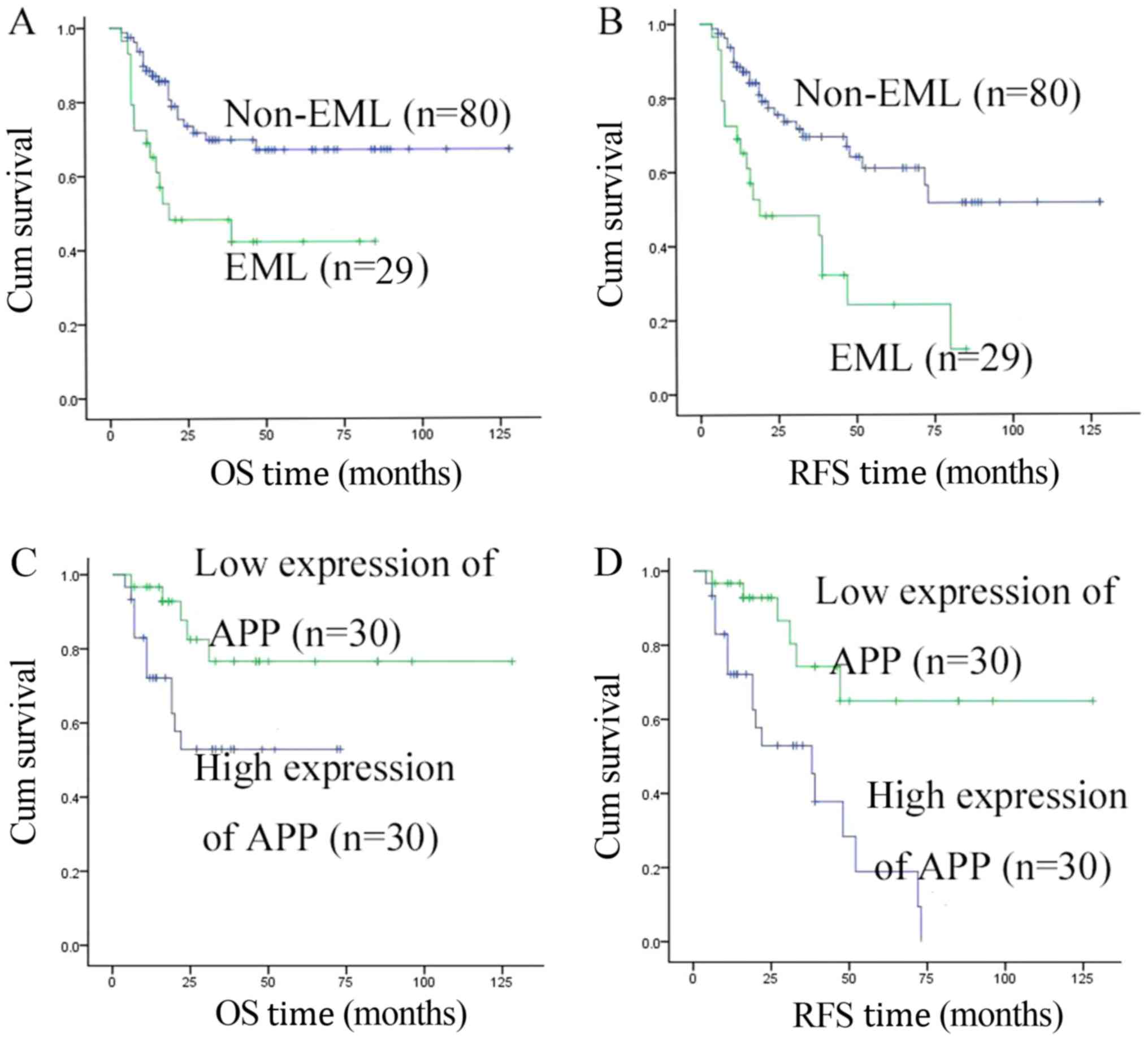
treatments following CR in these two groups (Table I). At the median follow-up time of 46
months (range, 6–141 months), the RFS and OS rates in the EML group
were significantly lower than those in the control group (34.5 vs.
68.8%, P<0.001; 48.3 vs. 73.8%, P=0.003, respectively; Table I). These results were consistent with
Fig. 1A and B.
Overall, 60 out of 65 patients with APP mRNA
achieved a CR and finished the follow-up treatment after the CR.
When we followed up for 35 months (6–96 months), the RFS rate in
the high expression group was 40.0%, which was significantly lower
compared with the low expression group with 80.0% (P=0.001). In
addition, a similar result was observed for the OS rate (60.0 vs.
83.3%; P=0.029; Table II). These
results were consistent with Fig. 1C and
D.
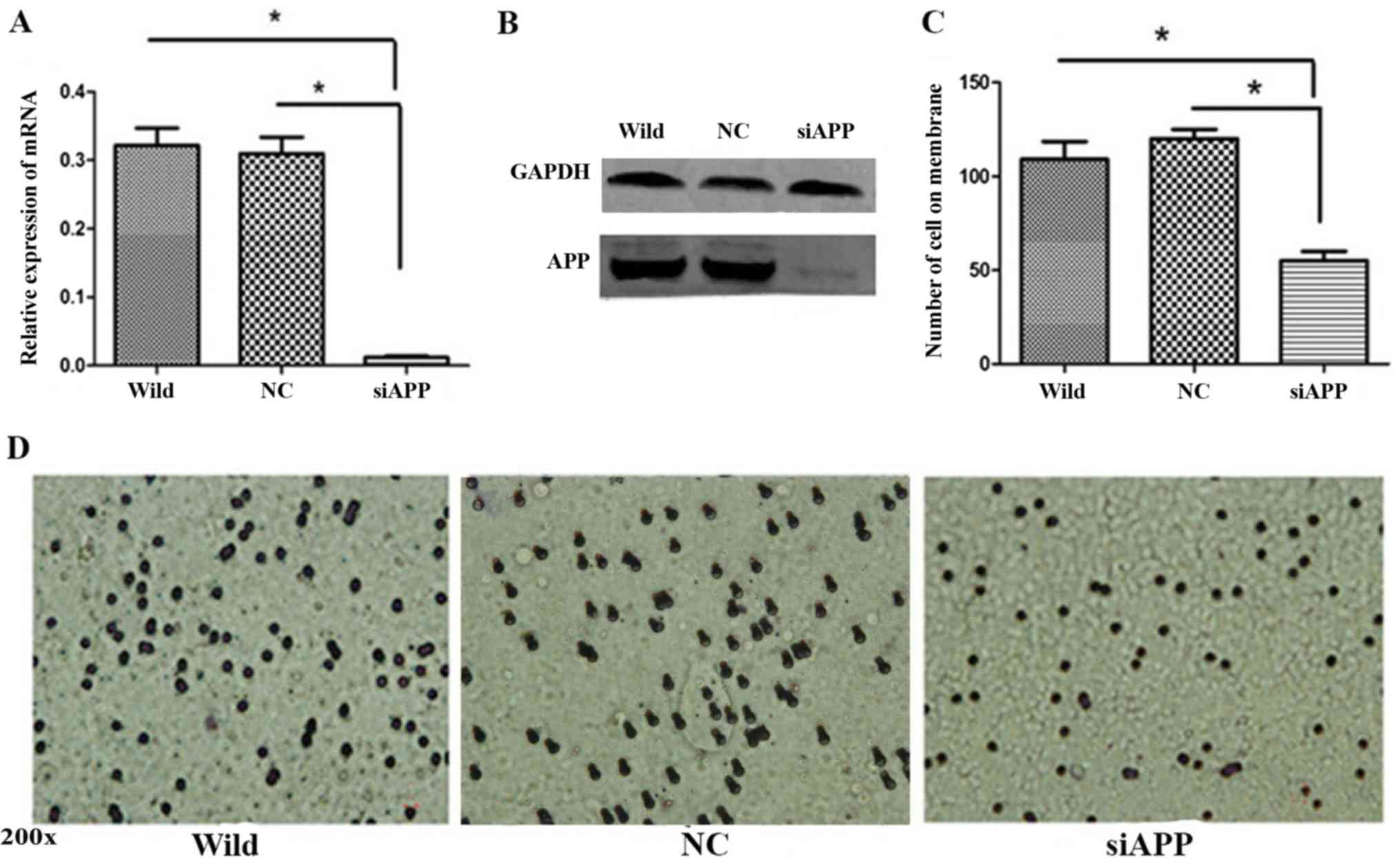
Effect of APP on cell migration
Kasumi-1 cells were transfected with siRNA against
APP (siAPP) and scramble siRNA (NC) and WT Kasumi-1 cells were used
as a control. RT-qPCR analysis demonstrated that the mRNA of APP
was significantly reduced in the siAPP group (Fig. 2A, P<0.01), and western blotting
revealed that the protein expression was markedly inhibited in the
siAPP group (Fig. 2B).
A Transwell assay was performed to evaluate cell
migration following the interference in APP. A significant
reduction in migrated cells in the siAPP group was observed when
compared with the control groups. Following the assessment of five
randomly-selected fields of view, the number of cells in the siAPP
group was observed to be significantly lower than that in the
control groups (P<0.05; Fig. 2C and
D).
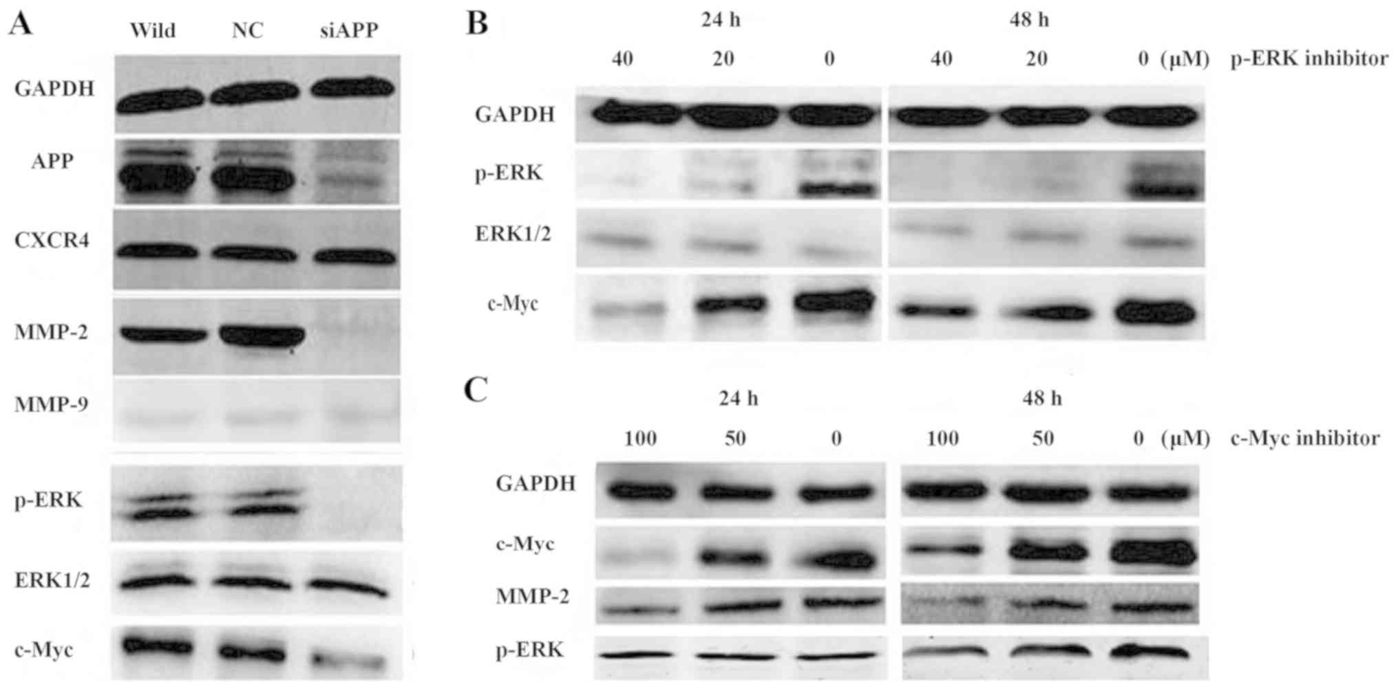
Mechanisms of APP regulate Kasumi-1
cell migration in vitro
The expression of MMP-2, p-ERK and c-Myc was
markedly decreased following the interference of APP expression;
however, there was no evident change in the expression of MMP-9 or
CXCR4 (Fig. 3A). Furthermore, WT
Kasumi-1 cells were treated with the p-ERK inhibitor PD98059 at
concentrations of 20 and 40 µM for 24 and 48 h. The western
blotting assay suggested that the expression levels of p-ERK and
c-Myc were markedly reduced (Fig.
3B). Similarly, when the c-Myc inhibitor 10058-F4 was
co-cultured with Kasumi-1 cells at concentrations of 50 and 100 µM
for 24 and 48 h, the expression levels of MMP-2 and c-Myc were
evidently decreased (Fig. 3C);
however, there was no evident change in p-ERK expression. These
results suggested that c-Myc was regulated by p-ERK.
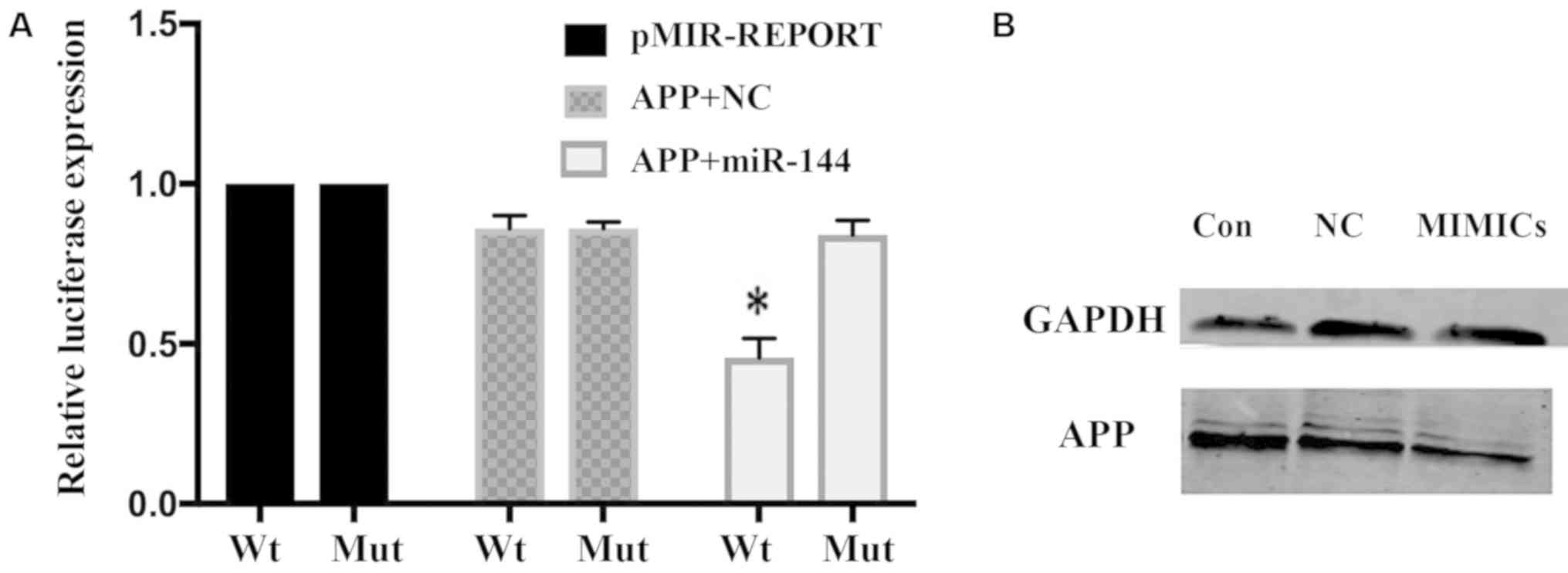
miR-144 negatively regulates APP
expression
To examine whether miR-144 repression of APP is
mediated by the direct interaction of miR-144 with the APP 3′-UTR,
a WT or MUT miR-144/IL-6R response element was cloned into a
pMIR-REPORT plasmid downstream of luciferase. miR-144-MIMIC was
transfected into Kasumi-1 cells. The reporter activities of WT, but
not MUT, were inversely associated with miR-144 expression. The
relative luciferase expression in the cells that were
co-transfected with psi-CHECK-APP-UTR and miR-144 oligonucleotide
was significantly reduced compared with the NC (P<0.01; Fig. 4A). Western blotting demonstrated that
the expression of the APP protein was decreased in the
miR-144-MIMIC group (Fig. 4B).
Discussion
Previous studies have demonstrated that the 5-year
OS rate of A/E+ AML patients is ~61% (25,26), and
EMI rates among A/E+ AML patients have been reported to
be as high as 15–26.7% (27). In our
previous study, it was demonstrated that the APP gene was involved
in the EMI of leukemia (10). The
process of EMI includes the escape of the leukemia cell from the
bone marrow, adhesion, degradation of the matrix and migration to
other areas. High expression of MMPs in patients with acute
leukemia is closely associated with invasion (16,28,29).
The present study examined the expression of other
migration-associated proteins, which may assist in investigating
the underlying mechanisms of APP in regulating leukemic EMI. The
mechanisms of EMI are complicated, with previous studies reporting
that leukemia cells that escaped from the bone marrow to the
surrounding organs were associated with different signal molecules,
including MMPs, CXCR4 and EZH2 (30–32). The
SDF1-CXCR4 axis serves a crucial role in hematopoiesis and cell
migration, and a number of studies have suggested that CXCR4 is
closely associated with metastasis in leukemia and solid tumors
(33–35). Möhle et al (36) reported that CXCR4 was highly
expressed in Kasumi-1 cells. We consequently hypothesized that the
central nervous system and other organs may attract leukemia cells
from the bone marrow to their locations via CXCR4. However, in the
present study, CXCR4 expression was not altered when the expression
of APP was reduced by siRNA. Therefore, it may be concluded that
the APP gene regulating Kasumi-1 cell migration is not directly
associated with CXCR4. In this study, MMP-2 expressuin was
significantly decreased after interference in APP, but there was no
change in MMP-9 or CXCR4, which are known as tumor metastasis
molecules. Therefore, the present study focused on the association
between APP and MMP-2.
The MAPK signaling pathway serves an important role
in tumor development, invasion, metastasis and chemo-resistance
(16,37). When cells are activated by external
stimuli, cell membrane receptors are triggered and the signals
transduce into the cytoplasm. In the nucleus, p-ERK binds to c-Myc
and other transcription factors to regulate cell proliferation,
differentiation and tumor development (38–41).
Numerous studies have demonstrated that MMPs are one of the
regulatory proteins downstream of the MAPK signaling pathway
(39,40). Therefore, we hypothesized that the
APP gene may regulate MMP-2 through the MAPK signaling pathway in
leukemia EMI. The present study also demonstrated that p-ERK and
c-Myc expression decreased at the same time, following the
interference in APP, suggesting that the APP gene activated the
MAPK/ERK signal transduction pathway. In order to understand the
details behind the function of APP in A/E+ leukemia cell
migration, wild-type Kasumi-1 cells were treated with the MEK
inhibitor PD98059 at concentrations of 20 and 40 µM for 24 and 48
h. The western blot assay suggested that the expression levels of
p-ERK and c-Myc were significantly reduced. Similarly, when the
c-Myc inhibitor 10058-F4 was co-cultured with Kasumi-1 cells at
concentrations of 50 and 100 µM for 24 and 48 h, the expression
levels of MMP-2 and c-Myc were significantly decreased; however,
there was no change in p-ERK expression. These results suggested
that c-Myc was regulated by p-ERK. Based on this, it was concluded
that APP regulated Kasumi-1 cell migration via the
p-ERK/c-Myc/MMP-2 pathway.
An increasing number of studies have reported that
miRNAs overexpressed in tumors may be considered as oncogenes, but
only a few oncogenic miRNAs have been well characterized. These
oncogenic miRNAs are called ‘oncomirs’. Several studies and
clinical analyses suggest that miRNAs may function as a novel class
of oncogenes or tumor suppressor genes (42–44).
TargetScan (Massachusetts Institute of Technology, Cambridge, MA,
USA), an online software used to predict binding sites on mRNAs,
predicted that the 3′-UTR of the APP gene incorporated a target
site of miR-144 with six complement oligo-nucleotide bases. In a
study by Garcia et al (45)
on human embryonic trophoblast cell invasion, it was found that
miR-144 could inhibit cell migration. Furthermore, the present
study identified that the expression of MMP-2 and MMP-9 was
significantly decreased by the inhibition of p-ERK expression. This
is consistent with the present results showing that miR-144
negatively regulates the APP gene.
In summary, miR-144 negatively regulates the APP
gene and then regulates EMI in patients with A/E+ AML.
The pathological molecular mechanism may be via the
APP/p-ERK/c-Myc/MMP-2 pathway. These results provide a novel
insight into the mechanism of EMI and could render a potential
therapeutic method to delay or prevent EMI, and may be useful in
preventing and treating A/E+ AML EMI.
Acknowledgements
The authors would like to acknowledge Dr Jie Yu Ye
(Department of Hematology, Nanfang Hospital, Southern Medical
University) for their assistance with revising the language of this
manuscript.
Funding
This work was supported by the grants from National
High Technology Research and Development Program of China (863)
(grant no. 2012AA02A505), National Natural Science Foundation of
China (grant no. 81400103), Natural Science Foundation of Guangzhou
province (grant no. 2014A030310061).
Availability of data and materials
All data generated or analyzed during this study are
included in this published article.
Authors' contributions
LJ wrote the article and performed the majority of
the experiments. WM performed the luciferase reporter assay and
knockdown of APP by lentiviral transfection. GY analyzed the
clinical data. CY and LL performed the polymerase chain reaction
experiments. ZW performed western blot analysis. FM designed the
all experiments. All authors read and approved the final
manuscript.
Ethics approval and consent to
participate
All patients provided written informed consent. The
study was approved by the Ethics Committee of Nanfang Hospital
(Guangzhou, China).
Patient consent for publication
Not applicable.
Competing interests
The authors declare that they have no competing
interests.
References
|
1
|
Wang L, Xu J, Tian X, Lv T and Yuan G:
Analysis of efficacy and prognostic factors of CLAG treatment in
chinese patients with refractory or relapsed acute myeloid
leukemia. Acta Haematol. 141:43–53. 2019. View Article : Google Scholar : PubMed/NCBI
|
|
2
|
Low M, Lee D, Coutsouvelis J, Patil S,
Opat S, Walker P, Schwarer A, Salem H, Avery S, Spencer A and Wei
A: High-dose cytarabine (24 g/m2) in combination with
idarubicin (HiDAC-3) results in high first-cycle response with
limited gastro- intestinal toxicity in adult acute myeloid
leukaemia. Intern Med J. 43:294–297. 2013. View Article : Google Scholar : PubMed/NCBI
|
|
3
|
Sasine JP and Schiller GJ: Emerging
strategies for high-risk and relapsed/refractory acute myeloid
leukemia: Novel agents and approaches currently in clinical trials.
Blood Rev. 29:1–9. 2015. View Article : Google Scholar : PubMed/NCBI
|
|
4
|
Pui CH: Central nervous system disease in
acute lymphoblastic leukemia: Prophylaxis and treatment. Hematology
Am Soc Hematol Educ Program. 2006:142–146. 2006. View Article : Google Scholar
|
|
5
|
Boissel N, Leroy H, Brethon B, Philippe N,
de Botton S, Auvrignon A, Raffoux E, Leblanc T, Thomas X, Hermine
O, et al: Incidence and prognostic impact of c-Kit, FLT3 and Ras
gene mutations in core binding factor acute myeloid leukemia
(CBF-AML). Leukemia. 20:965–970. 2006. View Article : Google Scholar : PubMed/NCBI
|
|
6
|
Prébet T, Boissel N, Reutenauer S, Thomas
X, Delaunay J, Cahn JY, Pigneux A, Quesnel B, Witz F, Thépot S, et
al: Acute myeloid leukemia with translocation (8;21) or inversion
(16) in elderly patients treated with conventional chemotherapy: A
collaborative study of the French CBF-AML intergroup. J Clin Oncol.
27:4747–4753. 2009. View Article : Google Scholar : PubMed/NCBI
|
|
7
|
Byrd JC, Mrózek K, Dodge RK, Carroll AJ,
Edwards CG, Arthur DC, Pettenati MJ, Patil SR, Rao KW, Watson MS,
et al: Pretreatment cytogenetic abnormalities are predictive of
induction success, cumulative incidence of relapse and overall
survival in adult patients with de novo acute myeloid leukemia:
Results from cancer and leukemia group B. Blood. 100:4325–4336.
2002. View Article : Google Scholar : PubMed/NCBI
|
|
8
|
Slovak ML, Kopecky KJ, Cassileth PA,
Harrington DH, Theil KS, Mohamed A, Paietta E, Willman CL, Head DR,
Rowe JM, et al: Karyotypic analysis predicts outcome of pre-
remission and postremission therapy in adult acute myeloid leuke-
mia: A Southwest Oncology Group/Eastern Cooperative Oncology Group
Study. Blood. 96:4075–4083. 2000.PubMed/NCBI
|
|
9
|
Grimwade D, Walker H, Oliver F, Wheatley
K, Harrison C, Harrison G, Rees J, Hann I, Stevens R, Burnett A and
Goldstone A: The importance of diagnostic cytogenetics on outcome
in AML: Analysis of 1,612 patients entered into the MRC AML 10
trial. Blood. 92:2322–2333. 1998.PubMed/NCBI
|
|
10
|
Jiang L, Yu G, Meng W, Wang Z, Meng F and
Ma W: Overexpression of amyloid precursor protein in acute myeloid
leukemia enhances extramedullary infiltration by MMP-2. Tumor Biol.
34:629–636. 2013. View Article : Google Scholar
|
|
11
|
Campbell JJ, Qin S, Bacon KB, Mackay CR
and Butcher EC: Biology of chemokine and classical chemoattractant
receptors: Differential requirements for adhesion-triggering versus
chemotactic responses in lymphoid cells. J Cell Biol. 134:255–266.
1996. View Article : Google Scholar : PubMed/NCBI
|
|
12
|
Liu SH, Gu Y, Pascual B, Yan Z, Hallin M,
Zhang C, Fan C, Wang W, Lam J, Spilker ME, et al: A novel CXCR4
antagonist IgG1 antibody (PF-06747143) for the treatment of
hematologic malignancies. Blood Adv. 1:1088–1100. 2017. View Article : Google Scholar : PubMed/NCBI
|
|
13
|
Tavor S, Petit I, Porozov S, Goichberg P,
Avigdor A, Sagiv S, Nagler A, Naparstek E and Lapidot T: Motility,
proliferation, and egress to the circulation of human AML cells are
elastase dependent in NOD/SCID chimeric mice. Blood. 106:2120–2127.
2005. View Article : Google Scholar : PubMed/NCBI
|
|
14
|
Klein G, Vellenga E, Fraaije MW, Kamps WA
and de Bont ES: The possible role of matrix metalloproteinase
(MMP)-2 and MMP-9 in cancer, e.g. acute leukemia. Crit Rev Oncol
Hematol. 50:87–100. 2004. View Article : Google Scholar : PubMed/NCBI
|
|
15
|
Suminoe A, Matsuzaki A, Hattori H, Koga Y,
Ishii E and Hara T: Expression of matrix metalloproteinase (MMP)
and tissue inhibitor of MMP (TIMP) genes in blasts of infant acute
lymphoblastic leukemia with organ involvement. Leuk Res.
31:1437–1440. 2007. View Article : Google Scholar : PubMed/NCBI
|
|
16
|
Egeblad M and Werb Z: New functions for
the matrix metalloproteinases in cancer progression. Nat Rev
Cancer. 2:161–174. 2002. View
Article : Google Scholar : PubMed/NCBI
|
|
17
|
Su Z, Si W, Li L, Zhou B, Li X, Xu Y, Xu
C, Jia H and Wang QK: MiR-144 regulates hematopoiesis and vascular
development by targeting meis1 during zebrafish development. Int J
Biochem Cell Biol. 49:53–63. 2014. View Article : Google Scholar : PubMed/NCBI
|
|
18
|
Papapetrou EP, Korkola JE and Sadelain M:
A genetic strategy for single and combinatorial analysis of miRNA
function in mammalian hematopoietic stem cells. Stem cells.
28:287–296. 2010.PubMed/NCBI
|
|
19
|
Li B, Zhu X, Ward CM, Starlard-Davenport
A, Takezaki M, Berry A, Ward A, Wilder C, Neunert C, Kutlar A and
Pace BS: MiR-144-mediated NRF2 gene silencing inhibits fetal
hemoglobin expression in sickle cell disease. Exp Hematol.
70:85–96.e5. 2019. View Article : Google Scholar : PubMed/NCBI
|
|
20
|
Trecul A, Morceau F, Gaigneaux A,
Schnekenburger M, Dicato M and Diederich M: Valproic acid regulates
erythro-megakaryocytic differentiation through the modulation of
transcription factors and microRNA regulatory micro-networks.
Biochem Pharmacol. 92:299–311. 2014. View Article : Google Scholar : PubMed/NCBI
|
|
21
|
Liu L, Wang S, Chen R, Wu Y, Zhang B,
Huang S, Zhang J, Xiao F, Wang M and Liang Y: Myc induced
miR-144/451 contributes to the acquired imatinib resistance in
chronic myelogenous leukemia cell K562. Biochem biophys Res Commun.
425:368–373. 2012. View Article : Google Scholar : PubMed/NCBI
|
|
22
|
Liang Y, Lin Q, Luo F, Wu W, Yang T and
Wan S: Requirement of miR-144 in CsA induced proliferation and
invasion of human trophoblast cells by targeting titin. J cell
Biochem. 115:690–696. 2014. View Article : Google Scholar : PubMed/NCBI
|
|
23
|
Vardiman JW, Thiele J, Arber DA, Brunning
RD, Borowitz MJ, Porwit A, Harris NL, Le Beau MM,
Hellström-Lindberg E, Tefferi A and Bloomfield CD: The 2008
revision of the World Health Organization (WHO) classification of
myeloid neoplasms and acute leukemia: Rationale and important
changes. Blood. 114:937–951. 2009. View Article : Google Scholar : PubMed/NCBI
|
|
24
|
Livak KJ and Schmittgen TD: Analysis of
relative gene expression data using real-time quantitative PCR and
the 2(-Delta Delta C(T)) method. Methods. 25:402–408. 2001.
View Article : Google Scholar : PubMed/NCBI
|
|
25
|
Grimwade D, Hills RK, Moorman AV, Walker
H, Chatters S, Goldstone AH, Wheatley K, Harrison CJ and Burnett
AK; National Cancer Research Institute Adult Leukaemia Working
Group, : Refinement of cytogenetic classification in acute myeloid
leukemia: Determination of prognostic significance of rare
recurring chromosomal abnormalities among 5876 younger adult
patients treated in the United Kingdom Medical Research Council
trials. Blood. 116:354–365. 2010. View Article : Google Scholar : PubMed/NCBI
|
|
26
|
Zhen T, Wu CF, Liu P, Wu HY, Zhou GB, Lu
Y, Liu JX, Liang Y, Li KK and Wang YY: Targeting of AML1-ETO in
t(8;21) leukemia by oridonin generates a tumor suppressor-like
protein. Sci Transl Med. 4:127ra382012. View Article : Google Scholar : PubMed/NCBI
|
|
27
|
Abdel Rahman H, Farrag SA and EI-Attar IA:
AML1/ETO fusion gene in de novo pediatric acute myeloid leukemia:
Clinical significance and prognostic implications. J Egypt Natl
Canc Inst. 19:39–47. 2007.PubMed/NCBI
|
|
28
|
Pan YX, Yang L, Wen SP, Liu XJ and Luo JM:
Expression and clinical significance of MMP-2 and MMP-9 in B acute
lymphoblastic leukemia. Zhongguo Shi Yan Xue Ye Xue Za Zhi.
22:640–643. 2014.(In Chinese). PubMed/NCBI
|
|
29
|
Itoh Y, Takamura A, Ito N, Maru Y, Sato H,
Suenaga N, Aoki T and Seiki M: Homophilic complex formation of
MT1-MMP facilitates proMMP-2 activation on the cell surface and
promotes tumor cell invasion. EMBO J. 20:4782–4793. 2001.
View Article : Google Scholar : PubMed/NCBI
|
|
30
|
Takayama K, Tsutsumi S, Suzuki T,
Horie-Inoue K, Ikeda K, Kaneshiro K, Fujimura T, Kumagai J, Urano
T, Sakaki Y, et al: Amyloid precursor protein is a primary androgen
target gene that promotes prostate cancer growth. Cancer Res.
69:137–142. 2009. View Article : Google Scholar : PubMed/NCBI
|
|
31
|
Krause K, Karger S, Sheu SY, Aigner T,
Kursawe R, Gimm O, Schmid KW, Dralle H and Fuhrer D: Evidence for a
role of the amyloid precursor protein in thyroid carcinogenesis. J
Endocrinol. 198:291–299. 2008. View Article : Google Scholar : PubMed/NCBI
|
|
32
|
Iannetti A, Ledoux AC, Tudhope SJ, Sellier
H, Zhao B, Mowla S, Moore A, Hummerich H, Gewurz BE, Cockell SJ, et
al: Regulation of p53 and Rb links the alternative NF-kB pathway to
EZH2 expression and cell senescence. PLoS Genet. 10:e10046422014.
View Article : Google Scholar : PubMed/NCBI
|
|
33
|
Nakahara F, Kitaura J, Uchida T, Nishida
C, Togami K, Inoue D, Matsukawa T, Kagiyama Y, Enomoto Y, Kawabata
KC, et al: Hes1 promotes blast crisis in chronic myelogenous
leukemia through MMP-9 upregulation in leukemic cells. Blood.
123:3932–3942. 2014. View Article : Google Scholar : PubMed/NCBI
|
|
34
|
Ugarte-Berzal E, Bailón E, Amigo-Jiménez
I, Albar JP, García-Marco JA and García-Pardo A: A novel
CD44-binding peptide from the pro-matrix metalloproteinase-9
hemopexin domain impairs adhesion and migration of chronic
lymphocytic leukemia cells. J Biol Chem. 289:15340–15349. 2014.
View Article : Google Scholar : PubMed/NCBI
|
|
35
|
Douglass S, Meeson AP, Overbeck-Zubrzycka
D, Brain JG, Bennett MR, Lamb CA, Lennard TW, Browell D, Ali S and
Kirby JA: Breast cancer metastasis: Demonstration that FOXP3
regulates CXCR4 expression and the response to CXCL12. J Pathol.
234:74–85. 2014. View Article : Google Scholar : PubMed/NCBI
|
|
36
|
Möhle R, Bautz F, Rafii S, Moore MA,
Brugger W and Kanz L: The chemokine receptor CXCR-4 is expressed on
CD34+ hematopoietic progenitors and leukemic cells and
mediates transendothelial migration induced by stromal cell-derived
factor-1. Blood. 91:4523–4530. 1998.PubMed/NCBI
|
|
37
|
Vlad A, Deglesne PA, Letestu R,
Saint-Georges S, Chevallier N, Baran-Marszak F, Varin-Blank N,
Ajchenbaum-Cymbalista F and Ledoux D: Down-regulation of CXCR4 and
CD62 L in chronic lymphocytic leukemia cells is triggered by B-cell
receptor ligation and associated with progressive disease. Cancer
Res. 69:6387–6395. 2009. View Article : Google Scholar : PubMed/NCBI
|
|
38
|
Sarkissyan S, Sarkissyan M, Wu Y, Cardenas
J, Koeffler HP and Vadgama JV: IGF-1 regulates cyr61 induced breast
cancer cell proliferation and invasion. PLoS One. 9:e1035342014.
View Article : Google Scholar : PubMed/NCBI
|
|
39
|
Jin W, Lu Y, Li Q, Wang J, Zhang H, Chang
G, Lin Y and Pang T: Down-regulation of the P-glycoprotein relevant
for multidrug resistance by intracellular acidification through the
crosstalk of MAPK signaling pathways. Int J Biochem Cell Biol.
54:111–121. 2014. View Article : Google Scholar : PubMed/NCBI
|
|
40
|
Zhang D, Liu D, Zhang J, Fong C and Yang
M: Gold nanoparticles stimulate differentiation and mineralization
of primary osteoblasts through the ERK/MAPK signaling pathway.
Mater Sci Eng C Mater Biol Appl. 42:70–77. 2014. View Article : Google Scholar : PubMed/NCBI
|
|
41
|
Verykokakis M, Papadaki C, Vorgia E, Le
Gallic L and Mavrothalassitis G: The RAS-dependent ERF control of
cell proliferation and differentiation is mediated by c-Myc
repression. J Biol Chem. 282:30285–942. 2007. View Article : Google Scholar : PubMed/NCBI
|
|
42
|
Gao Z, Zhang P, Xie M, Gao H, Yin L and
Liu R: miR-144/451 cluster plays an oncogenic role in esophageal
cancer by inhibiting cell invasion. Cancer Cell Int. 18:1842018.
View Article : Google Scholar : PubMed/NCBI
|
|
43
|
Witten LW, Cheng CJ and Slack FJ: miR-155
drives oncogenesis by promoting and cooperating with mutations in
the c-kit oncogene. Oncogene. 38:2151–2161. 2019. View Article : Google Scholar : PubMed/NCBI
|
|
44
|
Shirafkan N, Shomali N, Kazemi T,
Shanehbandi D, Ghasabi M, Baghbani E, Ganji M, Khaze V, Mansoori B
and Baradaran B: microRNA-193a-5p inhibits migration of human HT-29
colon cancer cells via suppression of metastasis pathway. J Cell
Biochem. Dec 2–2018.(Epub ahead of print). doi:
10.1002/jcb.28164.
|
|
45
|
Garcia TY, Gutierrez M, Reynolds J and
Lamba DA: Modeling the dynamic AMD-associated chronic oxidative
stress changes in human ESC and iPSC-derived RPE cells. Invest
Ophthalmol Vis Sci. 56:7480–7482. 2015. View Article : Google Scholar : PubMed/NCBI
|