Introduction
Rectal cancer occurs between the dentate line and
the recto-sigmoid junction and is one of the most common
malignancies in the digestive system (1). With the rapid development of the
economic level, people's mental pressure on life is also
increasing, the life pace is getting increasingly faster, the
incidence of colorectal cancer (CRC) has increased year by year,
and the incidence in youth has gradually increased (2). According to statistics, the top three
new cancers in women in the United States in 2018 are breast
cancer, lung cancer and CRCs, of which CRC accounts for 7% of new
cancer cases (3). Globally CRC is
the third most common cancer, accounting for 10% of all cancer
cases (4). In many cancers,
metastasis that is the main cause of death in patients begins with
the spread of tumor cells at the primary site and leads to
secondary tumors at distant anatomic site (5). Although significant progress has been
made in understanding the molecular nature of metastasis, there are
still many problems in the intracellular mechanism of controlling
metastasis process (6).
Runx3, a member of the Runx family, functions as a
transcription factor, and its encoded protein is an α subunit that
can bind to CBFβ/PEBP2β to form a heterodimer in the enhancer or
promoter of a target gene, so as to regulate the gene expression
(7). It is the first member of the
Runx family and plays a role as tumor suppressor gene in various
cancers such as gastric, lung and liver cancers (8,9). Runx3,
a downstream effector of the TGFβ signaling pathway, can
participate in the inhibition of Wnt signal transduction, thereby
exerting a carcinogenic effect (10). Studies have found that Runx3
expression is also downregulated in CRC, and Runx3 is associated
with the prognosis, clinical stage, differentiation degree and
lymph node metastasis of CRC (11,12).
However, the Runx3-mediated mechanism that inhibits tumor
metastasis is still unclear, and the effect of this gene on cell
proliferation, invasion and migration of CRC has not been studied.
This study explores potential new targets for targeted treatment of
rectal cancer by investigating the effect of Runx3 gene on the cell
proliferation and invasion of rectal cancer.
Materials and methods
Materials and reagents
HRC-9698 cells (Shanghai Zishi Biotechnology Co.,
Ltd.); Runx3 overexpression plasmid (Shandong Weizhen Biotechnology
Co., Ltd.); fetal bovine serum, RPMI-1640 medium, 25% trypsin and
0.02% EDTA (Procell Life Science & Technology Co., Ltd.);
TransScript Green Two-Step qRT-PCR SuperMix kit (TransGen
BiotechBiotechnology Co., Ltd.); Lipofectamine® 3000
transfection reagent and TRIzol reagent (Thermo Fisher Scientific,
Inc.); qPCR primer sequences (Shanghai Shenggong Biology
Engineering Technology Service, Ltd.); CCK-8 kit (Shanghai Yisheng
Biotechnology Co. Ltd.); Transwell chamber (Beijing Yiming Fuxing
Biotechnology.).
The study was approved by the Ethics Committee of
China-Japan Union Hospital of Jilin University (Changchun,
China).
Culture method for HRC-9698 cell
strain
HRC-9698 cells are suspension cells. The cells were
aspirated with a pipette into a RPMI-1640 medium with 9 ml of 10%
fetal bovine serum; centrifuged at 800 × g for 5 min at 4°C and
resuspended; the resuspended cells were aspirated with a pipette
into a new medium and incubated in an incubator at 37°C and 5%
CO2. After the cells grew to ~70% confluence, they were
digested with 0.25% trypsin and 0.02% EDTA mixture, inoculated into
a six-well plate, and divided into 4 groups (OE, blank plasmid
control, negative control and blank groups), 3 complex wells per
group.
Liposome transfection
Transfection was performed when the cell confluence
reached 80%. The Runx3 overexpression vector and liposome
Lipofectamine® 3000 were added to OE group, the empty
plasmid and liposome Lipofectamine 3000 to blank plasmid control
group, only liposome Lipofectamine 3000 to negative control group,
and equal amounts of complete medium to blank group but it was not
transfected. Operation was performed strictly in accordance with
Lipofectamine 3000 transfection reagent instruction and the cells
were incubated at room temperature for 5 min. The prepared plasmid
DNA and liposome complexes were added to the cells that were
incubated at 37°C and 5% CO2 for 2 days, and then the
transfected cells were detected and analyzed.
RT-qPCR detection of Runx3 gene
expression
RNA extraction and cDNA synthesis: TRIzol and
chloroform reagent were used for extraction of total RNA from each
group of cells, and the UV spectrophotometer for detection of RNA
concentration and purity, A260/A280.
The ratio is considered as qualified between
1.8–2.0. The reverse transcription reaction system was prepared
according to Table I, mixed gently,
incubated at 42°C for 15 min, heated at 85°C for 5 sec to
inactivate TransScript® RT/RI (TransGen
BiotechBiotechnology Co., Ltd.) and gDNA remover.
 | Table I.Preparation of reaction system. |
Table I.
Preparation of reaction system.
| Reverse
transcription | PCR |
|---|
| Total RNA 1 µg | Upstream and
downstream sequences (10 µM) 0.4 µl |
| 5X
TransScript® All-in-One | Passive reference
Dve |
| SuperMix for qPCR 4
µl | (50X) (optional) 0.4
µl |
| gDNA remover 1
µl | 2X
TransStart® Tin Green |
|
| aPCR SuperMix 0.4
µl |
| RNase-free water to
20 µl | cDNA 2 µl |
|
| RNase-free water to
20 µl |
qPCR reaction with TransScript Green Two-Step
qRT-PCR SuperMix kit (TransGen BiotechBiotechnology Co., Ltd.): the
qPCR reaction system was prepared according to Table I, and two-step method was used for
amplification, pre-denaturation at 94°C for 30 sec, at 94°C for 5
sec, and at 60°C for 30 sec for 40 cycles. GAPDH was used as an
internal reference, and the upstream and downstream sequences were
5′-GGTGGTGCTAAGCGTGTTA-3′ and 5′-CCCTCCACAATGCCAA-3′, respectively;
the upstream and downstream sequences of the target gene Runx3 were
5′-GGCGGTACCATGGCATCGAACAGCATCTTC-3′ and
5′-GCGCTCGAGTCAGTAGGGCCGCCACACGG-3′, respectively. The experimental
results were analyzed using the2−ΔΔCq method (13).
CCK-8 test of proliferation
Cell suspensions (1×10⁵) were prepared
and placed in a 96-well plate, 100 µl per well; 3 complex wells per
group; pre-incubated in an incubator at 37°C and 5% CO2
for 1 day; incubated continually in incubator and 10 µl of CCK-8
solution was added at 12 h. After the incubation for 3 h, a
microplate reader (Bio-Rad Laboratories, Inc.) was used for
determination of OD450 nm as the initial value. Then, 10 µl of
CCK-8 solution was added to each well every 24 h (24, 48, 72 and 96
h). After incubated at 37°C and 5% CO2 for 3 h, a
microplate reader was used for determination of the absorbance at
450 nm.
Transwell invasion experiment
Preparation of cell suspension: serum-free 1640
medium was used to culture cells for 12 h to remove serum influence
and prepare cell suspension. The cells were digested with 0.25%
trypsin and 0.02% EDTA mixture, the culture solution was discarded
by centrifugation at 3,000 × g for 8 min at 4°C after digestion,
and the cells were washed 1–2 times with PBS, resuspend with
serum-free medium, and counted to adjust cell density to
5×10⁵/ml; 100 µl of cell suspensions were added into the
Transwell upper chamber of a 24-well plate; 1640 medium with 600 µl
of 20% fetal bovine serum was added to the lower chamber; incubated
at 37°C and 5% CO2 for 12 h; the upper non-migrated
cells were gently wiped off with a cotton swab and washed 3 times
with PBS; fixed with 4% paraformaldehyde for 10 min; stained with
0.1% crystal violet for 20 min. The counts with five fields under a
microscope (Olympus Corporation) at 400 times were observed, and
five field counts were randomly selected in each well to obtain the
average value. The invasion experiment was performed on
Matrigel-coated cabs purchased from BD Company and diluted at the
ratio of 1:8.
Statistical analysis
SPSS20.0 (IBM Corp.) software statistical package
was used for test and analysis of the data. The measurement data
are expressed as mean ± standard deviation, and whether there was a
difference in results of the four groups was analyzed using ANOVA
single factor, then LSD was used to test whether there was a
difference between the two. The counting data was expressed as rate
and tested using Chi-square. The comparison among different
time-points in the same group was analyzed using repeated
measurement variance. The 2−ΔΔCq method was used to
statistically analyze the RT-qPCR results. The significance level
is α=0.05.
Results
RT-qPCR detection of Runx3 gene
expression
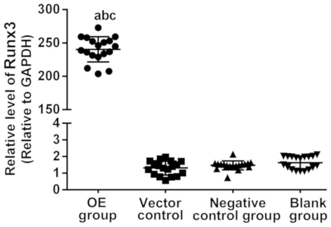
RT-qPCR was used for detection of the Runx3 gene
expression in the four groups of cells. The results of OE group,
blank plasmid control group, negative control group and blank group
were 242.55±13.97, 1.31±0.10, 1.45±0.06 and 1.55±0.12,
respectively. The difference was not statistically significant
among blank plasmid control, negative control and blank groups
(P>0.05). They were significantly higher in OE group than those
in blank plasmid control, negative control and blank groups (all
P<0.01) (Fig. 1).
CCK-8 test
The cell proliferation in each group was determined
at 12, 24, 48, 72 and 96 h using the CCK-8 method. The OD values
measured at the five time-points in each group by the CCK-8 test
are shown in Table II. At the same
time-point, pairwise comparison in each group found that OE group
was significantly lower than blank plasmid control, negative
control and blank groups (all P<0.01), and the difference was
not statistically significant among the latter three groups
(P>0.05). CCK-8 proliferation curve is shown in Fig. 2. Comparison among different
time-points in the same group found that the proliferation curves
of the four groups all showed an upward trend, and the OD values at
24 and 72 h were significantly higher than those at previous
time-point in blank plasmid control, negative control and blank
groups, and the OD values at 24 h were significantly higher than
those at previous time-point in OE group (all P<0.01).
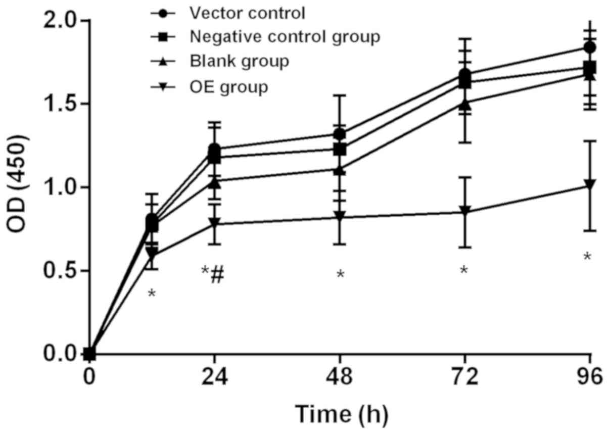 | Figure 2.CCK-8 test. The cell proliferation in
each group was determined at 12, 24, 48, 72 and 96 h. The OD values
measured at the five time-points in each group by the CCK-8 test
are shown in Table I. At the same
time-point, pairwise comparison in each group found that OE group
was significantly lower than blank plasmid control, negative
control and blank groups (all P<0.01), and the difference was
not statistically significant among the latter three groups
(P>0.05). Comparison among different time-points in the same
group found that the proliferation curves of the four groups all
showed an upward trend, and the OD values at 24 and 72 h were
significantly higher than those at previous time-point in blank
plasmid control, negative control and blank groups, and the OD
values at 24 h were significantly higher than those at previous
time-point in OE group (all P<0.01). *P<0.05, at the same
time-point, compared to blank plasmid control group, negative
control group and blank group. #P<0.05, compared to
previous time-point in the same group. OE group, overexpression
group. |
| Table II.CCK-8 test results of each group at
different time-points. |
Table II.
CCK-8 test results of each group at
different time-points.
| Items | Blank plasmid control
group | Negative control
group | Blank group | OE group | F-value | P-value |
|---|
| 12 h | 0.88±0.15 | 0.78±0.12 | 0.77±0.13 |
0.59±0.08a | 5.96 | 0.02 |
| 24 h |
1.23±0.16b |
1.18±0.18b |
1.04±0.11b |
0.78±0.12a,b | 17.34 | <0.001 |
| 48 h | 1.32±0.23 | 1.23±0.14 | 1.11±0.19 |
0.82±0.16a | 12.72 | <0.001 |
| 72 h |
1.68±0.21b |
1.63±0.19b |
1.51±0.24b |
0.85±0.21a | 29.34 | <0.001 |
| 96 h | 1.84±0.29 | 1.72±0.22 | 1.68±0.21 |
1.01±0.27a | 20.24 | <0.001 |
| F-value | 32.03 | 42.84 | 36.6 | 6.26 |
|
|
| P-value | <0.001 | <0.001 | <0.001 | <0.001 |
|
|
Transwell invasion experiment
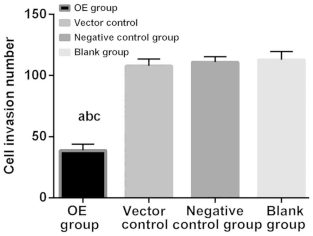
The number of invasion cells is shown in Table III. In OE group, blank plasmid
control, negative control and blank groups were 38.63±9.33,
107.87±5.66, 110.93±4.33 and 112.86±6.66, respectively. The
percentage of invasion was calculated as the mean number of
invasion in blank group of 100%. As shown in Fig. 3, the number of invasion cells in OE
group was significantly lower than that in blank plasmid control,
negative control and blank groups, and there was no significant
difference among the latter three groups. The experimental results
were statistically significant (P<0.01).
| Table III.Comparison of number of invasion
cells. |
Table III.
Comparison of number of invasion
cells.
| Groups | Number of invasion
cells | (%) |
|---|
| OE |
38.63±9.33a–c | 34.23a–c |
| Blank plasmid
control | 107.87±5.66 | 95.58 |
| Negative
control | 110.93±4.33 | 98.23 |
| Blank | 112.86±6.66 | 100.00 |
Discussion
CRC is one of the ‘four killers’ that threaten human
health (14). After the age of 50,
the incidence of CRC rapidly increases. It generally undergoes
precancerous lesions for several decades, so precancerous screening
before the age of 50 can early detect and treat it, so as to reduce
its mortality (15). Early radical
surgery can greatly reduce the mortality of rectal cancer, but due
to the particularity of the anatomical position of it, the
operation is difficult to complete, and it is difficult to retain
the anus and function after operation, bringing great inconvenience
to the patient's postoperative life (16). Runx3 that is downregulated in a
variety of tumor tissues has been found to be closely related to
the differentiation degree and clinical stage of rectal cancer
(17,18). The study of the effect of Runx3 on
the cell proliferation, invasion and migration of CRC can help us
to understand the antitumor effect and the manner of action of
Runx3, and provide more theoretical basis for the clinical study
and drug development of CRC cells.
In this study, the Runx3 gene expression in each
group was detected by RT-qPCR and was significantly higher in OE
group than that in blank plasmid control, negative control and
blank groups, indicating that Runx3 overexpression vector
successfully transfected into OE group of cells in this experiment.
CCK-8 proliferation experiment found that at the same time-point,
pairwise comparison in each group found that OE group was
significantly lower than blank plasmid control, negative control
and blank groups. Comparison among different time-points in the
same group found that the proliferation curves of the four groups
all showed an upward trend, suggesting that the Runx3
overexpression inhibited cell proliferation. Transwell invasion
experiment revealed that the number of invasion cells in OE group
was significantly lower than that in blank plasmid control,
negative control and blank groups, indicating that the Runx3
overexpression inhibited the invasion of OE group. In a study of
the effect of Runx3 on CRC metastasis and angiogenesis, Kim et
al (19) found that compared to
normal paracancerous colonic tissues, Runx3 expression decreased in
CRC tissues and was significantly associated with TNM stage. The
Runx3 overexpression inhibits the migration and invasion of CRC
cells due to high expression of matrix metalloproteinase-2 and −9,
similar to the results of this study. Whether Runx3 also inhibits
cell invasion by matrix metalloproteinase-2 and −9 in rectal cancer
requires further validation. Wang et al (20) also found that miR-301a promoted the
cell growth, migration and invasion of gastric cancer by
downregulating the Runx3 expression. However, only one HRC-9698
rectal cancer cell strain was used in this study, and it does not
truly represent the most natural occurrence and progression of
rectal cancer. What is the Runx3 expression in normal rectal cancer
cell strains? How does it function? Both need to be further
validated with several cell lines.
At the same time, studies (8) have found that Runx3 not only has the
ability to inhibit the invasion and metastasis of various cancer
cells, but also has many other potential functions and clinical
effects. Bauer et al (21)
found that the Runx3 expression in leukocytes is the key to affect
DMBA/TPA two-step method, thus inducing the occurrence of skin
tumors. The loss of Runx3 in leukocytes significantly increased the
content of antitumor cytokine thymic stromal lymphopoietin (TSLP),
and significantly decreased the content of pro-tumor cytokine
interleukin-17a and osteopontin. This unique combination of
cytokines enabled the tumor microenvironment to be polarized to an
effective antitumor state, which means that Runx3 expression
facilitates inflammation-mediated tumor progression in skin tumors.
It is contrary to this study and other literature reports that
Runx3 has antitumor effect, which may be related to the biological
function of TSLP. TSLP, a multifunctional IL-7-like cytokine, can
act on lymphocytes, DC cells and tumor cells. On the one hand, TSLP
plays an antitumor role in colon, skin early breast and pancreatic
cancers; on the other hand, it exerts tumor-promoting effects in
gastric cancer, lung cancer, cervical cancer and acute
lymphoblastic leukemia (22). In
addition, Runx3 has an inhibitory effect on various tumors such as
gastric, colon and breast cancers, but has a carcinogenic effect on
basal cell carcinoma, head and neck cancer and ovarian cancer
(23)
In conclusion, overexpression of Runx3 gene in
vitro inhibits the cell proliferation of rectal cancer and
blocks the cell invasion and metastasis. This study provides a new
idea and a new molecular therapeutic target for molecular targeted
therapy of rectal cancer.
Acknowledgements
Not applicable.
Funding
This study was supported by Jilin Province High
Technology Industrialization Program (2014Y083), Jilin Provincial
Science and Technology Development Program (20140204028XY), Jilin
Provincial Direct Health Special Program (2016, 2017), Jilin
Natural Science Foundation Program (20160101115JC) and Jilin
Provincial Youth Science Technology Innovation Leader and Team
Program (20180519025JH).
Availability of data and materials
The datasets used and/or analyzed during the present
study are available from the corresponding author on reasonable
request.
Authors' contributions
YF drafted the manuscript. YF and SG were mainly
devoted to cell culture and transfection. YG and DS performed PCR.
XW and ZC were responsible for the CCK-8 assay. All authors read
and approved the final study.
Ethics approval and consent to
participate
The study was approved by the Ethics Committee of
China-Japan Union Hospital of Jilin University (Changchun,
China).
Patient consent for publication
Not applicable.
Competing interests
The authors declare that they have no competing
interests.
References
|
1
|
Maingon P and Huguet F: Rectal Cancer: The
Radiation Oncologist: The Great Watchmaker. Semin Radiat Oncol.
26:173–175. 2016. View Article : Google Scholar : PubMed/NCBI
|
|
2
|
Cienfuegos JA, Rotellar F, Baixauli J,
Beorlegui C, Sola JJ, Arbea L, Pastor C, Arredondo J and
Hernández-Lizoáin JL: Impact of perineural and lymphovascular
invasion on oncological outcomes in rectal cancer treated with
neoadjuvant chemoradiotherapy and surgery. Ann Surg Oncol.
22:916–923. 2015. View Article : Google Scholar : PubMed/NCBI
|
|
3
|
Siegel RL, Miller KD and Jemal A: Cancer
statistics, 2015. CA Cancer J Clin. 65:5–29. 2015. View Article : Google Scholar : PubMed/NCBI
|
|
4
|
Suárez-Villanueva S, Ayala-Madrigal ML,
Peregrina-Sandoval J, Macías-Gómez N, Ramírez-Ramírez R,
Muñiz-Mendoza R, Moreno-Ortiz JM, Centeno-Flores M,
Maciel-Gutiérrez V, Cabrales E, et al: RUNX3 gene polymorphisms and
haplotypes in Mexican patients with colorectal cancer. Genet Mol
Res. 14:15505–15510. 2015. View Article : Google Scholar : PubMed/NCBI
|
|
5
|
Almaraz RT, Tian Y, Bhattarcharya R, Tan
E, Chen SH, Dallas MR, Chen L, Zhang Z, Zhang H, Konstantopoulos K,
et al: Metabolic flux increases glycoprotein sialylation:
implications for cell adhesion and cancer metastasis. Mol Cell
Proteomics. 11:M112.017558. 2012. View Article : Google Scholar : PubMed/NCBI
|
|
6
|
Su SC, Hsieh MJ, Yang WE, Chung WH, Reiter
RJ and Yang SF: Cancer metastasis: Mechanisms of inhibition by
melatonin. J Pineal Res. 62:622017. View Article : Google Scholar
|
|
7
|
Slattery ML, Lundgreen A, Herrick JS, Caan
BJ, Potter JD and Wolff RK: Associations between genetic variation
in RUNX1, RUNX2, RUNX3, MAPK1 and eIF4E and riskof colon and rectal
cancer: Additional support for a TGF-β-signaling pathway.
Carcinogenesis. 32:318–326. 2011. View Article : Google Scholar : PubMed/NCBI
|
|
8
|
Barghout SH, Zepeda N, Vincent K, Azad AK,
Xu Z, Yang C, Steed H, Postovit LM and Fu Y: RUNX3 contributes to
carboplatin resistance in epithelial ovarian cancer cells. Gynecol
Oncol. 138:647–655. 2015. View Article : Google Scholar : PubMed/NCBI
|
|
9
|
Kang KA, Zhang R, Kim GY, Bae SC and Hyun
JW: Epigenetic changes induced by oxidative stress in colorectal
cancer cells: Methylation of tumor suppressor RUNX3. Tumour Biol.
33:403–412. 2012. View Article : Google Scholar : PubMed/NCBI
|
|
10
|
Chen F, Liu X, Bai J, Pei D and Zheng J:
The emerging role of RUNX3 in cancer metastasis (Review). Oncol
Rep. 35:1227–1236. 2016. View Article : Google Scholar : PubMed/NCBI
|
|
11
|
Moon JW, Lee SK, Lee JO, Kim JH, Kim N,
Kim J, Kim HS and Park SH: Demethylation of RUNX3 by vincristine in
colorectal adenocarcinoma cells. Anticancer Res. 34:133–140.
2014.PubMed/NCBI
|
|
12
|
Nishina S, Shiraha H, Nakanishi Y, Tanaka
S, Matsubara M, Takaoka N, Uemura M, Horiguchi S, Kataoka J,
Iwamuro M, et al: Restored expression of the tumor suppressor gene
RUNX3 reduces cancer stem cells in hepatocellular carcinoma by
suppressing Jagged1-Notch signaling. Oncol Rep. 26:523–531.
2011.PubMed/NCBI
|
|
13
|
Livak KJ and Schmittgen TD: Analysis of
relative gene expression data using real-time quantitative PCR and
the 2 (-Delta Delta C(T)) method. Methods. 25:402–408. 2001.
View Article : Google Scholar : PubMed/NCBI
|
|
14
|
Slattery ML, Lundgreen A, Herrick JS,
Wolff RK and Caan BJ: Genetic variation in the transforming growth
factor-β signaling pathway and survival after diagnosis with colon
and rectal cancer. Cancer. 117:4175–4183. 2011. View Article : Google Scholar : PubMed/NCBI
|
|
15
|
Nie K, Shi L, Chen Q, Hu X, Jabbour SK,
Yue N, Niu T and Sun X: Rectal cancer: Assessment of neoadjuvant
chemoradiation outcome based on radiomics of multiparametric MRI.
Clin Cancer Res. 22:5256–5264. 2016. View Article : Google Scholar : PubMed/NCBI
|
|
16
|
Acuna SA, Chesney TR and Baxter NN:
Pathologic outcomes of laparoscopic vs open mesorectal excision for
rectal cancer. JAMA Surg. 152:986–987. 2017. View Article : Google Scholar : PubMed/NCBI
|
|
17
|
Kim YS, Chae YK, Choi YS, Min JH, Ahn SW,
Yoon JW, Lee SE and Lee YK: A comparative study of emergence
agitation between sevoflurane and propofol anesthesia in adults
after closed reduction of nasal bone fracture. Korean J
Anesthesiol. 63:48–53. 2012. View Article : Google Scholar : PubMed/NCBI
|
|
18
|
Slattery ML and Lundgreen A: The influence
of the CHIEF pathway on colorectal cancer-specific mortality. PLoS
One. 9:e1161692014. View Article : Google Scholar : PubMed/NCBI
|
|
19
|
Kim BR, Kang MH, Kim JL, Na YJ, Park SH,
Lee SI, Kang S, Joung SY, Lee SY, Lee DH, et al: RUNX3 inhibits the
metastasis and angiogenesis of colorectal cancer. Oncol Rep.
36:2601–2608. 2016. View Article : Google Scholar : PubMed/NCBI
|
|
20
|
Wang M, Li C, Yu B, Su L, Li J, Ju J, Yu
Y, Gu Q, Zhu Z and Liu B: Overexpressed miR-301a promotes cell
proliferation and invasion by targeting RUNX3 in gastric cancer. J
Gastroenterol. 48:1023–1033. 2013. View Article : Google Scholar : PubMed/NCBI
|
|
21
|
Bauer O, Hantisteanu S, Lotem J and Groner
Y: Carcinogen-induced skin tumor development requires leukocytic
expression of the transcription factor Runx3. Cancer Prev Res
(Phila). 7:913–926. 2014. View Article : Google Scholar : PubMed/NCBI
|
|
22
|
Milford TA, Su RJ, Francis OL, Baez I,
Martinez SR, Coats JS, Weldon AJ, Calderon MN, Nwosu MC, Botimer
AR, et al: TSLP or IL-7 provide an IL-7Rα signal that is critical
for human B lymphopoiesis. Eur J Immunol. 46:2155–2161. 2016.
View Article : Google Scholar : PubMed/NCBI
|
|
23
|
Lee CW, Chuang LS, Kimura S, Lai SK, Ong
CW, Yan B, Salto-Tellez M, Choolani M and Ito Y: RUNX3 functions as
an oncogene in ovarian cancer. Gynecol Oncol. 122:410–417. 2011.
View Article : Google Scholar : PubMed/NCBI
|