Introduction
Colon cancer is the most common malignancy and the
third leading cause of cancer-related mortality in China. Recently,
the incidence and mortality of colon cancer have increased
gradually in China (1). Therefore,
a deeper understanding of the molecular pathology of colon cancer
is critical for establishing novel therapeutic and diagnostic
strategies against this potentially lethal disease.
The PI3K/Akt pathway is crucially involved in
mammalian cell survival and resistance to apoptosis (2). PI3K/Akt signaling has been shown to be
overactivated in a variety of cancers, thereby promoting cancer
initiation and progression (3).
However, angiogenesis plays a significant role in tumor metastasis
and vascular endothelial growth factor (VEGF) is one of the most
effective factors that promotes tumor neovessel angiogenesis. The
high expression level of VEGF has been shown to be correlated with
the metastasis of several cancers, including esophageal and
pancreatic cancer (4,5). However, few studies have examined the
correlation between the activation of PI3K/Akt signaling and VEGF
expression in colon cancer tissue samples. Therefore, in the
present study we mainly employed immunohistochemical methods to
detect the protein expression of PI3K, Akt and VEGF in colon cancer
tissues and then analyzed the correlations of the expression levels
of these proteins with the clinicopathological features of colon
cancer.
Materials and methods
Sample collection and case data
From April 1990 to February 2007, samples from 60
cases of colon cancer were collected from the Department of Surgery
at the First Affiliated Hospital of Liaoning Medical University,
Jinzhou, China. The clinical samples were obtained from surgically
removed, pathologically confirmed colon cancer. The patients
included 34 males and 26 females and their ages ranged from 39 to
74 years (median age, 53.6 years). None of the patients accepted
preoperative radiotherapy or chemotherapy. Among the 60 patients, 4
succumbed to the disease and the remaining 56 patients were
followed up for 10 to 204 months following surgery. The
pathological classifications of colon cancer were based on tumor
size, infiltration, differentiation and lymph node metastasis.
Normal colon tissue samples adjacent to the colon cancer were also
collected from each case and used as negative controls. The
collection of clinical samples was approved by the Ethics Committee
of Liaoning Medical University, and all patients gave their written
informed consent.
Immunohistochemical staining
Tumor tissues and normal tissues were fixed in 10%
formaldehyde and were paraffin wax-embedded. Subsequently, the
tissues were cut into 5-mm thick serial sections, which were washed
carefully with 0.01 M phosphate-buffered saline (PBS) three times,
and then blocked with 2% goat serum in 0.01 M PBS containing 0.3%
Triton X-100 for 1 h at room temperature. The sections were
incubated at 4°C overnight with rabbit antibody against human PI3K,
pAkt or VEGF (Santa Cruz Biotechnology, Inc., Santa Cruz, CA, USA).
Subsequently, the slides were subjected to immunohistochemical
staining using a streptavidin-biotinperoxidase SP kit (Boster,
Wuhan, China). After visualizing the reaction with
3,3′-Diaminobenzidine (DAB), the slides were counterstained with
haematoxylin. For negative controls, the primary antibodies were
replaced with PBS.
Evaluation of immunohistochemical
staining
Following immunohistochemical staining, the tissue
sections were evaluated independently by two blinded investigators
who provided a consensus opinion of the staining patterns observed
under light microscopy. Five fields were randomly selected and
immunohistochemical staining of the cells was assessed according to
the proportion of cells stained and the staining intensity. The
proportion of cells stained was assessed using a semiquantitative
4-point scale: 0, <10% staining; 1, 10–20% staining; 2, 21–50%
staining; and 3, >50% staining. The staining intensity was also
graded using a 4-point scale: 0, no staining; 1, light yellow; 2,
brown; and 3, dark brown. The combined score was calculated by
multiplying the individual scale of the proportion and intensity
(range 0–9) and was assessed as follows: 0–2, negative staining
(−); 3–6, positive staining (+); and ≥6, strong positive staining
(++).
Statistical analysis
The differences between groups were tested for
significance using the χ2 test. The Pearson test was
performed for correlation analysis. Statistical analyses were
performed using SPSS version 15.0 (SPSS Inc., Chicago, IL, USA).
P<0.05 was considered to indicate a statistically significant
result.
Results
PI3K, pAkt and VEGF are highly expressed
in colon cancer tissues
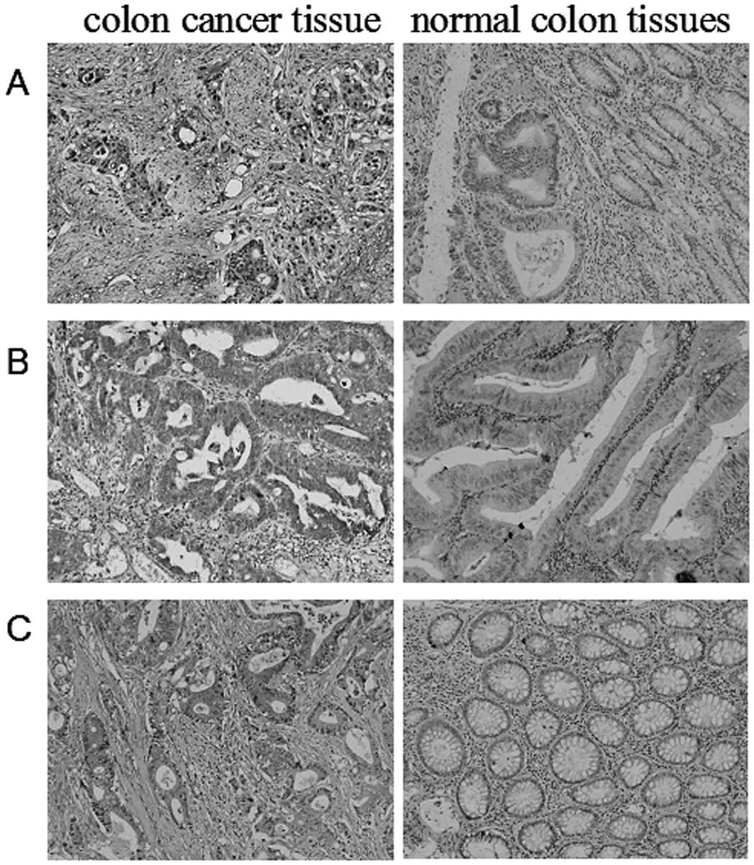
Immunohistochemical staining showed that the
positive rates of PI3K, pAkt and VEGF expression were 71.7%
(43/60), 68.3% (41/60) and 61.7% (37/60), respectively, among 60
colon cancer tissue samples. In the control group of adjacent
normal colon tissues, the positive rates of PI3K, pAkt and VEGF
expression were clearly lower than in the colon cancer group
(Fig. 1). The differences between
the two groups were statistically significant (P<0.05; Table I).
 | Table I.Expression of PI3K, pAkt and VEGF in
colon cancer and normal colon tissues. |
Table I.
Expression of PI3K, pAkt and VEGF in
colon cancer and normal colon tissues.
| Group (n) | PI3K(+) | % | pAkt(+) | % | VEGF(+) | % |
|---|
| Colon cancer
(60) | 43 | 71.7 | 41 | 65 | 37 | 61.7 |
| Normal colon
(20) | 9 | 45 | 7 | 35 | 6 | 30 |
Expression of PI3K, pAkt and VEGF have a
positive correlation in colon cancer tissues
Among the 43 colon cancer tissues in which PI3K was
highly expressed, pAkt was highly expressed in 32 cases and VEGF
protein was highly expressed in 31 cases. Furthermore, among the 17
colon cancer tissues with negative staining for PI3K, pAkt staining
was negative in 12 cases and VEGF staining was negative in 13
cases. The Pearson analysis showed that PI3K expression had a
positive correlation with pAkt and VEGF expression in colon cancer
tissues (P<0.05, Table II).
| Table II.Correlation between PI3K, pAkt and
VEGF expression in colon cancer tissues. |
Table II.
Correlation between PI3K, pAkt and
VEGF expression in colon cancer tissues.
| pAkt
| VEGF
|
|---|
| Group (n) | + | − | + | − |
|---|
| PI3K(+) (43) | 34 | 9 | 31 | 12 |
| PI3K(−) (17) | 7 | 10 | 6 | 11 |
| P=0.004 | P=0.008 |
Correlation between the expression of
PI3K, pAkt and VEGF and clinicopathological parameters of colon
cancer
To investigate the clinical significance of the
expression of PI3K, pAkt and VEGF in colon cancer, we analyzed the
clinicopathological parameters of the patients. Statistical
analysis showed that PI3K expression was not significantly
associated with the gender or age of the patients, tumor size or
differentiation (P>0.05), but was closely associated with serous
coat infiltration and lymphatic metastasis of colon cancer
(P<0.05). Neither pAkt nor VEGF expression were significantly
associated with the gender or age of the patients, or tumor
differentiation (P>0.05), but were closely associated with tumor
size, serous coat infiltration and lymphatic metastasis of colon
cancer (P<0.05; Table III).
| Table III.Correlation between PI3K, pAkt and
VEGF expression in colon cancer and clinicopathological
features. |
Table III.
Correlation between PI3K, pAkt and
VEGF expression in colon cancer and clinicopathological
features.
| Clinicopathological
feature | n | PI3K(+) | % |
χ2/P-value | pAkt(+) | % |
χ2/P-value | VEGF(+) | % |
χ2/P-value |
|---|
| Gender |
| Male | 28 | 21 | 75 | 0.287/0.592 | 19 | 67.9 | 0.188/0.664 | 19 | 67.9 | 0.851/0.356 |
| Female | 32 | 22 | 68.8 | | 20 | 62.5 | | 18 | 56.3 | |
| Age (years) |
| <65 | 46 | 34 | 73.9 | 0.490/0.484 | 29 | 63 | 0.066/0.798 | 26 | 56.5 | 1.373/0.241 |
| ≥65 | 14 | 9 | 64.3 | | 10 | 71.4 | | 11 | 78.6 | |
| Tumor size
(cm) |
| <5 | 26 | 16 | 61.5 | 2.318/0.128 | 13 | 50 | 4.538/0.033 | 10 | 38.5 | 10.452/0.001 |
| ≥5 | 34 | 27 | 79.4 | | 26 | 76.5 | | 27 | 79.4 | |
| Serous coat
infiltration |
| Yes | 39 | 33 | 84.6 | 11.613/0.001 | 29 | 74.4 | 4.290/0.038 | 28 | 71.8 | 4.835/0.028 |
| No | 21 | 10 | 47.6 | | 10 | 47.6 | | 9 | 42.9 | |
|
Differentiation |
| High/medium | 45 | 31 | 68.9 | 0.318/0.528 | 26 | 57.8 | 0.178/0.604 | 26 | 57.8 | 0.151/0.656 |
| Low | 15 | 12 | 80 | | 13 | 86.7 | | 11 | 73.3 | |
| Lymphatic
metastasis |
| Yes | 36 | 30 | 83.3 | 4.602/0.032 | 28 | 77.8 | 6.459/0.011 | 26 | 72.2 | 4.242/0.039 |
| No | 24 | 14 | 58.3 | | 11 | 45.8 | | 11 | 45.8 | |
Discussion
Despite the progress in the development of effective
diagnostic and therapeutic strategies for colon cancer, treatment
of the disease remains a challenge due to the overall poor
prognosis of colon cancer patients. Consequently, great efforts
have been taken recently to identify novel prognostic and
predictive biomarkers for colon cancer and recent studies reported
that several biomarkers were able to predict the survival of colon
cancer patients (6–8).
The PI3K/Akt pathway is involved in cellular
survival by inhibiting apoptotic processes and stimulating cell
growth and proliferation. The PI3K/Akt pathway is commonly
activated in human cancers and has been recognized as an important
target for anti-cancer therapy. In addition, the negative
prognostic value of the PI3K/Akt pathway has been revealed in
several types of cancer (9–11). However, the clinical significance of
the activation of PI3K/Akt in colon cancer remains uncertain. VEGF
is a potent angiogenic factor that promotes tumor metastasis and
angiogenesis (12). A recent study
demonstrated that the phosphorylation of Akt and ERK1/2 is required
for VEGF-induced proliferation and migration of lymphatic
endothelium, suggesting that PI3K/Akt signaling could cross-talk
with VEGF signaling to promote tumor angiogenesis (13). The phosphorylation of Akt is
established as a hallmark for the activation of PI3K/Akt signaling
but few studies have examined the correlation between the
phosphorylation of Akt and VEGF expression in colon cancer tissue
samples. Therefore, in the present study we aimed to detect the
protein levels of PI3K, pAkt and VEGF in colon cancer tissues and
investigate the correlations of the expression levels of these
proteins with the clinicopathological variables of colon
cancer.
By immunohistochemical staining, we showed that the
positive rates of PI3K, pAkt and VEGF expression were 71.7%
(43/60), 68.3% (41/60) and 61.7% (37/60), respectively, in colon
cancer, which were significantly higher than in adjacent normal
colon tissues (P<0.001). The correlation analysis showed that
the expression level of PI3K was closely associated with serous
coat infiltration and lymphatic metastasis of colon cancer
(P<0.05), and the expression levels of pAkt and VEGF were
closely associated with tumor size, serous coat infiltration and
lymphatic metastasis in colon cancer (P<0.05). In addition, the
expression levels of Pl3K and pAkt were positively correlated with
that of VEGF in colon cancer tissues (P<0.05). These results
suggest that PI3K/Akt signaling and VEGF signaling cross-talk with
each other to promote the progression, metastasis and angiogenesis
of colon cancer. In addition, our findings of the positive
correlation between PI3K/Akt activation and VEGF expression in
colorectal cancer tissues are largely consistent with the results
of other studies that investigated the roles of PI3K/Akt and VEGF
in other types of cancers, particularly epithelial cancers.
In conclusion, our data suggest that pAkt is an
independent prognostic marker for colon cancer patients. Further
large-scale and long-term follow-up studies may be useful in
comfirming the prognostic value of pAkt for colon cancer.
Acknowledgements
This study was supported by a Province
Key Lab grant (No. LS2010102) awarded by the Department of
Education of Liaoning Province, China.
References
|
1.
|
L YangDM ParkinL LiTime trends in cancer
mortality in China: 1987–1999Int J Cancer1067717832003
|
|
2.
|
V DuronioThe life of a cell: apoptosis
regulation by the PI3K/PKB pathwayBiochem
J415333344200810.1042/BJ2008105618842113
|
|
3.
|
A HlobilkovaJ EhrmannE SedlakovaV KrejciP
KnizetovaCould changes in the regulation of the PI3K/PKB/Akt
signaling pathway and cell cycle be involved in astrocytic tumor
pathogenesis and progression?Neoplasma54334341200717822324
|
|
4.
|
C HuangR HuangW ChangT JiangK HuangThe
expression and clinical significance of pSTAT3, VEGF and VEGF-C in
pancreatic
adenocarcinomaNeoplasma595261201210.4149/neo_2012_00722082308
|
|
5.
|
M KozlowskiW NaumnikJ NiklinskiR MilewskiP
DziegielewskiJ LaudanskiVascular endothelial growth factor C and D
expression correlates with lymph node metastasis and poor prognosis
in patients with resected esophageal
cancerNeoplasma58311319201110.4149/neo_2011_04_31121520987
|
|
6.
|
F WangP ZhangC ShiImmunohistochemical
detection of HSP27 and hnRNP K as prognostic and predictive
biomarkers for colorectal cancerMed OncolAug232011(Epub ahead of
print)
|
|
7.
|
QL LiangZY LiGQ ChenPrognostic value of
serum soluble Fas in patients with locally advanced unresectable
rectal cancer receiving concurrent chemoradiotherapyJ Zhejiang Univ
Sci B11912917201010.1631/jzus.B1000277
|
|
8.
|
QL LiangBR WangGH LiDcR3 and survivin are
highly expressed in colorectal carcinoma and closely correlated to
its clinicopathologic parametersJ Zhejiang Univ Sci
B10675682200910.1631/jzus.B0920077
|
|
9.
|
S HasselblomU HanssonM OlssonL TorenA
BergstromHigh immunohistochemical expression of p-AKT predicts
inferior survival in patients with diffuse large B-cell lymphoma
treated with immunochemotherapyBr J
Haematol149560568201010.1111/j.1365-2141.2010.08123.x20201946
|
|
10.
|
A YoshiokaH MiyataY DokiT YasudaM
YamasakiThe activation of Akt during preoperative chemotherapy for
esophageal cancer correlates with poor prognosisOncol
Rep1910991107200818425364
|
|
11.
|
A ValkovTK KilvaerSW SorbyeT DonnemE
SmelandThe prognostic impact of Akt isoforms, PI3K and PTEN related
to female steroid hormone receptors in soft tissue sarcomasJ Transl
Med9200201110.1186/1479-5876-9-20022107784
|
|
12.
|
AK OlssonA DimbergJ KreugerL
Claesson-WelshVEGF receptor signalling - in control of vascular
functionNat Rev Mol Cell Biol7359371200610.1038/nrm191116633338
|
|
13.
|
MT DellingerRA BrekkenPhosphorylation of
Akt and ERK1/2 is required for VEGF-A/VEGFR2-induced proliferation
and migration of lymphatic endotheliumPLoS
One6e28947201110.1371/journal.pone.002894722174934
|