Introduction
Large gallbladders are commonly present in biliary
diseases, but reports of giant gallbladder are rare (1–4).
Previous studies have revealed that a giant gallbladder was always
accompanied with a tumor or gallstones. However, extremely large
gallbladder with no marked obstructive factors such as biliary
tumors and gallstones is considerably rarer. In this case report we
present a rare case of a giant gallbladder that contained nothing
but extremely large hydrops. This unique case may be useful in
exploring the mechanisms involved in the development of giant
gallbladders. Written informed consent was obtained from the
patient’s family.
Case report
The patient was a 55-year-old female who complained
of a mass in the right upper abdomen that had first appeared over
six months previously. Prior to this, the patient had a 5-year-long
history of abdominal distension. Until admission, the patient was
well; she experienced epigastric discomfort due to compression, but
did not experience a fever, vomiting, abdominal ache or jaundice. A
physical examination revealed a large lump in the liver area on the
right side of the rib cage. This continued down as far as the hip
bone, but not past the mid-section, and the patient did not
experience any notable tenderness. Routine blood test results,
including hepatic function and renal function, were found to be
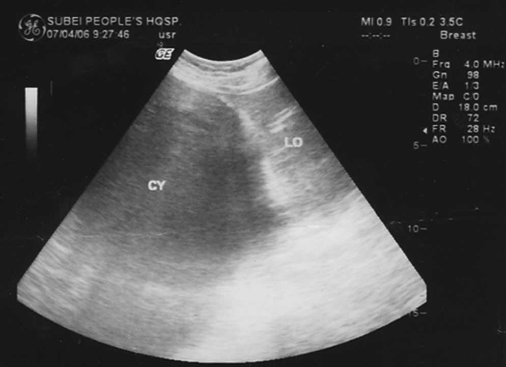
normal. An abdominal ultrasound scan revealed a giant, liquid, dark
area measuring 30.0×18.0 cm, visible in the hepatic region
(Fig. 1). No abnormalities were
revealed in the laboratory data, including the tumor markers that
are listed in Table I.
 | Table ILaboratory findings on admission. |
Table I
Laboratory findings on admission.
| Blood markers | Index | Normal range |
|---|
| WBC
(×109/l) | 10.1 | 4.0–10.0 |
| RBC
(×1012/l) | 3.7 | 3.5–5.0 |
| BUN (mmol/l) | 5.6 | 3.2–6.0 |
| Hemoglobin (g/l) | 113 | 110–150 |
| Hematocrit
(vol%) | 40 | 37–48 |
| Platelets
(×109/l) | 210 | 100–300l |
| Total protein
(g/l) | 67 | 60–80 |
| Albumin/globulin
ratio | 1.2 | 1.1–2.5 |
| Total bilirubin
(μmol/l) | 11.2 | 5.7–23.5 |
| Direct bilirubin
(μmol/l) | 2.3 | 1.7–7.8 |
| SGOT (U/l) | 37 | 0–50 |
| SGPT (U/l) | 42 | 0–50 |
| Total cholesterol
(mmol/l) | 1.30 | 1.29–1.55 |
| Serum amylase
(U/l) | 61 | 25–125 |
| Lactic dehydrogenase
(U/l) | 147.0 | 135.0–215.0 |
| Alkaline phosphatase
(U/l) | 76 | 25–150 |
| CA19-9 (KU/l) | 5.47 | <35.00 |
| CA242 (KU/l) | 1.31 | <20.00 |
| CA125 (KU/l) | 2.27 | <35.00 |
| CA15-3 (KU/l) | 2.28 | <35.00l |
| NSE (ng/ml) | <1.0 | <13.00 |
| CEA (ng/ml) | 1.21 | <5.00 |
| Ferritin (ng/ml) | 27.13 | <219.00 |
| Bta-HCG (MIU/ml) | <0.02l | <3.00 |
| AFP (ng/ml) | 0.88 | <20.00 |
| Free-PSA (ng/ml) | <0.22 | <1.00 |
| PSA (ng/ml) | <0.04 | <5.00 |
| HGH (ng/ml) | 2.15 | <7.50 |
A laparotomy was performed, during which an incision
was made in the middle line, below the umbilicus. Initially, it was
not possible to determine the point at which the sac originated,
due to its large size of approximately 30.0×31.0×18.0 cm. The sac
constituted almost half of the total epigastric region.
Additionally, it was not possible to discern the layers of
peritoneum and sac, owing to its very firm adhesion. As the
uncertainty concerning the sac was so great, it was decided that
the sac be emptied with a large trocar; approximately 3,800 ml of
fluid was drained, leaving at least 200 ml in the bottom of the
cyst. The diagnosis was corrected to be a giant gallbladder when a
4 mm slender cystic duct connecting to an 8 mm choledochus was
discovered during the operation. As no obstructive matter, such as
a tumor or gallstones were observed, this particular giant
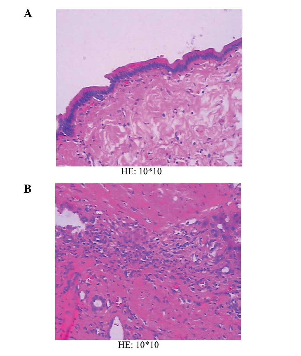
gallbladder was considered to be congenital. The pathological
section revealed that columnar epithelium was present in the lining
of the gallbladder wall along with inflammatory cell infiltration
(Fig. 2A and B). Following the
operation, there were no complications. The patient was treated and
discharged after 10 days.
Discussion
A normal gallbladder is typically no more than
7.5–10 cm in length. However, it can present a considerably large
size under the rare condition of genoconstitution. However, most
gallbladders of extremely large size are correlated with
pathological states, particularly in obstructive biliary diseases.
The original study by Courvoisier et al stated that a
growing, large gallbladder was more frequently caused by a biliary
obstructive tumor such as a pancreatic malignancy, rather than
gallstones, which may attribute to intraluminal hypertension over a
shorter period of time (5–7). According to Courvoisier et al,
a gallstone does not result in an enlarged gallbladder, as it is
formed over an extended period of time, resulting in a shrunken,
fibrotic and scarred gallbladder that does not allow extended
enlargement. However, an exception to Courvoisier’s law supports
the theory that the stones may be responsible for a growing, large
gallbladder. This theory suggests that the stones may dislodge and
acutely block the duct distally to the hepatic/cystic duct
junction, contributing to the action of check valves. In a review
of published work (1), such cases
of a giant gallbladder combined with gallstones can be further
explained by this theory of etiology.
The case we present here was unique, owing to the
unusually large size of the gallbladder, being amongst the largest
found in a review of the literature (Table II). Moreover, no obstructive
factors, such as a tumor or stones, were present. However, the
biliary duct was fluent, which enabled bile to freely enter the
intestinal tract. As neither marked biliary inflammation nor
biliary obstruction was observed, this particular giant gallbladder
was considered to be congenital. With the progression of the
disease, the giant gallbladder became increasingly filled with
bile, which lead to chronic inflammation that subsequently damaged
the contractile function of the gallbladder and contributed to
further growth of the congenital large gallbladder.
| Table IIOverview of the reported cases of
giant gallbladder. |
Table II
Overview of the reported cases of
giant gallbladder.
| Authors (Ref.) | Gender | Age (years) | Size (cm) | Obstruction | Post-operative
diagnosis |
|---|
| Grosberg (1) | Female | 95 | 14×5.5 | Stone | Acute gangrenous
cholecystitis, cholelithiasis |
| Maeda et
al(2) | Female | 36 | 18×4 | No | Chronic
cholecystitis, cholelithiasis |
| Hsu et
al(3) | Female | 87 | 16.4×13.6×7.8 | No | Acute cholecystitis,
gall bladder adenocarcinoma |
| Panaro et
al(4) | NA | 17 | 43×21×20 | No | Byler’s disease |
References
|
1
|
Grosberg SJ: Giant gallbladder. Am J Dig
Dis. 7:1039–1040. 1962. View Article : Google Scholar
|
|
2
|
Maeda Y, Setoguchi T, Yoshida T, et al: A
giant gallbladder. Gastroenterol Jpn. 14:621–624. 1979.
|
|
3
|
Hsu KF, Yeh CL, Shih ML, et al: Giant
gallbladder: adenocarcinoma complicated with empyema. J Trauma.
70:2612011. View Article : Google Scholar : PubMed/NCBI
|
|
4
|
Panaro F, Chastaing L and Navarro F:
Education and imaging. Hepatobiliary and pancreatic: giant
gallbladder associated with Byler’s disease. J Gastroenterol
Hepatol. 27:6202012.PubMed/NCBI
|
|
5
|
Hagege A: Case of retention jaundice;
importance of Courvoisier-Terrier law. Tunis Med. 45:4391957.(In
French).
|
|
6
|
Watts GT: Courvoisier’s law. Lancet.
2:1293–1294. 1985.
|
|
7
|
Verghese A and Berk SL: Courvoisier’s law.
Lancet. 1:991986.
|