Introduction
Hamartomas of the breast are rare, benign lesions
(1). These growths are also
referred to as lipofibroadenomas, fibroadenolipomas or
adenolipomas, based on their predominant components (1–3).
Hamartomas are composed of glandular, adipose and fibrous tissue
(3) often in abnormal proportions
(as malformations) (4).
Mammographic examination usually describe these lesions as a
circumscribed, non-homogeneous tumor; however, the sonographic
apperance may vary (2). The current
study presents two cases of breast hamartoma. Written informed
consent was obtained from the patients.
Case reports
Case 1
A 46-year-old premenopausal female was admitted to
Karaman State Hospital, Karaman, Turkey with a lump in the left
breast that had been detected by autodiagnosis. A physical
examination revealed a round, mobile, regular mass in the upper
outer quadrant of the breast. There was no axillar lymphadenopathy.
Mammography identified a well-circumscribed mass in the left breast
that was ∼8 cm in diameter and consistent with a lipoma. An
ultrasonographic examination revealed that the breast mass was
composed of isoechoic fat tissue, consistent with lipoma, and also
revealed an intramammarian lymph node. The patient was treated by
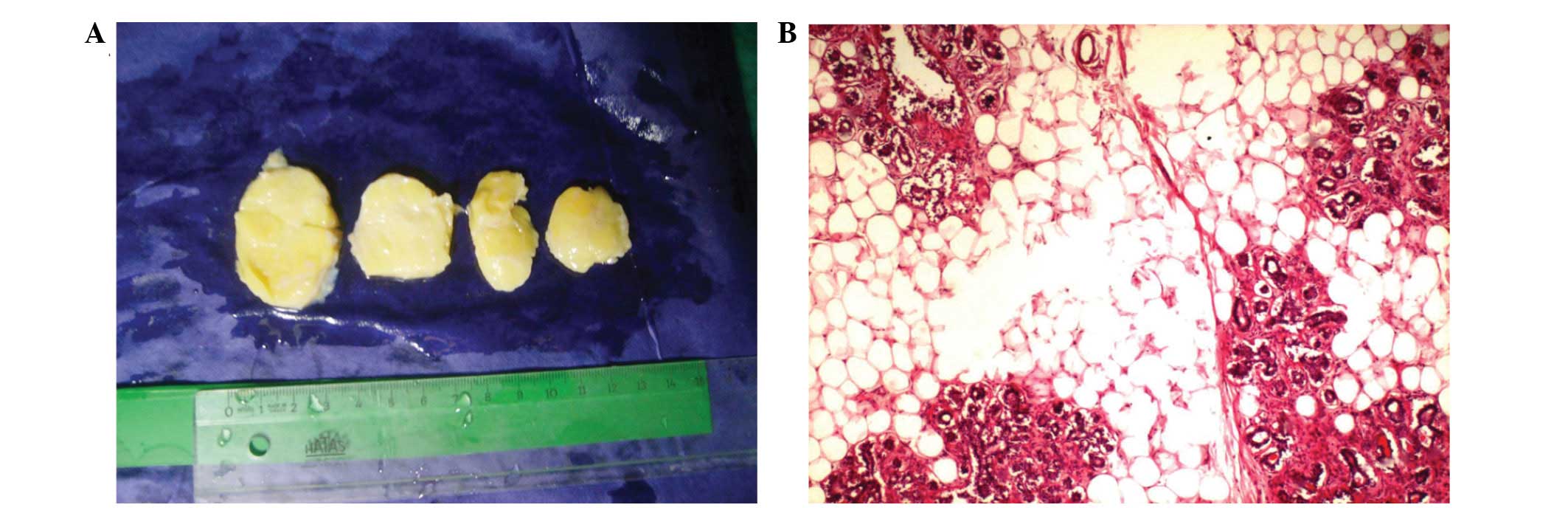
simple excision. Macroscopically, the tumor was a well-defined oval
that was encapsulated, yellow in color and soft, measuring
5.2×5×2.5 cm in size. The cut surface was lobulated and yellow,
with small greyish-white areas (Fig.
1A). The gross apperance of the tumor resembled that of a
lipoma. Microscopic observations were also characteristic of a
lipoma; the tumor was surrounded by a fibrous pseudocapsule and
consisted of mature fat and islands of structurally normal
glandular tissue with lobular arrangement (Fig. 1B). A fibrous stroma was found around
the glandular tissue, however, in specific areas, lobular
aggregates had direct contact with the fat cells without
interference by the fibrous tissue. No proliferative changes in
lobules and ducts were detected within the lesion. This case was
consequently diagnosed as an adenolipoma.
Case 2
A 41-year-old female was admitted to the same
hospital with a palpable right breast mass. A physical examination
revealed a round mass of ∼7 cm in diameter. No palpable lymph nodes
or other masses of the contralateral breast were detected. The
ultrasonography of the lesion revealed a solid, heterogeneous
echogenic mass with smooth margins, measuring 5×2 cm. A excisional
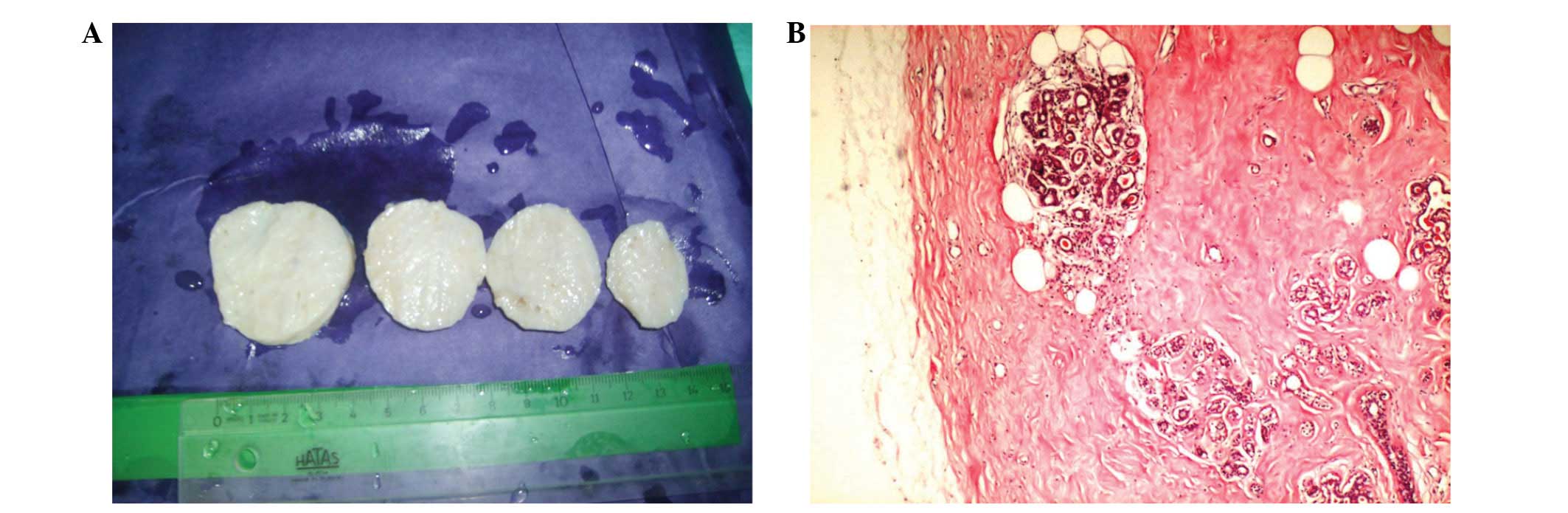
biopsy was performed on the right breast, and a 6.7×4.6×4.5-cm,
greyish-white colored, oval-shaped, encapsulated mass was found
upon gross examination (Fig. 2A).
In addition, small cystic spaces were detected on the cut surface.
Histopathologically, the tumor consisted of mammary glandular
tissue in hyalinized fibrous stroma, interspersed with islands of
mature fatty tissue (Fig. 2B).
Cystic ducts with apocrine metaplasia were evident in specific
areas as fibrocystic changes. All lobules were structurally normal.
The lesion was well-defined and had a pseudocapsule of compressed
adjacent breast tissue. This case was consequently diagnosed as a
fibroadenolipoma.
Discussion
Hamartomas of the breast are uncommon benign
tumor-like nodules, also known as fibroadenolipoma,
lipofibroadenoma or adenolipomas (3). The reported incidence of breast
hamartomas is 0.7% of all benign breast tumors in females (5). Hamartomas were first described in 1971
by Arrigoni et al in a study of 10 patients whose breast
tumors clinically and grossly resembled fibroadenomas (6). The majority of these lesions occur in
females >35 years old. At clinical examination, hamartomas are
usually occult, but they may manifest as large, mobile, soft to
firm masses (7). Tumors as large as
17 cm have been reported (8).
Breast hamartomas have become more frequently diagnosed due to the
increased use of mammography, but they may be mistaken for
neoplasms (9). During mammography
scans, hamartomas are identified as typically well-circumscribed,
round to oval masses containing fat and soft tissue densities with
a thin, radiopaque pseudocapsule (7). The sonographic appearance of breast
hamartoma has been reported to be variable and non-specific
(8,9). Upon gross examination, hamartomas are
typically well-demarcated, occasionally lobulated lesions with
smooth contours and an often rubbery greyish-white to yellow cut
surface, resembling a fibroadenoma or lipoma (3,4). The
two common variants of breast hamartoma are adenolipoma and
chondrolipoma (8). Adenohibernoma
and myoid hamartoma are rare variants of hamartoma (4,10).
Upon gross examination, adenolipomas are soft, circumscribed,
occasionally lobulated masses, bordered by a thin fibrous
pseudocapsule. The cut surface reveals a variegated pattern of fat
and fibrous breast parenchyma. Lesions with abundant fat resemble
lipomas (8).
Upon microscopic examination, Arrigoni et al
identified ‘mammary glanduler tissue with a prominent lobuler
arrangement, fibrous stroma and fat in varible proportions’
(6). Pathological observations in
previous studies are varied and include circumscribed fibrocystic
disease, adenolipoma, fibroadenoma with fat and fibroadenoma with
lobules, as reported by Jones et al. In the same case
report, heterologous elements were identified as cartilage and
smooth muscle (11). This
circumscribed mass of breast tissue may reveal fibrocystic and
atrophic changes; pseudoangiomatous hyperplasia is frequently
observed (10). The lesion
generates the impression of a ‘breast within a breast’ (4,10).
Usual ductal hyperplasia, apocrine metaplasia, calcification,
stromal giant cells and adenosis may be associated with hamartoma
(4). Lobular intraepithelial
neoplasms and ductal intraepithelial neoplasms have also been
reported to occur within the hamartoma in rare cases (4,12)
Although hamartomas are usually benign, malignant transformation is
possible (2,5).
Adenolipomas are composed of mature fat and mammary
parenchyma with pseudocapsules. Lobules and ducts are structurally
normal, with little or no proliferative change. Adenoliomas differ
from other mammary lesions that contain fat (4).
Surgical removal is the curative method for breast
hamartomas (2,3). If there is a coincidental epithelial
malignancy in the lesion, there is a potential for recurrence
(3). There was no recurrence or
other problems in the 18-month follow-up of the patients in the
present study. Excision and histological examination is necessary
for a differential diagnosis and also for any epithelial lesions of
the hamartoma.
References
|
1.
|
Altermatt HJ, Gebbers JO and Laissue JA:
Hamartoma of the breast. Schweiz Med Wochenschr. 117:365–368.
1987.(In German).
|
|
2.
|
Barbaros U, Deveci U, Erbil Y and Budak D:
Breast hamartoma: a case report. Acta Chir Belg. 105:658–659.
2005.
|
|
3.
|
Guray M and Sahin AA: Benign breast
diseases: classification, diagnosis, and management. Oncologist.
11:435–449. 2006. View Article : Google Scholar : PubMed/NCBI
|
|
4.
|
Moinfar F: Mesenchymal lesions/tumors In:
Essentials of Diagnostic Breast Pathology. Springer-Verlag;
Heidelberg: pp. 3832007
|
|
5.
|
Lee EH, Wylie EJ, Barke AG and Bastiaan De
Boer W: Invasive ductal carcinoma arising in a breast hamartoma:
two case reports and a review of the literature. Clin Radiol.
58:80–83. 2003. View Article : Google Scholar : PubMed/NCBI
|
|
6.
|
Arrigoni MG, Dockerty MB and Judd ES: The
identification and treatment of mammary hamartoma. Surg Gynecol
Obstet. 133:577–582. 1971.PubMed/NCBI
|
|
7.
|
Feder JM, de Paredes ES, Hogge JP and
Wilken JJ: Unusual breast lesions: radiologic-pathologic
correlation. Radiographics. 19:S11–S26. 1999. View Article : Google Scholar : PubMed/NCBI
|
|
8.
|
Rosen PP: Rosen’s Breast Pathology. 2nd
edition. Lippincott Williams & Wilkins; Philadelphia: pp.
7792001
|
|
9.
|
Wu CY, Lin SH, Tu SH, Huang CS and Jeng
CM: Hamartoma of the breast. Zhonghua Fang She Xue Za Zhi.
28:143–148. 2003.(In Chinese).
|
|
10.
|
Tavassoli FA and Devilee P: World Health
Organization: Pathology & Genetics: Tumours of the Breast and
Female Genital Organs. IARC Press; Lyon: pp. 101–102. 2003
|
|
11.
|
Jones MW, Norris HJ and Wargotz ES:
Hamartomas of the breast. Surg Gynecol Obstet. 173:54–56. 1991.
|
|
12.
|
Mester J, Simmons RM, Vazquez MF and
Rosenblatt R: In situ and infiltrating ductal carcinoma arising in
a breast hamartoma. AJR Am J Roentgenol. 175:64–66. 2000.
View Article : Google Scholar : PubMed/NCBI
|