Spandidos Publications Logo
  • About
    • About Spandidos
    • Aims and Scopes
    • Abstracting and Indexing
    • Editorial Policies
    • Reprints and Permissions
    • Job Opportunities
    • Terms and Conditions
    • Contact
  • Journals
    • All Journals
    • Oncology Letters
      • Oncology Letters
      • Information for Authors
      • Editorial Policies
      • Editorial Board
      • Aims and Scope
      • Abstracting and Indexing
      • Bibliographic Information
      • Archive
    • International Journal of Oncology
      • International Journal of Oncology
      • Information for Authors
      • Editorial Policies
      • Editorial Board
      • Aims and Scope
      • Abstracting and Indexing
      • Bibliographic Information
      • Archive
    • Molecular and Clinical Oncology
      • Molecular and Clinical Oncology
      • Information for Authors
      • Editorial Policies
      • Editorial Board
      • Aims and Scope
      • Abstracting and Indexing
      • Bibliographic Information
      • Archive
    • Experimental and Therapeutic Medicine
      • Experimental and Therapeutic Medicine
      • Information for Authors
      • Editorial Policies
      • Editorial Board
      • Aims and Scope
      • Abstracting and Indexing
      • Bibliographic Information
      • Archive
    • International Journal of Molecular Medicine
      • International Journal of Molecular Medicine
      • Information for Authors
      • Editorial Policies
      • Editorial Board
      • Aims and Scope
      • Abstracting and Indexing
      • Bibliographic Information
      • Archive
    • Biomedical Reports
      • Biomedical Reports
      • Information for Authors
      • Editorial Policies
      • Editorial Board
      • Aims and Scope
      • Abstracting and Indexing
      • Bibliographic Information
      • Archive
    • Oncology Reports
      • Oncology Reports
      • Information for Authors
      • Editorial Policies
      • Editorial Board
      • Aims and Scope
      • Abstracting and Indexing
      • Bibliographic Information
      • Archive
    • Molecular Medicine Reports
      • Molecular Medicine Reports
      • Information for Authors
      • Editorial Policies
      • Editorial Board
      • Aims and Scope
      • Abstracting and Indexing
      • Bibliographic Information
      • Archive
    • World Academy of Sciences Journal
      • World Academy of Sciences Journal
      • Information for Authors
      • Editorial Policies
      • Editorial Board
      • Aims and Scope
      • Abstracting and Indexing
      • Bibliographic Information
      • Archive
    • International Journal of Functional Nutrition
      • International Journal of Functional Nutrition
      • Information for Authors
      • Editorial Policies
      • Editorial Board
      • Aims and Scope
      • Abstracting and Indexing
      • Bibliographic Information
      • Archive
    • International Journal of Epigenetics
      • International Journal of Epigenetics
      • Information for Authors
      • Editorial Policies
      • Editorial Board
      • Aims and Scope
      • Abstracting and Indexing
      • Bibliographic Information
      • Archive
    • Medicine International
      • Medicine International
      • Information for Authors
      • Editorial Policies
      • Editorial Board
      • Aims and Scope
      • Abstracting and Indexing
      • Bibliographic Information
      • Archive
  • Articles
  • Information
    • Information for Authors
    • Information for Reviewers
    • Information for Librarians
    • Information for Advertisers
    • Conferences
  • Language Editing
Spandidos Publications Logo
  • About
    • About Spandidos
    • Aims and Scopes
    • Abstracting and Indexing
    • Editorial Policies
    • Reprints and Permissions
    • Job Opportunities
    • Terms and Conditions
    • Contact
  • Journals
    • All Journals
    • Biomedical Reports
      • Information for Authors
      • Editorial Policies
      • Editorial Board
      • Aims and Scope
      • Abstracting and Indexing
      • Bibliographic Information
      • Archive
    • Experimental and Therapeutic Medicine
      • Information for Authors
      • Editorial Policies
      • Editorial Board
      • Aims and Scope
      • Abstracting and Indexing
      • Bibliographic Information
      • Archive
    • International Journal of Epigenetics
      • Information for Authors
      • Editorial Policies
      • Editorial Board
      • Aims and Scope
      • Abstracting and Indexing
      • Bibliographic Information
      • Archive
    • International Journal of Functional Nutrition
      • Information for Authors
      • Editorial Policies
      • Editorial Board
      • Aims and Scope
      • Abstracting and Indexing
      • Bibliographic Information
      • Archive
    • International Journal of Molecular Medicine
      • Information for Authors
      • Editorial Policies
      • Editorial Board
      • Aims and Scope
      • Abstracting and Indexing
      • Bibliographic Information
      • Archive
    • International Journal of Oncology
      • Information for Authors
      • Editorial Policies
      • Editorial Board
      • Aims and Scope
      • Abstracting and Indexing
      • Bibliographic Information
      • Archive
    • Medicine International
      • Information for Authors
      • Editorial Policies
      • Editorial Board
      • Aims and Scope
      • Abstracting and Indexing
      • Bibliographic Information
      • Archive
    • Molecular and Clinical Oncology
      • Information for Authors
      • Editorial Policies
      • Editorial Board
      • Aims and Scope
      • Abstracting and Indexing
      • Bibliographic Information
      • Archive
    • Molecular Medicine Reports
      • Information for Authors
      • Editorial Policies
      • Editorial Board
      • Aims and Scope
      • Abstracting and Indexing
      • Bibliographic Information
      • Archive
    • Oncology Letters
      • Information for Authors
      • Editorial Policies
      • Editorial Board
      • Aims and Scope
      • Abstracting and Indexing
      • Bibliographic Information
      • Archive
    • Oncology Reports
      • Information for Authors
      • Editorial Policies
      • Editorial Board
      • Aims and Scope
      • Abstracting and Indexing
      • Bibliographic Information
      • Archive
    • World Academy of Sciences Journal
      • Information for Authors
      • Editorial Policies
      • Editorial Board
      • Aims and Scope
      • Abstracting and Indexing
      • Bibliographic Information
      • Archive
  • Articles
  • Information
    • For Authors
    • For Reviewers
    • For Librarians
    • For Advertisers
    • Conferences
  • Language Editing
Login Register Submit
  • This site uses cookies
  • You can change your cookie settings at any time by following the instructions in our Cookie Policy. To find out more, you may read our Privacy Policy.

    I agree
Search articles by DOI, keyword, author or affiliation
Search
Advanced Search
presentation
Oncology Letters
Join Editorial Board Propose a Special Issue
Print ISSN: 1792-1074 Online ISSN: 1792-1082
Journal Cover
July-2014 Volume 8 Issue 1

Full Size Image

Sign up for eToc alerts
Recommend to Library

Journals

International Journal of Molecular Medicine

International Journal of Molecular Medicine

International Journal of Molecular Medicine is an international journal devoted to molecular mechanisms of human disease.

International Journal of Oncology

International Journal of Oncology

International Journal of Oncology is an international journal devoted to oncology research and cancer treatment.

Molecular Medicine Reports

Molecular Medicine Reports

Covers molecular medicine topics such as pharmacology, pathology, genetics, neuroscience, infectious diseases, molecular cardiology, and molecular surgery.

Oncology Reports

Oncology Reports

Oncology Reports is an international journal devoted to fundamental and applied research in Oncology.

Experimental and Therapeutic Medicine

Experimental and Therapeutic Medicine

Experimental and Therapeutic Medicine is an international journal devoted to laboratory and clinical medicine.

Oncology Letters

Oncology Letters

Oncology Letters is an international journal devoted to Experimental and Clinical Oncology.

Biomedical Reports

Biomedical Reports

Explores a wide range of biological and medical fields, including pharmacology, genetics, microbiology, neuroscience, and molecular cardiology.

Molecular and Clinical Oncology

Molecular and Clinical Oncology

International journal addressing all aspects of oncology research, from tumorigenesis and oncogenes to chemotherapy and metastasis.

World Academy of Sciences Journal

World Academy of Sciences Journal

Multidisciplinary open-access journal spanning biochemistry, genetics, neuroscience, environmental health, and synthetic biology.

International Journal of Functional Nutrition

International Journal of Functional Nutrition

Open-access journal combining biochemistry, pharmacology, immunology, and genetics to advance health through functional nutrition.

International Journal of Epigenetics

International Journal of Epigenetics

Publishes open-access research on using epigenetics to advance understanding and treatment of human disease.

Medicine International

Medicine International

An International Open Access Journal Devoted to General Medicine.

Journal Cover
July-2014 Volume 8 Issue 1

Full Size Image

Sign up for eToc alerts
Recommend to Library

  • Article
  • Citations
    • Cite This Article
    • Download Citation
    • Create Citation Alert
    • Remove Citation Alert
    • Cited By
  • Similar Articles
    • Related Articles (in Spandidos Publications)
    • Similar Articles (Google Scholar)
    • Similar Articles (PubMed)
  • Download PDF
  • Download XML
  • View XML
Case Report

Cerebellar, brainstem and spinal cord metastases from esophageal cancer following radiotherapy: A case report and literature review

  • Authors:
    • Peng Zhang
    • Wei Feng
    • Xiao Zheng
    • Yue‑Zhen Wang
    • Guo‑Ping Shan
  • View Affiliations / Copyright

    Affiliations: Department of Radiation Oncology, Zhejiang Key Laboratory of Diagnosis and Treatment Technology on Thoracic Oncology (Lung and Esophagus), Zhejiang Cancer Hospital, Hangzhou, Zhejiang 310022, P.R. China
  • Pages: 253-257
    |
    Published online on: May 9, 2014
       https://doi.org/10.3892/ol.2014.2130
  • Expand metrics +
Metrics: Total Views: 0 (Spandidos Publications: | PMC Statistics: )
Metrics: Total PDF Downloads: 0 (Spandidos Publications: | PMC Statistics: )
Cited By (CrossRef): 0 citations Loading Articles...

This article is mentioned in:


Abstract

Cerebellar, brainstem and spinal cord metastases from esophageal cancer following radiotherapy are extremely rare. The current study presents the case of a 74‑year‑old male who was admitted to the Zhejiang Cancer Hospital (Hangzhou, China) with a poorly‑differentiated neuroendocrine carcinoma of the esophagus. Following radiotherapy, multiple abnormal signals in the brainstem and spinal cord were found on magnetic resonance imaging (MRI). Following palliative radiochemotherapy, the clinical symptoms and abnormal MRI signals in the brainstem and spinal cord were found to improve. This case revealed that brain metastasis from esophageal carcinoma may occur simultaneously with brainstem and spinal cord metastases.

Introduction

The overall rate of brain metastasis from gastrointestinal malignant tumors, including colorectal, gastric, pancreatic, esophageal and gastrointestinal cancer, is reported as 3–8% (1–3). Notably, esophageal carcinomas account for 1–5% of these brain metastases (4,5). Neuroendocrine carcinoma of the esophagus is a rare malignant tumor originating from the neuroendocrine cells. Depending on the degree of differentiation, the tumors are classified as carcinoid (well-differentiated), atypical carcinoid (moderately-differentiated) and neuroendocrine small cell carcinoma (poorly-differentiated). Neuroendocrine carcinomas account for 0.15–2.8% of esophageal carcinomas (6,7). According to previous studies, the pathological types of brain metastases originating from esophageal carcinoma are generally squamous carcinoma in Asian countries and adenocarcinoma in Western countries (4,5,8). The incidence of esophageal cancer in China is high, and reports of brain metastasis of esophageal cancer in China in the international literature are rare. Due to the low incidence and lack of specific clinical manifestations, brain metastases from esophageal carcinoma usually remain undiagnosed and are treated incorrectly. Cases of simultaneous metastases occurring in the cerebellum, brainstem and spinal cord are extremely rare. The current study reports such a case. Patient provided written informed consent.

Case report

Primary tumor (esophageal cancer) diagnosis and treatment

A 74-year-old male presented to the Zhejiang Cancer Hospital (Hangzhou, China) with complaints of swallowing difficulties that had been apparent for three months. Gastroscopy revealed the presence of two nodules of 1.5 cm in diameter, with deep ulceration and eruption of the surrounding esophageal mucous membrane, at 22–25 and 27–35 cm from the incisor teeth. 
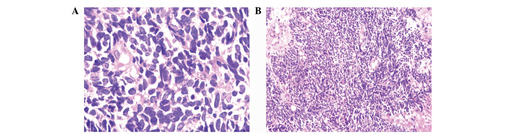
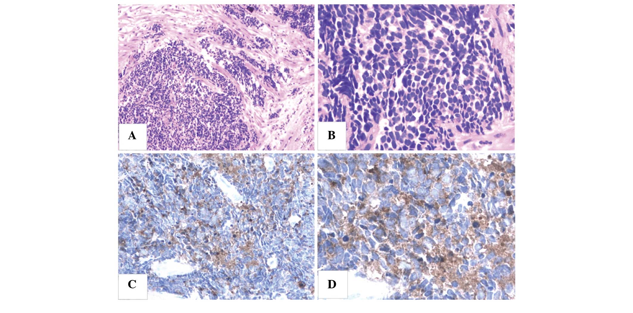
Pathological evaluation indicated malignant epithelial tumors described as poorly-differentiated neuroendocrine carcinoma or 
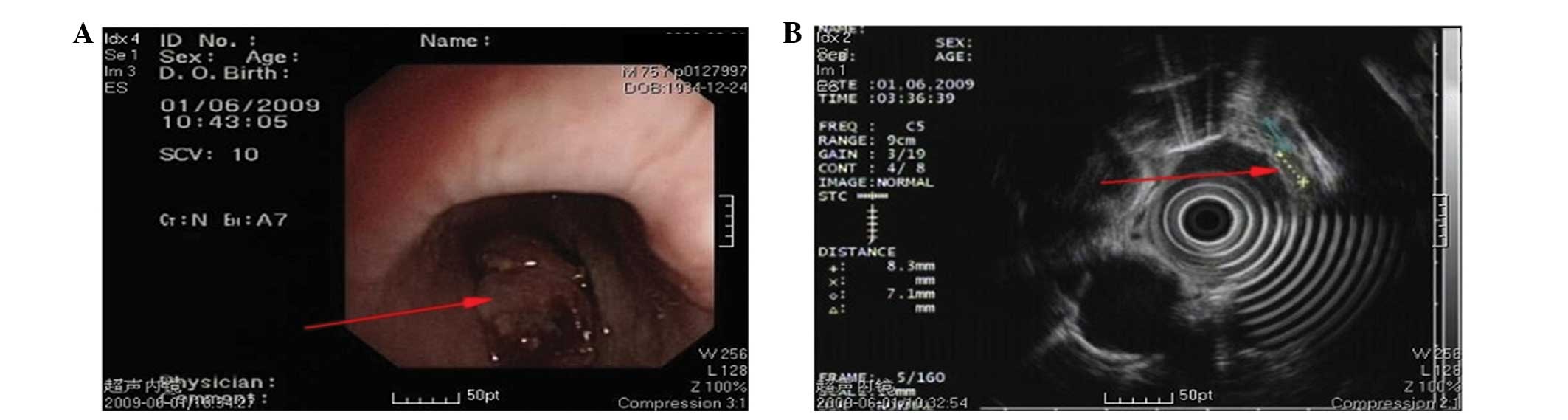
basal cell squamous carcinoma (Fig. 1). Endoscopic ultrasound revealed damage to five layers of the esophageal wall, with lesions invading the outer membrane and muscular layer; multiple enlarged lymph nodes were also observed (Fig. 2). Ultrasound revealed 
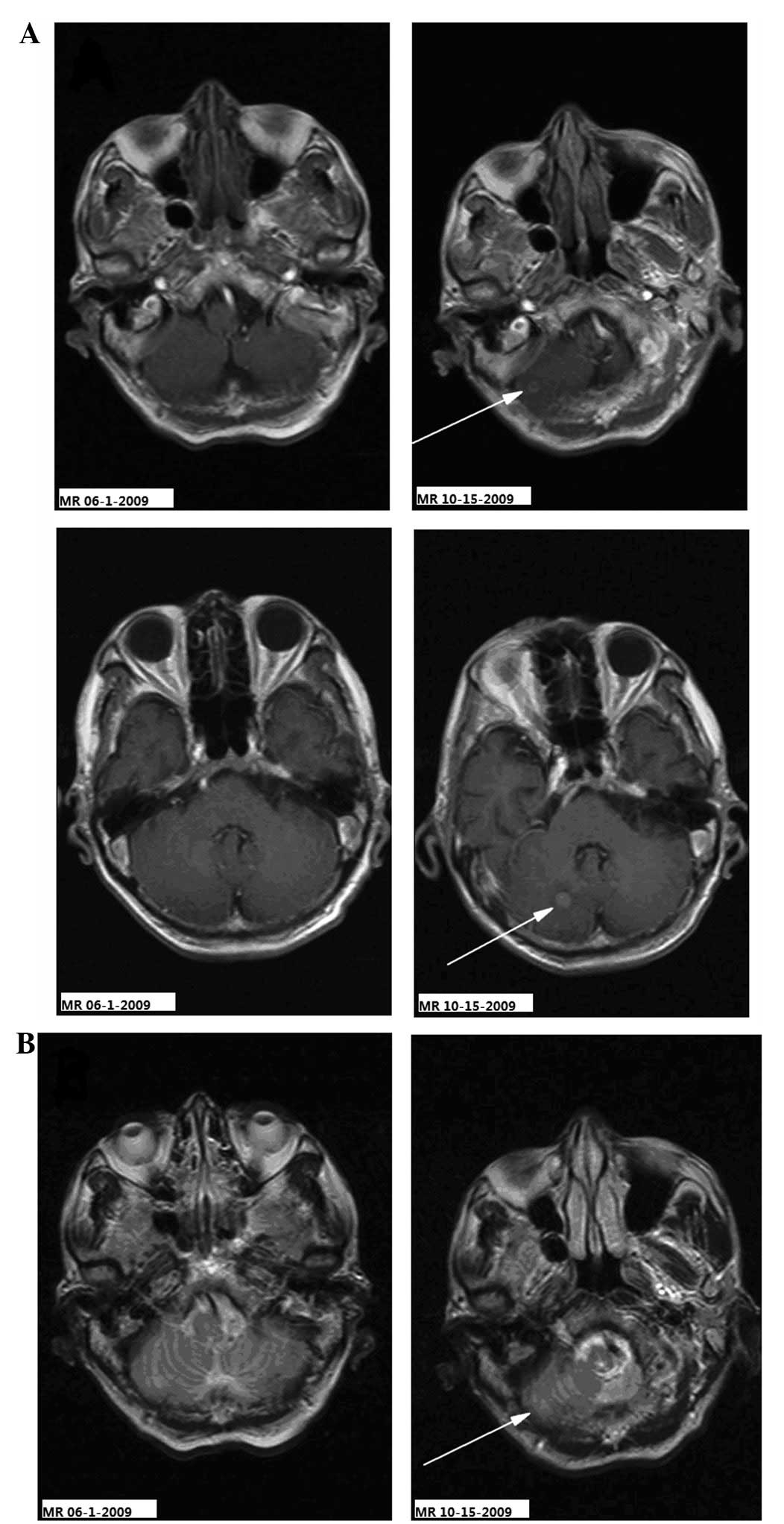
supraclavicular lymph node metastasis. No abnormal signals were observed by contrast-enhanced magnetic resonance imaging (MRI) of the brain and via whole body bone imaging (Figs. 3–5). The diagnosis was of esophageal carcinoma with supraclavicular metastasis. Radiotherapy was administered to the esophageal lesions and supraclavicular area, with a total dose of 64 Gy/32 fractions of 2 Gy per fraction. The swallowing difficulty symptom improved, and a follow-up enhanced chest computed tomography (CT) scan revealed slight thickening of the upper segment of the esophagus following radiotherapy.

Figure 1

Pathological appearance of the primary esophageal tumor cells exhibiting little cytoplasm and heavily stained nuclei. Magnifications of (A) ×400 and (B) ×100 (hematoxyling and eosin stain).

Figure 2

Endoscopic ultrasonography revealing (A) an esophageal neoplasm (indicated by the red arrow) and (B) multiple enlarged lymph nodes on the outside of the esophagus wall (indicated by the red arrow).

Figure 3

Comparison between the cerebral MR images of the esophageal cancer patient obtained prior to (MR 06-1-2009) and following (MR 10-15-2009) the metastasis. (A) Enhanced axial T1-weighted images and (B) axial T2-weighted images. Left (MR 06-1-2009), no abnormal signals were observed in the brain; and right (MR 10-15-2009), abnormal signals were observed in the right cerebellar hemisphere (indicated by the arrows). MR, magnetic resonance.

Figure 5

Comparison between the MR images of the cervical spinal cord prior to and following treatment. Sagittal T1-weighted images performed (A) prior to chemotherapy (MR 10-28-2009) showing abnormal signals in the medulla oblongata and cervical cord, considered to be the metastatic lesions (indicated by the two arrows), and (B) following chemotherapy (MR-11-23-2009) showing that the abnormal signals of the medulla oblongata and cervical cord had markedly improved (indicated by the two arrows). MR, magnetic resonance.

Diagnosis of central nervous system metastasis and treatment

The patient complained of constant occipital headache with slight pain in the bilateral upper arm and fatigue, but no nausea or vomiting, for 2.7 months after the treatment of the primary lesion. The Karnofsky performance status (KPS) score was 70 and no pathological reflexes were observed on physical examination. Brain MRI revealed two nodules (0.35 and 0.8 cm in diameter) in the right cerebellar hemisphere. The signal was slightly lower in the unenhanced T1-weighted images and slightly higher in the T2-weighted images. The lesions showed clear ring-like, uniform hardening on signal-enhanced scans, and mild edema was observed surrounding the larger lesion. The diagnosis was of a cerebellar metastasis from the esophageal cancer following treatment (Figs. 3–5). Following whole brain radiotherapy of a total dose of 30 Gy/20 fractions of 1.5 Gy per fraction by hyperfractionation (two fractions per day), the patient’s occipital 
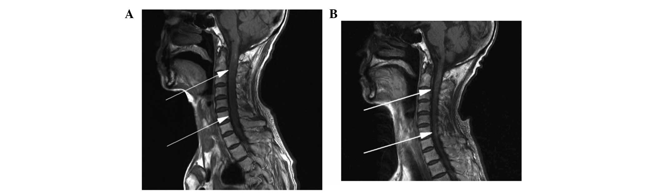
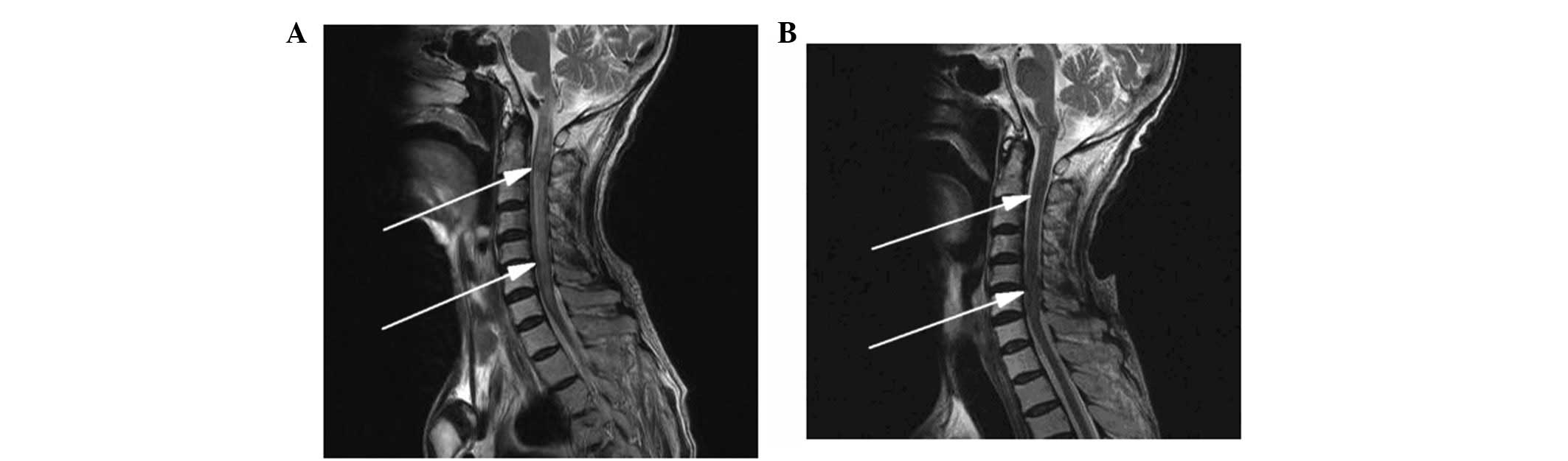
headache was relieved, however, the neck numbness was aggravated. A cervical MRI examination revealed enlargement of the medulla oblongata and spinal cord, with slightly increased T1 and T2 signals. A signal-enhanced scan showed heterogeneous enhancement in a region considered to be the metastasis. The patient continued with the planned whole brain radiotherapy, together with nedaplatin chemotherapy (40 mg on days one to three of the radiotherapy period). At the end of the first period of chemotherapy, the patient’s occipital headaches were alleviated and the neck numbness was markedly improved. One month later, the patient’s head and neck symptoms had further improved and a neck MRI revealed that the abnormal signal foci in the medulla oblongata and cervical cord had also markedly improved (Figs. 6 and 7). To strengthen the effects of the treatment, the patient was treated with palliative radiotherapy (total dose of 26 Gy/13 fractions of 2 Gy per fraction) to the cervical spinal cord. Following treatment, the patient’s head and neck pain and numbness symptoms had almost disappeared. The patient was discharged with oral etoposide (VP-16) capsules (50 mg daily for 20 days).

Figure 6

Comparison between the MR images of the cervical spinal cord prior to and following chemotherapy. Sagittal T2WI (A) prior to chemotherapy (MR 10-28-2009) showing increased T2WI signals and abnormal morphological augmentation in the medulla oblongata and cervical cord indicating metastatic lesions (indicated by the two arrows), and (B) following chemotherapy (MR 11-23-2009) showing decreased T2WI signals in the medulla oblongata and cervical cord consistent with the signals of normal tissues, and marked improvement in the abnormal morphology (as indicated by the two arrows). MR, magnetic resonance; T2WI, T2-weighted image.

Figure 7

(A) Tumor cells characteristically concentrated into canalicular and branching structures (H&E stain; magnification, ×100). (B) Tumor cells exhibiting little cytoplasm, heavily stained nuclei and lacking evident nucleoli (H&E stain; magnification, ×400). Tumor cells showing cytoplasmic positive staining for synaptophysin using the diaminobenzidine method at magnifications of (C) ×200 and (D) ×400. H&E, hematoxylin and eosin.

Prognosis of patient and follow-up

Once discharged, the patient refused to return to the hospital for further treatment and follow-up. The occipital headache symptoms recurred, and the patient’s condition gradually deteriorated. The patient succumbed to systemic organ failure on April 16, 2010, six months after the diagnosis of brain metastasis.

Discussion

A limitation of this case was the lack of autopsy tissue pathology and cerebrospinal fluid cytology to support the diagnosis. A review of the literature on the brain metastasis of esophageal cancer found that, with the exception of cases that underwent surgical resection of the brain metastatic lesions, the patients were treated with chemotherapy and diagnosed by imaging (4–6,8–12). Evidence from MRI and improvements in the clinical symptoms of the patient following radiotherapy determine the diagnosis of cerebellar, brainstem or spinal cord metastasis of esophageal cancer. The most common clinical signs and chief complaints for brain metastases of esophageal carcinoma are weakness (58%), headache (28%), epilepsy (22%) and cerebellar dysfunction (14%) (8).

In the present case, following the treatment of the primary esophageal tumor, the main positive central nervous system signs were fatigue, headaches and neck pain. Takeshima et al (11) reported on the characteristic cyst structure of metastatic brain tumors of squamous cell esophageal carcinoma. On T1-weighted images, following the administration of the gadolinium with diethylenetriaminepentaacetic acid contrast agent, the majority of lesions appear as thin-walled cysts with slightly higher signals than those of the brain tissues. Signals from the interior of the cysts are similar to those of the cerebrospinal fluid, however, the walls of the cystic tumors exhibit high signal reflection (11). In the present case, the brain MRI did not exhibit thin-walled cysts, but showed the appearance of solid nodules. Esophageal cancers may become necrotic, characterized by rapid growth, solid tumors that have an inadequate blood supply and the development of focal necrosis in the center of the tumor, resulting in a cyst-like appearance. Prior to the end of the 20th century, brain metastases of esophageal carcinoma were confirmed primarily by pathological examination at autopsy. Central nervous system metastasis of esophageal cancer is also extremely rare. Ogawa et al reported brain metastasis in 36 (1.4%) of 2,554 esophageal cancer patients (8). Gabrielsen et al (4) observed that the incidence of brain metastasis of esophageal cancer was 3.6% (4), and Weinberg et al (5) reported 27 cases of brain metastasis (1.7%) in 1,588 esophageal cancer patients. There are no literature reports of brainstem and spinal cord metastasis of esophageal cancer.

Due to the low incidence of brain metastasis, there is generally no requirement for an MRI examination of the brain and cervical spine of patients with esophageal cancer; Japanese studies have even opposed the routine brain CT examination of patients with esophageal cancer (4). Ogawa et al (8) suggested that with the rapid development of medical imaging technology, particularly the application of MR and the use of enhanced contrast agents in neuroimaging, faster and earlier detection of brain metastasis of esophageal carcinoma has become possible (5,8,9). Currently, for the detection and evaluation of tumor lesions, MRI is superior to CT. Therefore, the prompt use of MRI is necessary for the early detection of brainstem and spinal cord metastatic lesions. Certain studies have considered the use of adjuvant chemotherapy following the resection of esophageal carcinoma to increase the risk of brain metastasis (6,12).

It has been confirmed that the distant metastasis of esophageal cancer depends predominantly on the lymphatic and blood systems. The most common pathway for brain metastasis of esophageal cancer is hematogenous dissemination through the arterial circulation. In 1940, the Batson venous plexus was proposed as a pathway for the brain metastasis of esophageal carcinoma (13). However, the role of the vertebral venous system in the brain metastasis of esophageal cancers is supported by a more recent study (8). The present case may be more illustrative of the importance of the vertebral vascular system for cerebellar, brainstem and spinal cord metastases from esophageal cancer, as all three regions are anatomically associated with the same vertebral vascular system, which is distinct from the venous system of the thoracic and lumbar spine and brain.

Brain metastases are often treated with multiple therapies, including surgery, radiation and chemotherapy. Complete removal of the lesions is the goal of surgical treatment and may improve survival rates. However, this generally only applies to patients with a high KPS score or to those with single, solitary tumors (8,10).

Ogawa et al (8) reported that patients treated with surgery and radiation treatment have a median survival time of 9.6 months, while patients receiving only radiation treatment have a median survival time of only 1.8 months. In the present case, due to multiple metastases to the cerebellum, brainstem and spinal cord, a combined treatment of local irradiation and chemotherapy was used. The symptoms were significantly improved following treatment, with a survival time of 3.9 months.

Brain metastasis of esophageal cancer may also involve the brainstem and spinal cord, for which local and comprehensive treatments may improve the efficacy of therapy and therefore prolong survival time.

Acknowledgements

This study was supported by grants from the Zhejiang Province Science and Technology Project of Traditional Chinese Medicine (grant no. 2011ZB017) and the Talent Project of Medical and Health Sciences Fund of Zhejiang Province (grant no. 2012RCB005).

References

1 

Salvati M, Cervoni L, Paolini S and Delfini R: Solitary cerebral metastases from intestinal carcinoma. Acta Neurochir (Wien). 133:181–183. 1995.

2 

Flickinger JC, Kondziolka D, Lunsford LD, et al: A multi-institutional experience with stereotactic radiosurgery for solitary brain metastasis. Int J Radiat Oncol Biol Phys. 28:797–802. 1994.

3 

Hammoud MA, McCutcheon IE, Elsouki R, Schoppa D and Patt YZ: Colorectal carcinoma and brain metastasis: distribution, treatment, and survival. Ann Surg Oncol. 3:453–463. 1996.

4 

Gabrielsen TO, Eldevik OP, Orringer MB and Marshall BL: Esophageal carcinoma metastatic to the brain: clinical value and cost-effectiveness of routine enhanced head CT before esophagectomy. AJNR Am J Neuroradiol. 16:1915–1921. 1995.

5 

Weinberg JS, Suki D, Hanbali F, Cohen ZR, Lenzi R and Sawaya R: Metastasis of esophageal carcinoma to the brain. Cancer. 98:1925–1933. 2003.

6 

Pantvaidya GH, Pramesh CS, Deshpande MS, Jambhekar NA, Sharma S and Deshpande RK: Small cell carcinoma of the esophagus: the Tata Memorial Hospital experience. Ann Thorac Surg. 74:1924–1927. 2002.

7 

Linan Padilla A, Milla Sabas A, Abad Zamora JM, et al: Oat-cell carcinoma of the esophagus: presentation of two cases and literature review. Rev Esp Enferm Dig. 99:415–419. 2007.

8 

Ogawa K, Toita T, Sueyama H, et al: Brain metastases from esophageal carcinoma: natural history, prognostic factors, and outcome. Cancer. 94:759–764. 2002.

9 

Khuntia D, Sajja R, Chidel MA, et al: Factors associated with improved survival in patients with brain metastases from esophageal cancer: a retrospective review. Technol Cancer Res Treat. 2:267–272. 2003.

10 

Koga H, Mukawa J, Miyagi K, et al: Treatment of metastatic brain tumor from esophageal carcinoma--report of four cases. Neurol Med Chir (Tokyo). 31:518–522. 1991.

11 

Takeshima H, Kuratsu J, Nishi T, et al: Metastatic brain tumours from oesophageal carcinoma: neuro-imaging and clinicopathological characteristics in Japanese patients. Acta Neurochir (Wien). 143:31–36. 2001.

12 

Kawabata R, Doki Y, Ishikawa O, et al: Frequent brain metastasis after chemotherapy and surgery for advanced esophageal cancers. Hepatogastroenterology. 54:1043–1048. 2007.

13 

Batson OV: The function of the vertebral veins and their role in the spread of metastases. Ann Surg. 112:138–149. 1940.

Related Articles

  • Abstract
  • View
  • Download
  • Twitter
Copy and paste a formatted citation
Spandidos Publications style
Zhang P, Feng W, Zheng X, Wang YZ and Shan GP: Cerebellar, brainstem and spinal cord metastases from esophageal cancer following radiotherapy: A case report and literature review. Oncol Lett 8: 253-257, 2014.
APA
Zhang, P., Feng, W., Zheng, X., Wang, Y., & Shan, G. (2014). Cerebellar, brainstem and spinal cord metastases from esophageal cancer following radiotherapy: A case report and literature review. Oncology Letters, 8, 253-257. https://doi.org/10.3892/ol.2014.2130
MLA
Zhang, P., Feng, W., Zheng, X., Wang, Y., Shan, G."Cerebellar, brainstem and spinal cord metastases from esophageal cancer following radiotherapy: A case report and literature review". Oncology Letters 8.1 (2014): 253-257.
Chicago
Zhang, P., Feng, W., Zheng, X., Wang, Y., Shan, G."Cerebellar, brainstem and spinal cord metastases from esophageal cancer following radiotherapy: A case report and literature review". Oncology Letters 8, no. 1 (2014): 253-257. https://doi.org/10.3892/ol.2014.2130
Copy and paste a formatted citation
x
Spandidos Publications style
Zhang P, Feng W, Zheng X, Wang YZ and Shan GP: Cerebellar, brainstem and spinal cord metastases from esophageal cancer following radiotherapy: A case report and literature review. Oncol Lett 8: 253-257, 2014.
APA
Zhang, P., Feng, W., Zheng, X., Wang, Y., & Shan, G. (2014). Cerebellar, brainstem and spinal cord metastases from esophageal cancer following radiotherapy: A case report and literature review. Oncology Letters, 8, 253-257. https://doi.org/10.3892/ol.2014.2130
MLA
Zhang, P., Feng, W., Zheng, X., Wang, Y., Shan, G."Cerebellar, brainstem and spinal cord metastases from esophageal cancer following radiotherapy: A case report and literature review". Oncology Letters 8.1 (2014): 253-257.
Chicago
Zhang, P., Feng, W., Zheng, X., Wang, Y., Shan, G."Cerebellar, brainstem and spinal cord metastases from esophageal cancer following radiotherapy: A case report and literature review". Oncology Letters 8, no. 1 (2014): 253-257. https://doi.org/10.3892/ol.2014.2130
Follow us
  • Twitter
  • LinkedIn
  • Facebook
About
  • Spandidos Publications
  • Careers
  • Cookie Policy
  • Privacy Policy
How can we help?
  • Help
  • Live Chat
  • Contact
  • Email to our Support Team