Introduction
Gastric cancer is one of the most common malignant
diseases and contributes to a significant number of cancer-related
deaths throughout the world. The precise pathogenesis of gastric
cancer remains unclear, yet it has been correlated to many factors
such as eating habits, environmental factors, hereditary
predisposition, chronic gastritis, gastric polyps, gastric mucosa
obform hyperplasy and Helicobacter pylori infection. Owing
to the complicated pathogenesis, there are no specific strategies
for dealing with gastric cancer. Although efforts have been made to
introduce combination treatment strategies such as surgery combined
with chemotherapy or chemoradiotherapy, the control of advanced
gastric cancer remains extrememly difficult. Exploring new
effective ways to cure gastric cancer has become an imminent issue
to be solved. It has been shown that the grade of
tumor-infiltrating T lymphocytes is correlated with a favorable
outcome in cancer patients. Therefore, it is important to
understand the mechanisms of immunoregulation in gastric cancer, in
order to develop novel treatment strategies or to improve the
efficacy of standard therapy (1).
Recently, Th17 and regulatory T (Treg) cells have
been defined as two distinct CD4+ T subsets from Th1 and
Th2 cells, on the basis of their pattern of cytokine production and
functions (2). Th17 cells are
characterized as interleukin (IL)-17-producing CD4+ T
cells which also produce IL-21, IL-22 and IL-26 (3,4).
Retinoid orphan nuclear receptor (RORc), which encodes the human
ortholog of mouse RORγt, is a specific transcription factor for
Th17-cell lineage differentiation (5,6).
TGF-β, IL-6, IL-23 and IL-1β have been reported to collectively
mediate human Th17 cell differentiation in vitro(7–9).
Despite the important role of Th17 cells in host protection against
infectious pathogens and in the pathogenesis of various
inflammatory and autoimmune diseases, their prevalence and function
in human cancer is still under investigation (10–12).
Although several studies have shown the presence of Th17 cells in
several types of human cancers, the mechanism of accumulation of
Th17 cells or their generation and functional role in the tumor
microenvironment remain largely unknown.
Treg cells, which are characterized by expression of
Foxp3 in the nucleus, are functionally immunosuppressive subsets of
T cells. They play important roles in maintaining tolerance to self
components by contact-dependent suppression or by the release of
anti-inflammatory cytokines, IL-10 and TGF-β. The number or
functional abnormalities in Treg cells may lead to autoimmune
disease or tumor development. Accumulating evidence suggests that
increased number of Treg cells in tumor infiltrating lymphocytes
(TILs) or peripheral blood mononuclear cells (PBMCs) is one of the
reasons for impaired antitumor immunity in cancer-bearing hosts
(13). In addition, the high number
of Treg cells in several human tumors was reported to be correlated
with poor prognosis and decreased survival (14,15).
Although Th17 and Treg cells play different roles in
the pathogenesis of diseases, reciprocal developmental pathways for
their generation have been demonstrated. Naïve T cells exposed to
TGF-β upregulate Foxp3 and become induced Treg cells; however, when
cultured with TGF-β and IL-6, naïve T cells generate Th17 cells
with pathogenic activities (16).
It has been shown that the balance between Treg and Th17 cells is a
key factor that regulates helper T-cell function in autoimmune
disease and graft versus host disease (GVHD). However, there is
limited information regarding the balance between Treg and Th17
cells in cancer patients. To evaluate whether this balance is
disrupted in gastric cancer patients, and assess the possible role
of Th17/Treg in the pathogenesis and progression of gastric cancer,
we investigated the distribution of Th17 cells in relation to Treg
cells in gastric cancer patients, and evaluated how the imbalance
in Th17/Treg cells correlates with clinical and pathological
parameters.
Materials and methods
Patients
Peripheral blood was collected from 45 patients with
gastric cancer and 20 healthy volunteers. Lymph nodes and tumor
tissues of the stomach were collected during surgery. None of the
patients received radiotherapy, chemotherapy, or other medical
interventions before the study. The present study was approved by
the Ethics Committee of The Fourth Hospital of Hebei Medical
University and written informed consent was obtained from all
individuals.
Cell preparations
PBMCs were isolated by standard Ficoll-Hypaque
density centrifugation of heparinized peripheral blood from the
studied subjects.
Flow cytometric analysis of Th17
To analyze the prevalence of Th17 cells,
IL-17-producing CD4+ lymphocytes were detected. PBMCs
(2×10⁶) were incubated in the presence of 50 ng/ml PMA,
1 μg/ml ionomycin and 10 μg/ml brefeldin A (all from Sigma-Aldrich,
St. Louis, MO, USA) for 4 h at 37°C. The cells were stained with
FITC anti-human CD4 at 4°C for 30 min. After the surface staining,
the cells were stained with phycoerythrin (PE) anti-human IL-17A
for Th17 detection after fixation and permeabilization according to
the manufacturer’s directions. Isotype-matched antibodies were used
as controls. All of the Abs and reagents were purchased from
eBioscience (San Diego, CA, USA). The number of IL-17+
cells on the gating of CD4+ cells was evaluated, and the
frequency of Th17 cells was expressed as a percentage of the total
CD4+ cells.
Flow cytometric analysis of Treg
cells
CD4+CD25+FoxP3+
cells as Treg cells were measured using a human regulatory T-cell
staining kit (eBioscience) according to the manufacturer’s
protocol. Briefly, the single-cell suspension was incubated with a
cocktail of anti-CD4-FITC monoclonal antibody and anti-CD25-APC
monoclonal antibody for 30 min at 4°C. After
fixation/permeabilization, the cells were blocked by normal rat
serum and stained using anti-FoxP3-PE monoclonal antibody or
PE-conjugated rat IgG2a used as the isotype control for 45 min at
4°C. The number of FoxP3+ cells on the gating of
CD4+ cells was evaluated and the frequency of Th17 cells
was expressed as a percentage of the total CD4+
cells.
Immunohistochemical staining
Paraffin-embedded samples were cut into 4-μm
sections, which were then processed for double staining
immunohistochemistry. Briefly, the deparaffined and hydrated
sections were depleted of endogenous peroxide activity by adding
methanolic H2O2 for 20 min. After blocking
the samples with normal goat serum for 45 min, each section was
stained with mouse anti-human CD4 antibody (Santa Cruz
Biotechnology, Santa Cruz, CA, USA) at 4°C overnight. The samples
were incubated with biotinylated goat anti-mouse IgG at 37°C for 30
min, and then incubated with streptavidin-horseradish peroxidase
complex for 30 min followed by incubation with
3,3′-diaminobenzidine. Subsequently, the sections were incubated
with rabbit anti-human IL-17 antibody (Proteintech, Chicago, IL,
USA) or rabbit anti-human Foxp3 antibody (Santa Cruz
Biotechnology), respectively, followed by alkaline phosphatase
anti-rabbit IgG, and then the sections were stained with alkaline
phosphatase red. The sections were counterstained with
hematoxylin.
Quantitative real-time RT-PCR
Tumor samples were snap-frozen in liquid nitrogen.
Total RNA was extracted using TRIzol reagent (Invitrogen, Carlsbad,
CA, USA). Complementary DNA was synthesized from total RNA (0.5 μg)
using the ReverAid™ First Strand cDNA Synthesis kit (Takara
Biotechnology, Dalian, China). Subsequently, quantitative real-time
PCR was performed in an ABI PRISM 7500 Cycler (Applied Biosystems,
Warrington, UK) using the SYBR Premix Ex Taq™ kit (Takara
Biotechnology) for 40 cycles (95°C for 5 sec, 60°C for 31 sec)
after an initial 30-sec incubation at 95°C. The expression levels
of RORc, IL-17, IL-23, IL-21, IL-22, TGF-β, IL-1β, IL-6, Foxp3 and
IL-10 were determined from the cycle threshold values normalized to
β-actin using the relative standard curve method. Primers used in
the study are shown in Table I.
 | Table IPrimers for PCR. |
Table I
Primers for PCR.
| Gene | Sense primer | Antisense
primer |
|---|
| RORc |
5′-gcaacagcagcaacaggaa-3′ |
5′-ccaaggtgtaggtgagggtat-3′ |
| IL-17 |
5′-gccatagtgaaggcaggaat-3′ |
5′-gtgaggtggatcggttgtag-3′ |
| IL-23 |
5′-ctccctgatagccctgtgg-3′ |
5′-tgaagcggagaaggagacg-3′ |
| IL-21 |
5′-gattagaatgcgtcaactt-3′ |
5′-cctttcattgtttcctgta-3′ |
| IL-22 |
5′-aagaagtgctgttccctc-3′ |
5′-catgtgcttagcctgttg-3′ |
| FoxP3 |
5′-gctggtgctggagaaggag-3′ |
5′-cggatgatgccacagatgaa-3′ |
| IL-10 |
5′-tgagaaccaagacccagac-3′ |
5′-cattcttcacctgctccac-3′ |
| TGF-β |
5′-cccacaacgaaatctatgaca-3′ |
5′-gctgaggtatcgccaggaat-3′ |
| IL-1β |
5′-acgaatctccgaccacca-3′ |
5′-gcagggaaccagcatctt-3′ |
| IL-6 |
5′-atcatcactggtcttttggag-3′ |
5′-ctggcttgttcctcactactc-3′ |
| β-actin |
5′-taagaccttcaacaccccag-3′ |
5′-gccatctattgctcgaagtc-3′ |
Statistical analysis
Data analysis was performed using SPSS 13.0. Values
are expressed as means ± SD. The Student’s t-test and ANOVA
analysis were used to compare the significance of differences
between experimental groups. Correlations were evaluated using
linear correlation analysis. The adjusted partial regression
coefficient was determined using multiple linear regression.
P-values <0.05 were considered statistically significant.
Results
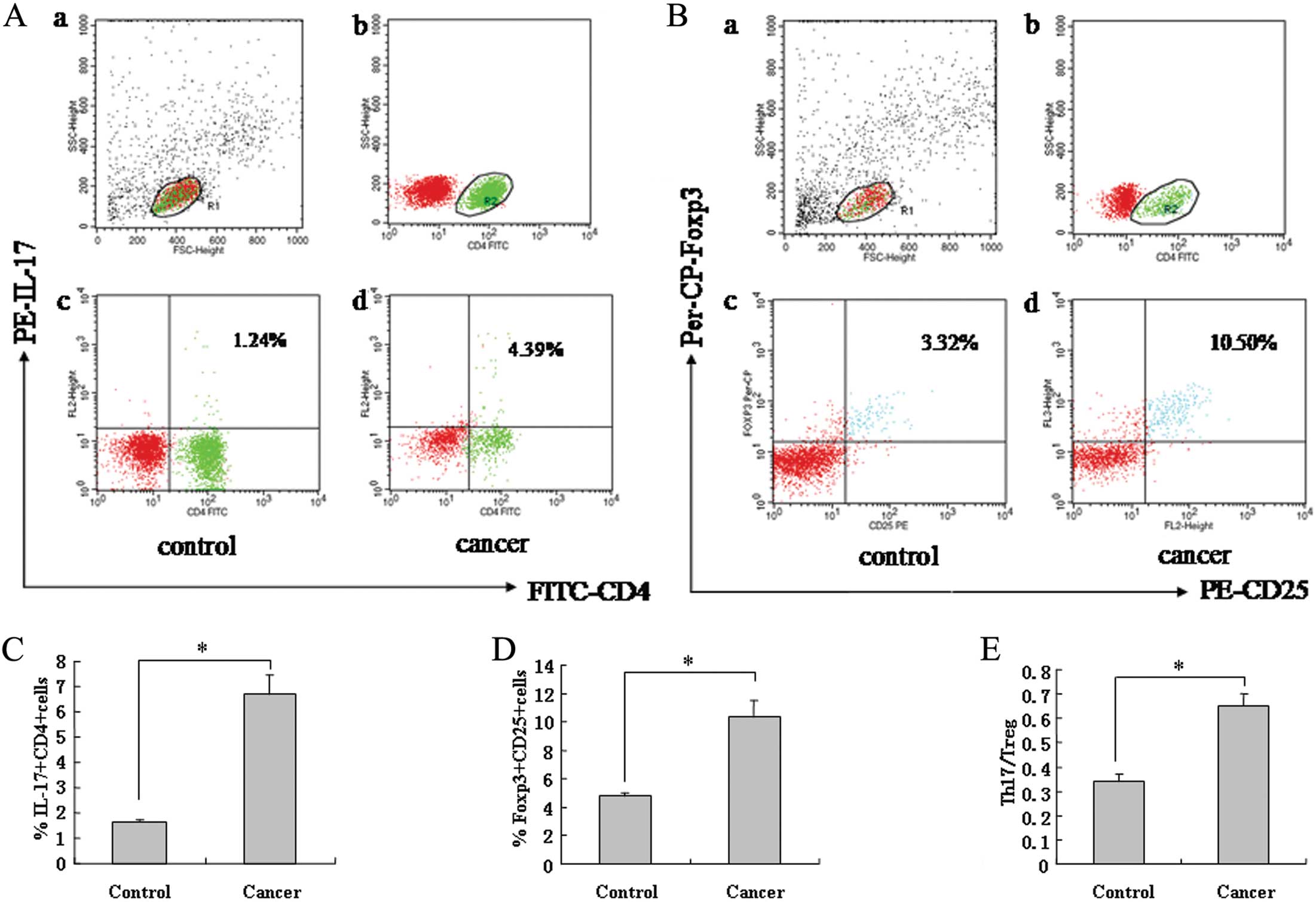
Increased frequency of circulating Th17
in gastric cancer patients
PBMCs in patients with gastric cancer and in healthy
donors were examined for the frequency of Th17 cells. The
population of Th17 cells as a percentage of total CD4+
cells was evaluated by flow cytometric analysis. Representative
dotplots are shown in Fig. 1A.
Summarized data from all individuals indicated that the percentage
of Th17 cells was significantly increased in patients with gastric
cancer when compared to healthy donors (Fig. 1C).
We next sought to determine whether this Th17
skewing was associated with clinicopathologic status including
gender, age, TNM stage, differentiation, metastatic lymph nodes,
tumor factors, invasive depth, blood vessel invasion, intravascular
cancer embolus and gastric cancer-related tumor antigens in gastric
cancer patients. As shown in Table
II, specifically, the frequency of Th17 cells was positively
correlated with TNM stage and lymph node metastases. There was no
association between Th17 frequency and other studied factors.
| Table IIAssociation between Th17, Treg cells
or Th17/Treg in peripheral blood and clinicopathologic factors of
gastric cancer patients. |
Table II
Association between Th17, Treg cells
or Th17/Treg in peripheral blood and clinicopathologic factors of
gastric cancer patients.
| | | P-value |
|---|
| | |
|
|---|
| Clinical
characteristic | Evaluation | N=45 | Th17 | Treg | Th17/Treg |
|---|
| Gender | | | 0.754 | 0.674 | 0.917 |
| Male | 0 | 36 | | | |
| Female | 1 | 9 | | | |
| Age, years | | | 0.963 | 0.884 | 0.890 |
| <60 | 0 | 24 | | | |
| ≥60 | 1 | 21 | | | |
| TNM stage | | | 0.002a | 0.034a | 0.110 |
| I | 1 | 15 | | | |
| II | 2 | 9 | | | |
| III | 3 | 9 | | | |
| IV | 4 | 12 | | | |
|
Differentiation | | | 0.176 | 0.015a | 0.165 |
| Well | 0 | 14 | | | |
| Poor | 1 | 31 | | | |
| Metastatic lymph
node | | | 0.017a | 0.042 | 0.036a |
| Yes | 1 | 18 | | | |
| No | 0 | 27 | | | |
| Size of tumor
(cm) | | | 0.262 | 0.279 | 0.476 |
| <4 | 0 | 19 | | | |
| ≥4 | 1 | 26 | | | |
| Invasive depth
(mm) | | | 0.507 | 0.572 | 0.291 |
| <15 | 0 | 17 | | | |
| ≥15 | 1 | 28 | | | |
| Blood vessel
invasion | | | 0.077 | 0.134 | 0.237 |
| Yes | 1 | 11 | | | |
| No | 0 | 34 | | | |
| Intravascular
cancer embolus | | | 0.508 | 0.519 | 0.623 |
| Yes | 1 | 9 | | | |
| No | 0 | 36 | | | |
| Gastric
cancer-related tumor antigens |
| CA50 (0.21–10
IU/ml) | | | 0.903 | 0.972 | 0.431 |
| <10 | 0 | 37 | | | |
| ≥10 | 1 | 8 | | | |
| CEA (0–5
ng/ml) | | | 0.321 | 0.465 | 0.936 |
| <5 | 0 | 40 | | | |
| ≥5 | 1 | 5 | | | |
| CA199 (0–27
U/ml) | | | 0.570 | 0.917 | 0.274 |
| <27 | 0 | 29 | | | |
| ≥27 | 1 | 6 | | | |
| CA724 (0–6.9
U/ml) | | | 0.309 | 0.274 | 0.268 |
| <6.9 | 0 | 31 | | | |
| ≥6.9 | 1 | 15 | | | |
Increased frequency of circulating Treg
cells in gastric cancer patients
To analyze the prevalence of Treg,
CD4+CD25+FoxP3+ cells were
examined by flow cytometry and expressed as a percentage of the
total CD4+ cells. Representative dotplots are shown in
Fig. 1B. Summarized data from all
individuals showed that the frequency of Treg cells in PBMCs of
gastric cancer patients was significantly higher than that in
healthy donors (Fig. 1D). The
association between expression of Treg and clinical/pathologic
parameters showed that the frequency of Treg cells was positively
correlated with TNM stage and tumor differentiation, and there was
no association between the frequency of Treg and other studied
factors (Table II).
Increased proportion of Th17/Treg cells
in gastric cancer patients
We also observed the proportion of Th17/Treg in
gastric cancer patients. As shown in Fig. 1E, the proportion of Th17/Treg cells
was significantly increased in gastric cancer patients when
compared to healthy donors. In addition, compared with the gastric
cancer patients without lymph node metastasis, a significantly
increased ratio of Th17/Treg was found in patients with lymph node
metastasis. There was no statistical correlation between the
Th17/Treg ratio and other clinical/pathologic parameters (Table II).
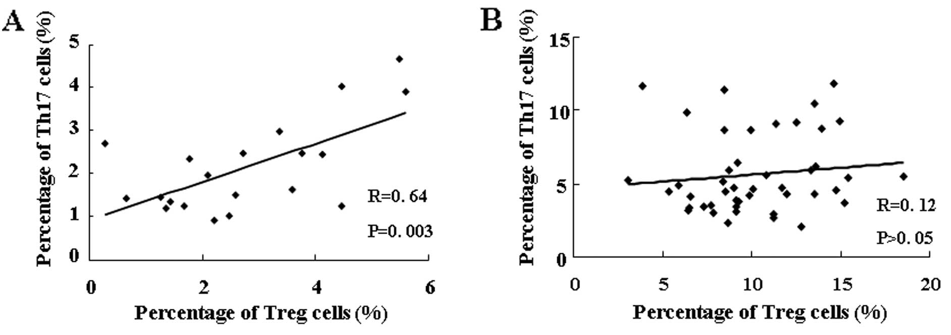
Correlation between Th17 and Treg
cells
Next, we evaluated the relationship between Th17 and
Treg cells in gastric cancer patients and in healthy donors. As
shown in Fig. 2, the frequencies of
Th17 and Treg cells were positively correlated in the healthy
donors, but not correlated in the gastric cancer patients.
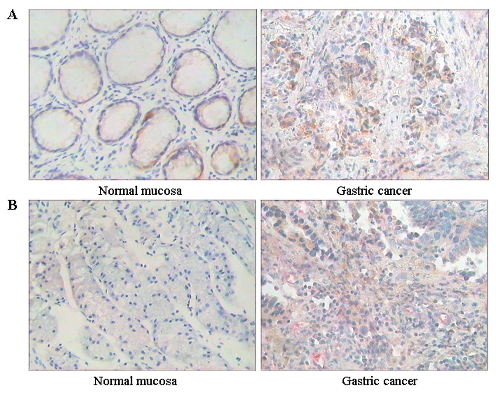
Distribution of Th17 and Treg cells in
gastric cancer tissue
To further evaluate the frequency of Th17 and Treg
cells in gastric cancer tissue, double staining
immunohistochemistry (CD4 and IL-17 for Th17, or CD4 and Foxp3 for
Treg) was performed. Notably, representative data revealed that
CD4+IL-17+ or
CD4+FoxP3+ cells were frequently observed in
gastric cancer in comparison to the controls (Fig. 3).
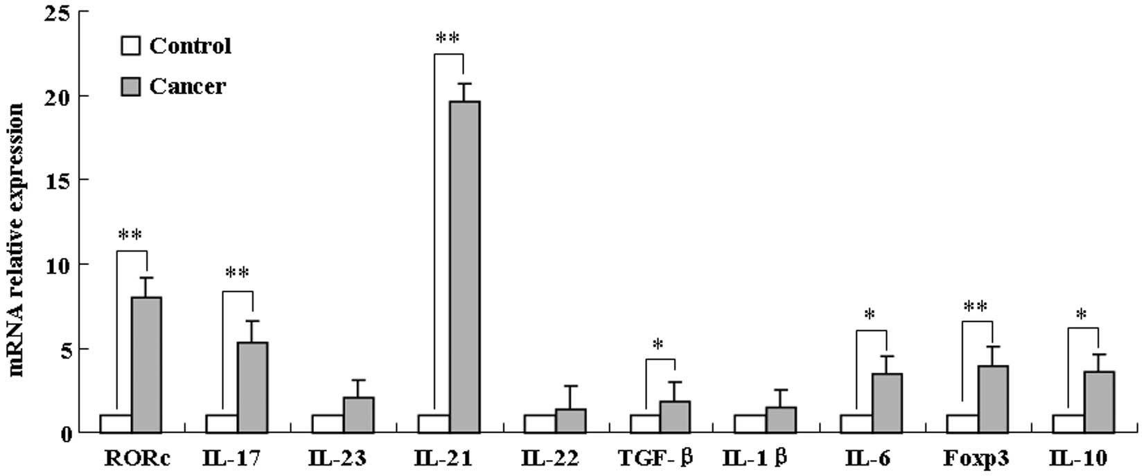
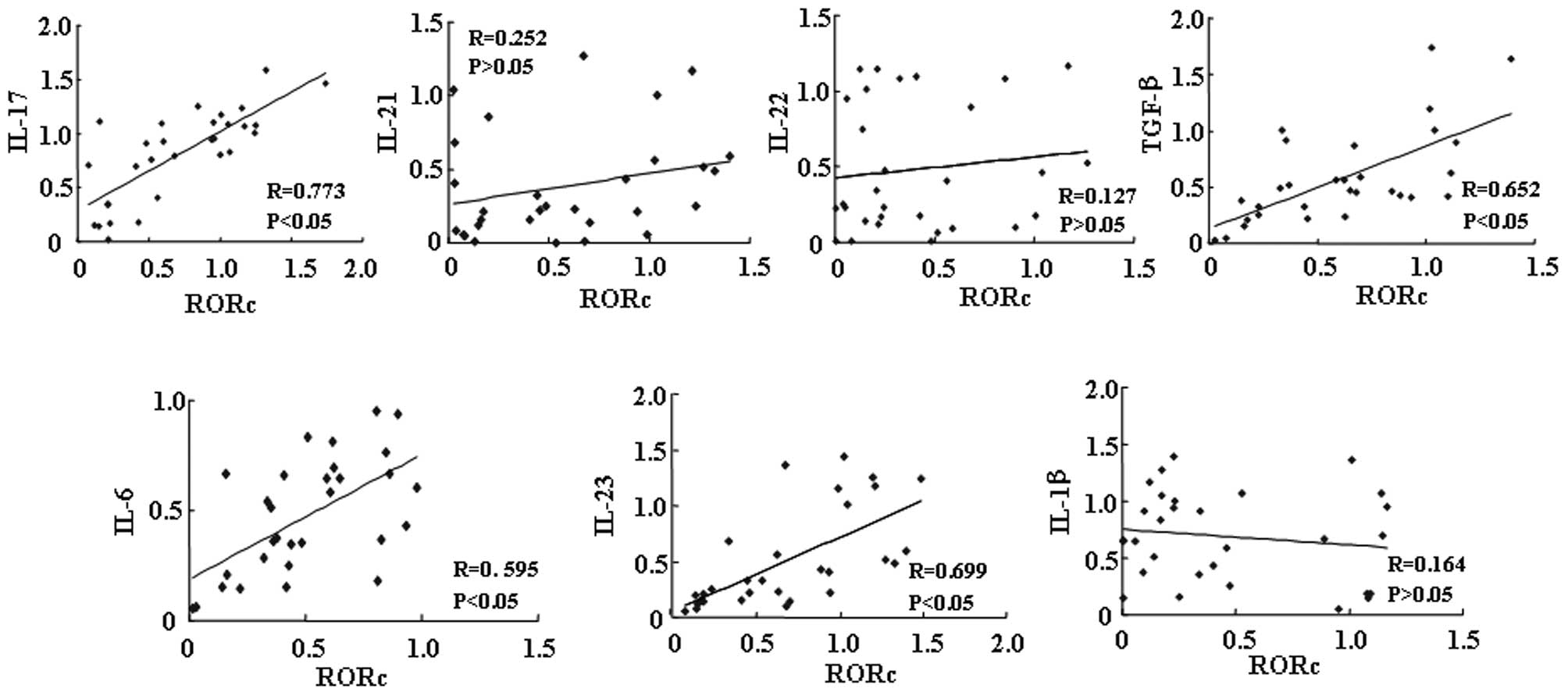
mRNA expression of RORc, IL-17, IL-23,
IL-21, IL-22, TGF-β, IL-1β, IL-6, Foxp3 and IL-10 in gastric cancer
tissue
To further analyze the mechanism of Th17 and Treg
accumulation in gastric cancer tissue or the relationship between
the immune-imbalance of Th17/Treg and their differentiation or
function in gastric cancer tissues, we also examined the mRNA
expression levels of RORc, IL-17, IL-23, IL-21, IL-22, TGF-β,
IL-1β, IL-6, Foxp3 and IL-10 in gastric cancer tissue by real-time
RT-PCR. As shown in Fig. 4, mRNA
expression levels of RORc, IL-17, IL-21, TGF-β, IL-6, Foxp3 and
IL-10 in gastric cancer were significantly increased in comparison
to the controls. The correlation between RORc and cytokines showed
that mRNA expression of RORc did correlate with mRNA expression of
IL-17, IL-6, TGF-β and IL-23, but did not correlate with mRNA
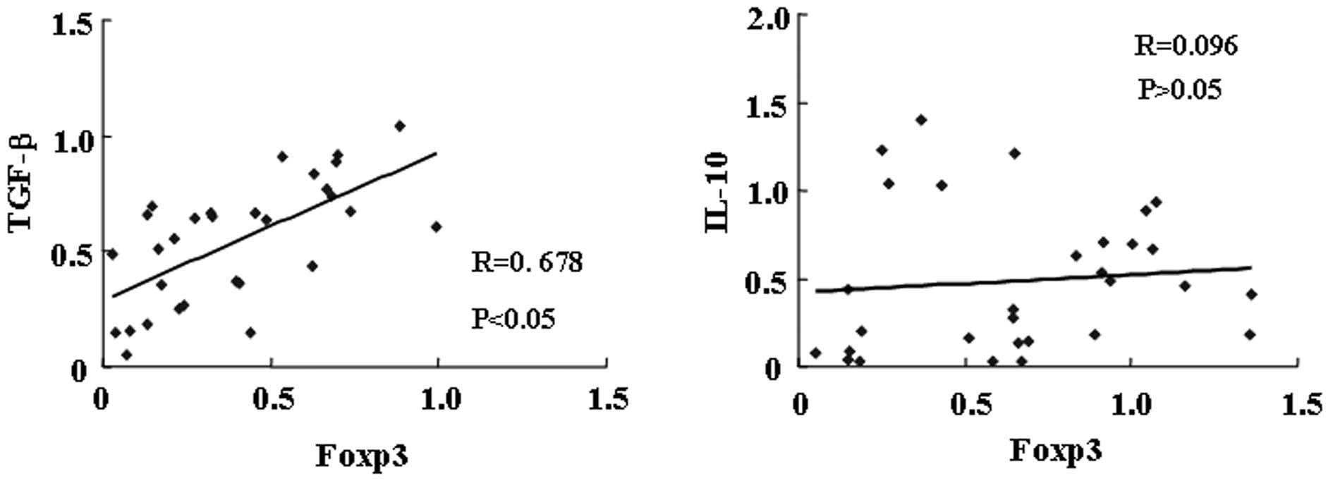
expression of IL-21, IL-22 and IL-1β (Fig. 5). The correlation between Foxp3 and
TGF-β or IL-10 showed that mRNA expression of Foxp3 did correlate
with mRNA expression of TGF-β, but did not correlate with mRNA
expression of IL-10 (Fig. 6).
Discussion
Th17 and Treg cells are two newly identified
important subsets of T helper cells. The Th17/Treg balance controls
autoimmunity and inflammation and has been found to play an
important role in the pathogenesis of tumor disease. To assess
whether this balance is disturbed in gastric cancer patients, we
determined the distribution of Th17 and Treg cells in gastric
cancer patients, and evaluated how the imbalance of Th17/Treg cells
correlates with clinical and pathological parameters.
There have been several reports describing Th17
cells in both murine and human tumors, but the nature and role of
Th17 cells in cancer immunity remains elusive. Previous
investigations demonstrated that the levels of Th17 cells were
significantly increased in peripheral blood, malignant ascites
fluid and tumor tissues in human ovarian, renal and pancreatic
malignancies (10,11,17,18).
In the present study, we reconfirmed an increased number of Th17
cells in the PBMCs of patients with gastric cancer. Moreover, an
increased population of Th17 cells was associated with TNM stage
and lymph node metastases. Compared with patients with early stage
disease, patients with advanced disease showed a significantly
higher percentage of Th17 cells. Compared with patients without
lymph node metastases, patients with lymph node metastases
exhibited a significantly higher percentage of Th17 cells. These
results are consistent with the findings of Zhang et
al(9), which indicated that
Th17 cells may contribute to the pathogenesis of gastric
cancer.
Zhang et al also demonstrated that the levels
of Th17 and Treg cells were higher in hepatocellular carcinoma than
in the corresponding non-tumor tissues. Our immunohistochemical
staining results showed that CD4/IL-17 double stained lymphocytes
were more frequently observed in gastric cancer tissues than in
their corresponding non-tumor tissues, which was consistent with
our flow cytometric results that the frequency of Th17 cells was
increased in PBMCs.
Since their discovery, Treg cells have gained much
attention in tumor immunity, due to their strong immune suppressive
activity on T cell responses. Accumulating evidence demonstrates
that the levels of Foxp3+ Treg cells are increased in
cancer patients, and the high number of Treg cells is associated
with poor survival (14,15,19–22).
In addition, research regarding Treg cells in ovarian and prostate
cancer has demonstrated that Treg cells exist in a markedly higher
proportion in PBMCs, and that a significant difference in the
prevalence of Treg cells between the early and advanced disease
stages was found (11,23). Consistently, our data showed that
the frequencies of Treg cells in PBMCs were prominently increased
in patients with gastric cancer. Moreover, the increased number of
Treg cells was associated with TNM stage and the degree of tumor
differentiation. Lymphocyte migration is modulated by chemokine
receptor interaction. Previous studies have reported that the
majority of tumor-associated Treg cells express lymphoid homing
receptor CD62L, CCR4 and CCR6, which induce a gradual increase in
Treg cells in tumors during disease progression (23,24).
This notion was supported by our study that the percentage of Treg
cells was obviously increased in gastric cancer tissue. In
addition, we also found that the frequency of Treg cells was
positively correlated with the degree of tumor differentiation.
These results suggest that suppression of immunity by Treg cells
may be an impediment to gastric cancer therapy.
Tumor progression has been considered to be the
result of an interaction between different cell types. According to
the reciprocal differentiation pathways of Th17 and Treg cells and
the regulatory action, Th17/Treg subsets may be involved in
immunomodulation, similar to the paradigm of Th1/Th2 subsets, and
the imbalance in Th17/Treg cells may lead to tumor progression.
This notion was supported by Zhang’s et al results (9) and our finding that the proportion of
Th17/Treg was significantly increased in gastric cancer patients
when compared to healthy donors. In addition, the frequencies of
Th17 and Treg cells were well correlated in the healthy donors, but
not in the gastric cancer patients, which may reflect that an
immune balance exists in healthy individuals but has been disrupted
in gastric cancer patients. Moreover, compared with patients
without lymph node metastases, an obviously increased ratio of
Th17/Treg cells was found in gastric cancer patients with lymph
node metastases. These data suggest that the immune imbalance
between Th17 and Treg cells may potentially play an important role
in the development and progression of gastric cancer.
To further analyze the mechanism of Th17 and Treg
accumulation in gastric cancer tissue or the relationship between
the immune-imbalance of Th17/Treg and their differentiation or
function in gastric cancer tissues, we examined the mRNA expression
levels of Th17- and Treg-related cytokines. Our results
demonstrated that the mRNA expression level of Th17-specific
transcript factor RORc was significantly elevated in gastric cancer
tissues, which was consistent with the elevation of Th17 in
peripheral blood. Moreover, the mRNA expression level of RORc was
positively correlated with that of IL-17, further elevated
prevalence of Th17 cells was associated with higher TNM stages. We,
therefore, conclude that increased expression of IL-17 in gastric
cancer tissue, most likely, originates from Th17 cells, and Th17
cells may promote tumor progression by promoting inflammation
through secretion of IL-17.
Recently, TGF-β, IL-1β and IL-6 have been recognized
as the most important cytokines for the initiation of Th17 cell
differentiation, whereas IL-23, originally thought to be the master
regulator, appears to be important for expansion and/or maintenance
of the Th17 response (25–30). In the present study, we found that
mRNA expression of both TGF-β and IL-6 was much higher in gastric
cancer tissues than in non-cancerous adjacent tissues. Importantly,
in tumor tissues, both TGF-β and IL-6 mRNA expression levels showed
a strong correlation with the RORc mRNA expression level. We also
examined IL-1β and IL-23, but we did not find a correlation between
them and RORc. These results suggest that TGF-β and IL-6 present in
the gastric cancer microenvironment may promote differentiation and
expansion of Th17 cells. Our results are consistent with data from
Manel et al(7) and Yang
et al(31).
Foxp3 is the characteristic transcription factor for
Treg cells. At the mRNA and protein levels, CD4+ T cells
expressing Foxp3 in the periphery are mainly
CD4+CD25+ Treg cells. Treg cells suppress
immune responses by a contact-dependent manner or modulate the
immune balance through the release of cytokines IL-10 and TGF-β.
The present study showed that mRNA expression of FoxP3, IL-10 and
TGF-β was much higher in gastric cancer tissues than in
non-cancerous adjacent tissues and that the high mRNA expression
levels of Foxp3 and TGF-β were associated with higher TNM stage.
Our data were consistent with the results from several studies
demonstrating that Treg cells are increased in blood, TDLN and
cancer specimens from patients with gastric cancer (32,33).
Previous studies have shown that Th17 and Treg cells are
synchronistically increased following tumor development, and both
populations reached maximal levels in advanced tumor (2,9,11,34,35).
Our data further reconfirmed this finding. Therefore, the dynamic
interaction between Th17 and Treg cells may be important in the
development of gastric cancer.
In conclusion, the accumulation of Th17 and Treg
cells in the gastric cancer tumor microenvironment was gradually
increased according to disease progression, leading to an imbalance
in Th17/Treg cells in gastric cancer patients. TGF-β and IL-6
present in the gastric cancer microenvironment promoted the
differentiation and expansion of Th17 cells, and increased Th17
cells promoted tumor progression through promotion of inflammation
by secretion of IL-17. Treg cells promoted tumor progression by
helping cancer cells escape from host immunosurveillance by
secreting TGF-β, and a high level of TGF-β in the tumor
microenvironment promoted differentiation and expansion of Treg
cells. A better understanding of the nature, regulation, and
function of Th17 and Treg cells in tumor immunity may aid in the
development of novel and effective immunotherapy for gastric
cancer.
Acknowledgements
The present study was supported by grants from the
Natural Science Foundation of Hebei Province (no. C2011206086); Key
Science and Technology Project of the Health Department of Hebei
Province (no. 20110456).
References
|
1
|
Maruyama T, Kono K, Mizukami Y, Kawaguchi
Y, Mimura K, Watanabe M, Izawa S and Fujii H: Distribution of Th17
cells and FoxP3(+) regulatory T cells in tumor-infiltrating
lymphocytes, tumor-draining lymph nodes and peripheral blood
lymphocytes in patients with gastric cancer. Cancer Sci.
101:1947–1954. 2010.
|
|
2
|
Zhang Y, Ma D, Zhang Y, Tian Y, Wang X,
Qiao Y and Cui B: The imbalance of Th17/Treg in patients with
uterine cervical cancer. Clin Chim Acta. 412:894–900. 2011.
View Article : Google Scholar : PubMed/NCBI
|
|
3
|
Korn T, Bettelli E, Oukka M and Kuchroo
VK: IL-17 and Th17 cells. Annu Rev Immunol. 27:485–517. 2009.
View Article : Google Scholar : PubMed/NCBI
|
|
4
|
Dong C: Diversification of T-helper-cell
lineages: finding the family root of IL-17-producing cells. Nat Rev
Immunol. 6:329–333. 2006. View
Article : Google Scholar : PubMed/NCBI
|
|
5
|
Ivanov II, McKenzie BS, Zhou L, Tadokoro
CE, Lepelley A, Lafaille JJ, Cua DJ and Littman DR: The orphan
nuclear receptor RORγt directs the differentiation program of
proinflammatory IL-17+ T helper cells. Cell.
126:1121–1133. 2006.
|
|
6
|
McGeachy MJ and Cua DJ: Th17 cell
differentiation: the long and winding road. Immunity. 28:445–453.
2008. View Article : Google Scholar : PubMed/NCBI
|
|
7
|
Manel N, Unutmaz D and Littman DR: The
differentiation of human T(H)-17 cells requires transforming growth
factor-beta and induction of the nuclear receptor RORγt. Nat
Immunol. 9:641–649. 2008.PubMed/NCBI
|
|
8
|
Volpe E, Servant N, Zollinger R, Bogiatzi
SI, Hupé P, Barillot E and Soumelis V: A critical function for
transforming growth factor-beta, interleukin 23 and proinflammatory
cytokines in driving and modulating human T(H)-17 responses. Nat
Immunol. 9:650–657. 2008. View
Article : Google Scholar : PubMed/NCBI
|
|
9
|
Zhang B, Rong G, Wei H, Zhang M, Bi J, Ma
L, Xue X, Wei G, Liu X and Fang G: The prevalence of Th17 cells in
patients with gastric cancer. Biochem Biophys Res Commun.
374:533–537. 2008. View Article : Google Scholar : PubMed/NCBI
|
|
10
|
Sfanos KS, Bruno TC, Maris CH, Xu L,
Thoburn CJ, DeMarzo AM, Meeker AK, Isaacs WB and Drake CG:
Phenotypic analysis of prostate-infiltrating lymphocytes reveals
TH17 and Treg skewing. Clin Cancer Res. 14:3254–3261. 2008.
View Article : Google Scholar : PubMed/NCBI
|
|
11
|
Kryczek I, Wei S, Zou L, Altuwaijri S,
Szeliga W, Kolls J, Chang A and Zou W: Cutting edge: Th17 and
regulatory T cell dynamics and the regulation by IL-2 in the tumor
microenvironment. J Immunol. 178:6730–6733. 2007. View Article : Google Scholar : PubMed/NCBI
|
|
12
|
Su X, Ye J, Hsueh EC, Zhang Y, Hoft DF and
Peng G: Tumor microenvironments direct the recruitment and
expansion of human Th17 cells. J Immunol. 184:1630–1641. 2010.
View Article : Google Scholar : PubMed/NCBI
|
|
13
|
Wang RF: Regulatory T cells and innate
immune regulation in tumor immunity. Springer Semin Immunopathol.
28:17–23. 2006. View Article : Google Scholar : PubMed/NCBI
|
|
14
|
Nakamura T, Shima T, Saeki A, Hidaka T,
Nakashima A, Takikawa O and Saito S: Expression of indoleamine
2,3-dioxygenase and the recruitment of Foxp3-expressing regulatory
T cells in the development and progression of uterine cervical
cancer. Cancer Sci. 98:874–881. 2007. View Article : Google Scholar : PubMed/NCBI
|
|
15
|
Wolf D, Wolf AM, Rumpold H, Fiegl H,
Zeimet AG, Muller-Holzner E, Deibl M, Gastl G, Gunsilius E and
Marth C: The expression of the regulatory T cell-specific forkhead
box transcription factor FoxP3 is associated with poor prognosis in
ovarian cancer. Clin Cancer Res. 11:8326–8331. 2005. View Article : Google Scholar : PubMed/NCBI
|
|
16
|
Vernal R and Garcia-Sanz JA: Th17 and Treg
cells, two new lymphocyte subpopulations with a key role in the
immune response against infection. Infect Disord Drug Targets.
8:207–220. 2008. View Article : Google Scholar : PubMed/NCBI
|
|
17
|
Kryczek I, Banerjee M, Cheng P, et al:
Phenotype, distribution, generation, and functional and clinical
relevance of Th17 cells in the human tumor environments. Blood.
114:1141–1149. 2009. View Article : Google Scholar : PubMed/NCBI
|
|
18
|
Miyahara Y, Odunsi K, Chen W, Peng G,
Matsuzaki J and Wang RF: Generation and regulation of human
CD4+ IL-17-producing T cells in ovarian cancer. Proc
Natl Acad Sci USA. 105:15505–15510. 2008. View Article : Google Scholar : PubMed/NCBI
|
|
19
|
Beyer M and Schultze JL: Regulatory T
cells in cancer. Blood. 108:804–811. 2006. View Article : Google Scholar : PubMed/NCBI
|
|
20
|
Byrne WL, Mills KH, Lederer JA and
O’Sullivan GC: Targeting regulatory T cells in cancer. Cancer Res.
71:6915–6920. 2011. View Article : Google Scholar : PubMed/NCBI
|
|
21
|
Shen S, Liang L, Peng BG, He Q, Kuang M
and Lai JM: Foxp3+ regulatory T cells and the formation
of portal vein tumor thrombus in patients with hepatocellular
carcinoma. Can J Surg. 54:89–94. 2011.
|
|
22
|
Xu L, Xu W, Qiu S and Xiong S: Enrichment
of CCR6+Foxp3+ regulatory T cells in the
tumor mass correlates with impaired CD8+ T cell function
and poor prognosis of breast cancer. Clin Immunol. 135:466–475.
2010.PubMed/NCBI
|
|
23
|
Miller AM, Lundberg K, Ozenci V, Banham
AH, Hellström M, Egevad L and Pisa P:
CD4+CD25high T cells are enriched in the
tumor and peripheral blood of prostate cancer patients. J Immunol.
177:7398–7405. 2006.
|
|
24
|
Curiel TJ, Coukos G, Zou L, et al:
Specific recruitment of regulatory T cells in ovarian carcinoma
fosters immune privilege and predicts reduced survival. Nat Med.
10:942–949. 2004. View
Article : Google Scholar : PubMed/NCBI
|
|
25
|
Acosta-Rodriguez EV, Napolitani G,
Lanzavecchia A and Sallusto F: Interleukins 1beta and 6 but not
transforming growth factor-beta are essential for the
differentiation of interleukin 17-producing human T helper cells.
Nat Immunol. 8:942–949. 2007. View
Article : Google Scholar : PubMed/NCBI
|
|
26
|
Wilson NJ, Boniface K, Chan JR, et al:
Development, cytokine profile and function of human interleukin
17-producing helper T cells. Nat Immunol. 8:950–957. 2007.
View Article : Google Scholar : PubMed/NCBI
|
|
27
|
Bettelli E, Carrier Y, Gao W, Korn T,
Strom TB, Oukka M, Weiner HL and Kuchroo VK: Reciprocal
developmental pathways for the generation of pathogenic effector
TH17 and regulatory T cells. Nature. 441:235–238. 2006. View Article : Google Scholar
|
|
28
|
Mangan PR, Harrington LE, O’Quinn DB,
Helms WS, Bullard DC, Elson CO, Hatton RD, Wahl SM, Schoeb TR and
Weaver CT: Transforming growth factor-beta induces development of
the T(H)17 lineage. Nature. 441:231–234. 2006. View Article : Google Scholar
|
|
29
|
Veldhoen M, Hocking RJ, Atkins CJ,
Locksley RM and Stockinger B: TGFbeta in the context of an
inflammatory cytokine milieu supports de novo differentiation of
IL-17-producing T cells. Immunity. 24:179–189. 2006. View Article : Google Scholar
|
|
30
|
Stritesky GL, Yeh N and Kaplan MH: IL-23
promotes maintenance but not commitment to the Th17 lineage. J
Immunol. 181:5948–5955. 2008. View Article : Google Scholar : PubMed/NCBI
|
|
31
|
Yang L, Anderson DE, Baecher-Allan C,
Hastings WD, Bettelli E, Oukka M, Kuchroo VK and Hafler DA: IL-21
and TGF-beta are required for differentiation of human T(H)17
cells. Nature. 454:350–352. 2008. View Article : Google Scholar : PubMed/NCBI
|
|
32
|
Ichihara F, Kono K, Takahashi A, Kawaida
H, Sugai H and Fujii H: Increased populations of regulatory T cells
in peripheral blood and tumor-infiltrating lymphocytes in patient
with gastric and esophageal cancers. Clin Cancer Res. 9:4404–4408.
2003.PubMed/NCBI
|
|
33
|
Sasada T, Kimura M, Yoshida Y, Kanai M and
Takabayashi A: CD4+CD25+ regulatory T cells
in patients with gastrointestinal malignancies: possible
involvement of regulatory T cells in disease progression. Cancer.
98:1089–1099. 2003.
|
|
34
|
Bouchliou I, Miltiades P, Nakou E, et al:
Th17 and Foxp3(+) T regulatory cell dynamics and distribution in
myelodysplastic syndromes. Clin Immunol. 139:350–359. 2011.
|
|
35
|
Hou F, Li Z, Ma D, Zhang W, Zhang Y, Zhang
T, Kong B and Cui B: Distribution of Th17 cells and
Foxp3-expressing T cells in tumor-infiltrating lymphocytes in
patients with uterine cervical cancer. Clin Chim Acta.
413:1848–1854. 2012. View Article : Google Scholar : PubMed/NCBI
|