Introduction
Although the prognosis of gastric cancer has
improved with the development of early diagnosis and new
therapeutic strategies, it remains one of the main causes of
cancer-related mortality worldwide (1), with peritoneal carcinomatosis, often
associated with malignant ascites, being the most frequent cause of
death in patients with advanced gastric cancer. Peritoneal
dissemination is considered to arise from free cancer cells in the
peritoneal cavity exfoliated from the serosal surface of the
stomach penetrated by the primary tumor (2). Therefore, cytologic examination of
peritoneal washes has been performed at laparotomy to detect free
cancer cells in patients with advanced gastric cancer, and it is
recognized as one of the most important prognostic factors
(3–7). Since 1999, it has been reported that
the presence of free cancer cells in the peritoneal cavity should
be considered as an independent prognostic factor in patients with
gastric cancer by the Japanese Gastric Cancer Association.
Moreover, in the International Union Against Cancer’s TNM
classification 7th edition for gastric cancer, positive peritoneal
cytology is defined as stage IV.
Therefore, detection of free cancer cells in
peritoneal washes is a standard method for the assessment of risk
for peritoneal carcinomatosis. However, patients who are diagnosed
as having no free cancer cells in the peritoneal cavity by
conventional cytology sometimes develop peritoneal recurrence after
curative resection. This may occur since cytology is considered to
have low sensitivity. In fact, recently, real-time RT-PCR
techniques have been developed in order to increase the sensitivity
of conventional peritoneal lavage cytology (8,9).
Concerning another cause for the development of recurrent
peritoneal disease, the cytokine network may play an important role
in the immunosuppressive and immunostimulatory properties of
cancer-related ascites fluid (10,11).
However, the role of cytokines produced by cells in the peritoneal
cavity on tumor growth in gastric cancer patients is still
unclear.
Th17 cells have recently been identified as having a
distinct Th cell lineage and were found in an experimental animal
model of cancer and in human cancers. Th17 cells have been found in
several types of human cancers, such as ovarian, prostate,
colorectal, and other malignancies, as well as gastric cancer
(12–17). Research has found that IL-17
promotes tumor growth through angiogenesis and inflammation
(18), and several other studies
have demonstrated that IL-17 contributes to reduced tumor growth by
promoting dendritic cell, cytotoxic T cell (CTL), and NK cell
trafficking to, and retention within, the tumor microenvironment
(19,20). Therefore, whether these cells
promote tumor growth or regulate antitumor responses remains
controversial. In the case of human gastric cancer, there are
several reports concerning the prevalence of Th17 cells in the
tumor microenvironment, tumor-draining lymph nodes and peripheral
blood (12,13). In our previous study, the expression
level of IL-17 mRNA in gastric tumors was associated with the depth
of tumor invasion, lymphovascular invasion and lymph node
involvement suggesting that IL-17 is clearly associated with tumor
progression (21). However, since
no previous studies have been carried out concerning the proportion
of IL-17 in the peritoneal cavity in human gastric cancer patients,
we hypothesized that the expression level of IL-17 mRNA in
peritoneal lavage may be involved in the development of peritoneal
carcinomatosis in gastric cancer.
In the present study, we quantitatively investigated
expression of IL-17 messenger RNA (mRNA) in the peritoneal lavage
of gastric cancer patients who underwent curative resection. The
association of IL-17 expression levels with clinicopathological
factors and prognosis was also assessed. Since IL-2 and IL-12 have
been used for intraperitoneal immunotherapy in patients with
various types of cancers, including gastric cancer (22–25),
the possibility of IL-17 as a therapeutic target for patients with
gastric cancer was also investigated.
Materials and methods
Patients
Included in the present study was a series of 114
patients (80 men and 34 women) with gastric cancer who underwent
surgery at Wakayama Medical University Hospital (WMUH) from 2003 to
2006. At the beginning of the operation, we examined tumor
metastases in the abdominal cavity. When it was thought that
curative resection was possible, we performed gastrectomy with
lymphadenectomy. We performed gastrectomy in 114 patients with
gastric cancer. Seventy-nine patients underwent surgically curative
resection and 35 underwent non-curative resection. None of the
patients received anticancer therapy prior to surgery. Individuals
with autoimmune disease, inflammatory bowel disease or viral
infections were excluded. The clinicopathological characteristics
of the 114 patients are summarized in Table IA. Clinical stages of the tumors
were determined according to the International Union Against
Cancer’s TNM classification for gastric cancer. After surgery, all
patients underwent a follow-up, with the median follow-up at
analysis being 61 months (range, 1.3–98.5) for all patients. Every
3 to 6 months, physical examination, blood chemistry, including
carcinoembryonic antigen (CEA) and cancer antigen (CA) 19-9, and
computed tomography were performed for each patients. Written
informed consent was obtained from all patients before
participation in the present study. In addition, the local ethics
committee of WMUH approved the study.
 | Table IClinicopathological data and IL-17
mRNA expression of the 114 patients and the 79 patients who
underwent curative resection. |
Table I
Clinicopathological data and IL-17
mRNA expression of the 114 patients and the 79 patients who
underwent curative resection.
| A, Data of the 114
patients |
|---|
|
|---|
| Factor | No. of patients | Expression of IL-17
mRNAa | P-value |
|---|
| Age (years) |
| ≤65 | 50 | 2.11±0.648 | 0.448 |
| >65 | 64 | 1.57±0.399 | |
| Gender |
| Male | 80 | 1.75±0.415 | 0.718 |
| Female | 34 | 1.93±0.724 | |
| Depth of tumor
invasion |
| T1 | 30 | 1.00±0.523 | 0.023c |
| T2 | 13 | 1.49±0.130 | |
| T3 | 31 | 2.44±0.809 | |
| T4 | 40 | 2.51±0.582 | |
| Lymph node
metastasis |
| N0 | 43 | 2.25±0.681 | 0.477 |
| N1 | 28 | 1.26±0.581 | |
| N2 | 21 | 1.61±0.708 | |
| N3 | 22 | 1.84±0.894 | |
| Histological
type |
|
Differentiated | 54 | 2.08±0.601 | 0.387 |
|
Undifferentiated | 60 | 1.55±0.423 | |
| Lymphatic
invasion |
| Negative | 37 | 1.90±0.659 | 0.338 |
| Positive | 77 | 1.76±0.432 | |
| Vessel
invasion |
| Negative | 57 | 1.83±0.544 | 0.292 |
| Positive | 57 | 1.78±0.479 | |
| Peritoneal
metastasis |
| Negative | 100 | 2.07±0.439 | 0.031d |
| Positive | 14 | 7.21±0.290 | |
| Cytologic
examination |
| Negative | 91 | 1.22±0.591 | 0.801 |
| Positive | 23 | 1.89±0.404 | |
| Stageb |
| I | 40 | 1.95±0.660 | 0.412 |
| II | 18 | 2.29±0.109 | |
| III | 21 | 1.38±0.784 | |
| IV | 33 | 1.64±0.531 | |
| Tumor size
(cm) |
| ≤5 | 60 | 1.75±0.516 | 0.059 |
| >5 | 54 | 1.87±0.506 | |
| B, Data of the 79
patients who underwent curative resection |
|---|
|
|---|
| Factor | No. of
patients | Expression of IL-17
mRNAa | P-value |
|---|
| Age (years) |
| ≤65 | 35 | 2.01±0.781 | 0.329 |
| >65 | 44 | 1.72±0.527 | |
| Gender |
| Male | 58 | 1.80±0.517 | 0.975 |
| Female | 21 | 1.93±0.724 | |
| Depth of tumor
invasion |
| T1 | 30 | 1.00±0.523 | 0.020c |
| T2 | 13 | 1.49±0.130 | |
| T3 | 23 | 2.39±0.927 | |
| T4 | 13 | 2.90±0.124 | |
| Lymph node
metastasis |
| N0 | 43 | 2.25±0.681 | 0.307 |
| N1 | 20 | 1.48±0.700 | |
| N2 | 10 | 1.90±0.106 | |
| N3 | 6 | 3.26±0.284 | |
| Histological
type |
|
Differentiated | 41 | 2.31±0.719 | 0.613 |
|
Undifferentiated | 38 | 1.33±0.524 | |
| Lymphatic
invasion |
| Negative | 34 | 1.72±0.690 | 0.118 |
| Positive | 45 | 1.95±0.610 | |
| Vessel
invasion |
| Negative | 49 | 1.72±0.597 | 0.087 |
| Positive | 30 | 2.05±0.708 | |
| Stageb |
| I | 40 | 1.95±0.660 | 0.497 |
| II | 18 | 2.29±0.109 | |
| III | 21 | 1.44±0.819 | |
| Tumor size
(cm) |
| ≤5 | 54 | 1.62±0.509 | 0.097 |
| >5 | 25 | 2.36±0.943 | |
Preoperative peritoneal wash
examination
At the beginning of each operation, 100 ml saline
was introduced into the Douglas cavity and aspirated by gentle
stirring. These washes were centrifuged at 1,800 rpm for 5 min to
collect intact cells. A part of each peritoneal wash was examined
cytopathologically after conventional Papanicolaou staining.
RNA extraction and DNA synthesis
Total RNA was extracted using an RNeasy Mini kit
(Qiagen, Hilden, Germany) followed by RNase-Free DNase Set
treatment (Qiagen). Complementary DNA was synthesized from 1 μg of
total RNA using a reverse transcription system (Promega, Madison,
WI, USA) according to the manufacturer’s instructions. Samples were
stored at −80°C until use.
Quantitative real-time RT-PCR
Quantitative real-time reverse
transcription-polymerase chain reaction (RT-PCR) was performed with
isolated total RNA (1 μg) on the LightCycler system (Roche
Molecular Biochemicals, Mannheim, Germany). The following
oligonucleotide primers and hybridization probes were used: human
IL-17 (GenBank accession no. NM 002190; 53–231 bp): sense,
5′-CTGGGAAGACCTCA TTGG-3′; antisense, 5′-CCTTTTGGGATTGGTATTGG-3′;
fluorescein-labeled probe, 5′-TCCTCAGAATTTGGGC ATCCTGGATTTC-3′; and
LC Red 640-labeled probe, 5′-TGGGATTGTGATTCCTGCCTTCACTATGG-3′;
human glyceraldehyde 3-phosphate dehydrogenase (GAPDH; GenBank
accession no. NM 002046; 746–1052 bp): sense,
5′-TGAACGGGAAGCTCACTGG-3′; antisense, 5′-TCC ACCACCCTGTTGCTGTA-3′;
fluorescein-labeled probe, 5′-TCAACAGCGACACCCACTCCT-3′; and LC Red
640-labeled probe, 5′-CACCTTTGACGCTGGGGCT-3′. Primers and probes
were designed by Nihon Gene Research Laboratories, Inc. (Miyagi,
Japan). After 10 min of initial denaturation at 95°C, the cycling
protocol entailed 40 cycles of denaturation at 95°C (10 sec),
annealing at 62°C (15 sec) and elongation at 72°C (8 sec). For
GAPDH, the thermocycling protocol was the same, except that
annealing was performed at 55°C (15 sec) and 50 cycles were run. On
each run, all samples were quantified according to the LightCycler
software program, version 3.8 (Roche Molecular Biochemicals). The
levels of mRNA for IL-17 were corrected with GAPDH housekeeping
control amplifications. We used the following for quantitative
RT-PCR analysis: IL-17 ratio = IL-17 value/GAPDH value ×
104.
Determination of the cut-off value
The cut-off value of the IL-17 mRNA ratio was
determined as the median value based on the quantified values of
114 samples in the present study. The cut-off value was 1.22.
Immunohistochemistry and quantitative
microscopy
Sections (4 μm) were prepared from paraffin-embedded
blocks derived from gastric tumors. Sections were deparaffinized in
xylene and graded alcohols, and rinsed in phosphate-buffered
saline. Antigen retrieval from the tissues was carried out by
autoclaving the tissues in 0.01 M citrate buffer (pH 6.0) at 100°C
for 10 min. The antibody used was goat anti-IL-17 (dilution at 10
μg/ml; R&D Systems, Minneapolis, MN, USA). The antibodies were
incubated overnight at 4°C. The immunocomplex was visualized by a
polymer envision method, EnVision™+ Kit (DakoCytomation, Glostrup,
Denmark). For quantification of IL-17-positive cells, highly
positive areas were initially identified by scanning tumor sections
using light microscopy. Data were obtained by manually counting
positively stained cells in five separate areas of intratumoral
regions under ×400 high power magnification. The regions were
counted by a pathologist who had no knowledge of the other
clinicopathological features and survival outcomes.
Flow cytometry
For intracellular molecule measurements, cells were
stimulated with PMA (10 ng/ml) and ionomycin (500 ng/ml)
(Sigma-Aldrich Chemie GmbH, Steinheim, Germany) for 5 h in the
presence of GolgiPlug (BD Biosciences, San Diego, CA, USA). Then,
the cells were harvested and stained with PerCP-Cy5.5-conjugated
anti-CD3 mAb, FITC-conjugated anti-CD4 mAb, FITC-conjugated
anti-CD8 mAb and FITC-conjugated anti-γδ TCR mAb (BD Biosciences)
for 30 min on ice. For intracellular staining, after fixation and
permeabilization using BD Cytofix/Cytoperm (BD Biosciences), the
cells were stained with PE-conjugated anti-IL-17 mAb (BD
Biosciences) for 30 min at 4°C. After washing, the cells were
analyzed by FACSCalibur, using CellQuest (BD Biosciences).
Statistical analysis
The Mann-Whitney test and the Kruskal-Wallis test
were used to determine statistical significance between the
covariates. The Cox proportional hazards model was used to compute
the univariate and multivariate hazards ratios for the study
parameters. Survival curves were computed using the Kaplan-Meier
method and compared by means of the log-rank test. The survival
curve was calculated from the date of surgery. In Fig. 3, we used the Spearman rank
correlation coefficient. All statistical analyses were performed
with StatView 6.0 (Abacus Concepts, Inc., Berkeley, CA, USA)
statistical software program. A value of P<0.05 was considered
to indicate a statistically significant result.
Results
Relationship between IL-17 mRNA and
clinicopathological factors
To evaluate the biological significance of IL-17
expression in peritoneal lavage from patients with gastric cancer,
the association between mRNA expression levels of IL-17 and
clinicopathological factors was investigated. In all patients, the
IL-17 mRNA expression level in the peritoneal lavage increased
according to the depth of tumor invasion and peritoneal metastases,
while the expression level was not associated with cytologic
examination (Table IA). In the
patients who underwent curative resection, IL-17 mRNA expression
levels in peritoneal lavage increased according to the depth of
tumor invasion (Table IB). On the
other hand, no significant association was recognized between the
expression level of IL-17 mRNA and histological type, lymph node
metastases, lymphatic invasion, vessel invasion, clinical stage or
tumor size.
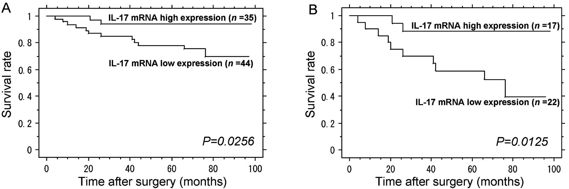
Correlation between patient survival and
IL-17 mRNA expression in peritoneal lavage
Kaplan-Meier survival curves indicated the overall
survival of gastric carcinoma patients stratified according to the
results of the IL-17 mRNA expression status in peritoneal lavage.
The survival curves of all 114 patients displayed no significant
difference between the IL-17 mRNA low expression group and the
IL-17 mRNA high expression group (data were not shown).
Importantly, however, based on the survival curves, among the 79
patients who underwent curative R0 resection, the patients in the
IL-17 mRNA low expression group (n=44) had a significantly poorer
prognosis when compared with the patients in the IL-17 mRNA high
expression group (n=35) (Fig. 1A;
P<0.05). During the median 61 months of postoperative
surveillance, 14 (17.8%) of the 79 patients who underwent curative
resection died, and 12 (85.7%) of these 14 patients developed
peritoneal metastasis. Regarding peritoneal recurrence, 10 (22.7%)
of the 44 cases in the IL-17 mRNA low expression group developed
peritoneal metastases, while 2 (5.7%) of the 35 cases in the IL-17
mRNA high expression group developed peritoneal metastases.
Correlation between survival and IL-17
mRNA in peritoneal lavage in advanced gastric cancer
Among the patients who underwent curative resection
with clinical stage II/III tumors, the prognosis of the IL-17 mRNA
low group was significantly poorer than that of the patients in the
IL-17 mRNA high group (Fig. 1B;
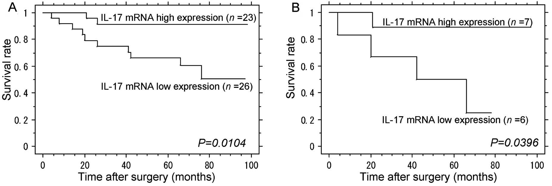
P<0.05). From the point of view of the depth of the invasion,
patients in the IL-17 mRNA low group had significantly poorer
outcome than those in the IL-17 mRNA high group for patients in the
T2/3/4 subgroups (Fig. 2A;
P<0.05). In the T4 subgroup, patients with IL-17 mRNA low
expression in peritoneal lavage had a significantly poorer survival
than those with IL-17 mRNA high expression (Fig. 2B; P<0.05).
Preoperative peritoneal wash assay as an
independent prognostic factor
We evaluated prognostic factors in the 79 patients
who underwent curative R0 resection. With the overall survival as
an endpoint, lymph node metastasis, serosal invasion, lymphatic
invasion, vessel invasion, tumor size and IL-17 mRNA expression
were found to be significant as prognostic factors by univariate
analysis. Moreover, when multivariate analysis was performed with
these six covariates and the same endpoint, IL-17 mRNA low
expression in peritoneal lavage and tumor size were found to be
independent significant predictive factors for prognosis (Table II; HR, 7.91; 95% CI, 1.65–38.03;
P=0.0098).
| Table IIUnivariate and multivariate analysis
of the overall survival for the 79 patients who underwent R0
curative resection. |
Table II
Univariate and multivariate analysis
of the overall survival for the 79 patients who underwent R0
curative resection.
| Univariate
analysis | Multivariate
analysis |
|---|
|
|
|
|---|
| Variables | Hazard ratio | 95% CI | P-value | Hazard ratio | 95% CI | P-value |
|---|
| Age (years) |
| ≤65 vs.
>65 | 1.62 | 0.542–4.840 | 0.388 | - | - | - |
| Gender |
| Male vs.
female | 1.07 | 0.335–3.438 | 0.906 | - | - | - |
| Lymph node
metastasis |
| Negative vs.
positive | 9.98 | 1.296–76.95 | 0.027 | 2.94 | 0.175–49.39 | 0.454 |
| Serosal
invasion |
| Negative vs.
positive | 9.04 | 2.014–40.53 | 0.004 | 1.56 | 0.215–11.35 | 0.659 |
| Lymphatic
invasion |
| Negative vs.
positive | 9.98 | 1.296–76.95 | 0.027 | 1.05 | 0.046–23.79 | 0.976 |
| Vessel
invasion |
| Negative vs.
positive | 10.38 | 2.320–46.42 | 0.0022 | 4.16 | 0.459–37.68 | 0.205 |
| Histological
type |
| Differentiated vs.
undifferentiated | 2.16 | 0.720–6.497 | 0.169 | - | - | - |
| Tumor size
(cm) |
| ≤5 vs. >5 | 7.32 | 2.289–23.40 | 0.0008 | 4.61 | 1.19–17.78 | 0.027 |
| IL-17 mRNA
expression |
| Low expression vs.
high expression | 4.69 | 1.049–20.99 | 0.043 | 7.91 | 1.65–38.03 | 0.0098 |
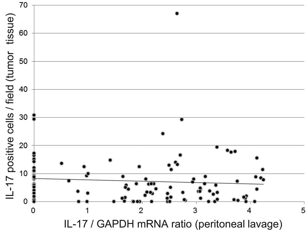
Correlation between IL-17-positive cells
in primary tumor tissues and IL-17 mRNA expression in peritoneal
lavage
To examine the correlation in IL-17 production
between the level in peritoneal lavage and in the primary tumor
tissues, we performed immunohistochemical staining for IL-17 in the
primary tumors in the same patient samples. In the primary tumor
tissues, IL-17 immunoreactive cells were detected in the cytoplasm
of mononuclear cells; however, none of the tumor cells were stained
for IL-17. IL-17-producing cells in the tumor tissues were
7.30±0.82 (mean ± SE) per field. There was no correlation between
the number of IL-17-positive cells in the tumor tissues and IL-17
mRNA expression in the peritoneal wash (r=0.092; P=0.329) (Fig. 3).
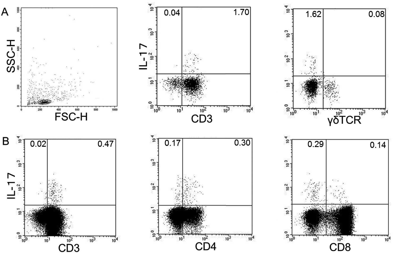
Analysis of IL-17-producing cells in
peritoneal lavage
Immunohistochemical staining of peritoneal wash
revealed that mononuclear cells were stained for IL-17. However,
neither tumor cells nor mesothelial cells were stained for IL-17.
To identify which mononuclear cells produced IL-17 in the
peritoneal lavage, we performed flow cytometric analysis using
anti-IL-17, -CD3, -CD4, -CD8 and -γδ TCR antibodies.
CD3+ T cells produced IL-17, while γδ T cells did not
produce IL-17 (Fig. 4A).
CD4+ T cells mainly produced IL-17, and a small
population of CD8+ T cells also produced IL-17. The mean
percentage of IL-17-positive CD8+ T cells among the
total IL-17-positive cell population was only 27.6±4.85% (n=5), and
in contrast, IL-17-positive CD4+ T cell population was
72.2±4.86% (n=5). Representative flow cytometry analysis is shown
in Fig. 4B.
Discussion
In the present study, we demonstrated that in
patients who underwent R0 resection, the prognosis of patients in
the IL-17 mRNA low expression group was significantly poorer than
those in the high expression group. This is the first study
evaluating the prognostic value of IL-17 detection by real-time
RT-PCR in peritoneal lavage as a valuable prognostic factor in
gastric cancer.
IL-17 was originally identified as a proinflammatory
cytokine that induces neutrophils, and previous studies also have
shown that inflammation is linked to cancer development and
progression. It has recently been reported that the levels of
IL-17-producing cells are significantly increased in tumor tissues,
peripheral blood, malignant ascites fluid, and malignant pleural
effusion from a variety of cancer patients (13,14,26–28).
Despite recent advances in our understanding of the function of
Th17 cells in humans, very little is known about their prevalence
and tumor immunosurveillance.
In mice, overexpression of IL-17 by gene
transduction into tumor cells promoted tumor growth through
angiogenesis (18), but seemingly
in contrast, IL-17 also suppressed tumor growth via a
T-cell-dependent mechanism (19).
Whether IL-17 promotes tumor growth or regulates antitumor
responses remains controversial.
In humans, there are several reports concerning
intratumoral expression of IL-17 and its prognostic role in several
cancer types such as hepatocellular carcinoma (HCC), colon,
esophageal and gastric cancer. In HCC, colon, and prostate cancer
patients, intratumoral IL-17-positive cells were found to be
correlated with poor survival (17,29,30).
Our previous study showed that Th17 cells infiltrated the tumor and
secreted IL-17 in the tumor microenvironment, leading to tumor
progression through angiogenesis and neutrophil infiltration in
patients with gastric cancer. In the present study, we hypothesized
that IL-17 promotes tumor progression in the peritoneal cavity,
based on our previous study suggesting that IL-17 is related to
tumor progression in the tumor microenvironment. We quantitatively
analyzed the expression levels of IL-17 mRNA in peritoneal lavage
from gastric cancer patients. Based on the survival curves, among
the 79 patients who underwent R0 resection, the patients in the
IL-17 mRNA low expression group had a significantly poorer
prognosis than the patients in the IL-17 mRNA high expression
group. This result was contradictory to our hypothesis. This
discrepancy may be explained by the difference in the impact of
IL-17 on tumor progression in the thoracoabdominal cavity and in
tumor tissue. In fact, in the present study, there were no
correlations noted between primary tumor tissues and peritoneal
wash in terms of IL-17 expression. In lung cancer, Ye et al
(26) reported that patients with a
higher proportion of Th17 cells in malignant pleural effusion
exhibited significantly longer overall survival than patients with
a lower proportion of Th17 cells. Similarly, in ovarian cancer, the
expression of IL-17 in ascites was analyzed, and patients with a
higher IL-17 expression in ascites had a significantly lower death
hazard than those with a lower IL-17 expression (28).
Most recently, it has been reported that
CD8+ T cells that produce IL-17 (Tc17 cells) are
abundant in gastric cancer tissue, and the percentage of Th17 cells
is relatively lower than that of Tc17 cells in tumors. The
intratumoral Tc17 cell percentage was significantly associated with
tumor progression and poor prognosis (31). In the present study, flow cytometric
analysis showed that CD4+ Th17 cells predominantly
produced IL-17 in the peritoneal lavage; however, the percentage of
Tc17 cells was lower than that of Th17. This suggests that
IL-17-producing T cells are different between tumor tissue and the
abdominal cavity, and the potential role of IL-17 could also be
different in the tumor microenvironment between tumor tissue and
the abdominal cavity.
There is another reason why the results of the
present study were in conflict with our expectations. This may be
because the role of IL-17 is different before and after the tumor
is established. In the present study, the expression levels of
IL-17 were significantly higher in peritoneal
carcinomatosis-positive cases than those of negative cases, while
they were not associated with cytologic examination (Table IA). Once tumor cells attach to the
peritoneum, IL-17 may play a role as a tumor growth cytokine
through angiogenesis to a greater exent than its role in regulating
antitumor responses. Our results suggest that endogenous IL-17
plays different roles before tumor attachment versus in established
tumor growth. Furthermore, in the abdominal cavity, previous
studies indicate that peritoneal mesothelial cells secrete various
cytokines and growth factors, such as IL-6, IL-8, IL-1α and β,
granulocyte colony stimulating factor (G-CSF), as well as vascular
endothelial growth factor (VEGF) and fibroblast growth factor
(FGF)-2. These results indicate that peritoneal mesothelial cells
are one of the central elements of the cytokine network controlling
disease processes in the abdominal cavity. Kryczek et al
suggested that, in the peritoneal cavity, IL-17 is positively
associated with INF-γ effector T cells and Th1-type chemokines,
CXCL9 and CXCL10, but not with Th2-type chemokines, CXCL12 and
CCL22, in ovarian cancer ascites. Mechanistically, Th17
cell-derived IL-17 and INF-γ were found to synergistically induce
the production of CXCL9 and CXCL10 and in turn promote effector
T-cell migration (28). Thus, IL-17
may function as a polyfunctional cytokine profile in human tumors.
There is no doubt that the role of IL-17 is highly complicated, and
it remains controversial whether IL-17 promotes tumor growth or
regulates the antitumor response.
In conclusion, IL-17 mRNA expression in peritoneal
lavage detected by real-time RT-PCR is a reliable prognostic factor
for patients with curative resection in gastric cancer. Low IL-17
gene expression in the peritoneal cavity may correlate with cancer
development in the peritoneal cavity and poor prognosis in patients
with gastric cancer.
References
|
1
|
Takahashi I, Matsusaka T, Onohara T, et
al: Clinicopathological features of long-term survivors of
scirrhous gastric cancer. Hepatogastroenterology. 47:1485–1488.
2000.PubMed/NCBI
|
|
2
|
Ito S, Nakanishi H, Kodera Y, Mochizuki Y,
Tatemastu M and Yamamura Y: Prospective validation of quantitative
CEA mRNA detection in peritoneal washes in gastric carcinoma
patients. Br J Cancer. 93:986–992. 2005. View Article : Google Scholar : PubMed/NCBI
|
|
3
|
Bonenkamp JJ, Songun I, Hermans J and van
de Velde CJ: Prognostic value of positive cytology findings from
abdominal washings in patients with gastric cancer. Br J Surg.
83:672–674. 1996. View Article : Google Scholar : PubMed/NCBI
|
|
4
|
Kodera Y, Nakanishi H, Yamamura Y, et al:
Prognostic value and clinical implications of disseminated cancer
cells in the peritoneal cavity detected by reverse
transcriptase-polymerase chain reaction and cytology. Int J Cancer.
79:429–433. 1998. View Article : Google Scholar
|
|
5
|
Fujii S, Kitayama J, Kaisaki S, et al:
Carcinoembryonic antigen mRNA in abdominal cavity as a useful
predictor of peritoneal recurrence of gastric cancer with serosal
exposure. J Exp Clin Cancer Res. 21:547–553. 2002.PubMed/NCBI
|
|
6
|
Tokuda K, Natsugoe S, Nakajo A, et al:
Clinical significance of CEA-mRNA expression in peritoneal lavage
fluid from patients with gastric cancer. Int J Mol Med. 11:79–84.
2003.PubMed/NCBI
|
|
7
|
Boku T, Nakane Y, Minoura T, et al:
Prognostic significance of serosal invasion and free
intraperitoneal cancer cells in gastric cancer. Br J Surg.
77:436–439. 1990. View Article : Google Scholar : PubMed/NCBI
|
|
8
|
Kodera Y, Nakanishi H, Ito S, et al:
Quantitative detection of disseminated free cancer cells in
peritoneal washes with real-time reverse transcriptase-polymerase
chain reaction: a sensitive predictor of outcome for patients with
gastric carcinoma. Ann Surg. 235:499–506. 2002. View Article : Google Scholar
|
|
9
|
Oyama K, Terashima M, Takagane A, Maesawa
C, et al: Prognostic significance of peritoneal minimal residual
disease in gastric cancer detected by reverse
transcription-polymerase chain reaction. Br J Surg. 91:435–443.
2004. View
Article : Google Scholar
|
|
10
|
Zeimet AG, Widschwendter M, Knabbe C, et
al: Ascitic interleukin-12 is an independent prognostic factor in
ovarian cancer. J Clin Oncol. 16:1861–1868. 1998.PubMed/NCBI
|
|
11
|
Majima T, Ichikura T, Seki S, Takayama E,
Hiraide H and Mochizuki H: Interleukin-10 and interferon-gamma
levels within the peritoneal cavity of patients with gastric
cancer. J Surg Oncol. 78:124–130. 2001. View Article : Google Scholar : PubMed/NCBI
|
|
12
|
Zhang B, Rong G, Wei H, et al: The
prevalence of Th17 cells in patients with gastric cancer. Biochem
Biophys Res Commun. 374:533–537. 2008. View Article : Google Scholar : PubMed/NCBI
|
|
13
|
Maruyama T, Kono K, Mizukami Y, et al:
Distribution of Th17 cells and FoxP3(+) regulatory T cells in
tumor-infiltrating lymphocytes, tumor-draining lymph nodes and
peripheral blood lymphocytes in patients with gastric cancer.
Cancer Sci. 101:1947–1954. 2010.
|
|
14
|
Miyahara Y, Odunsi K, Chen W, Peng G,
Matsuzaki J and Wang RF: Generation and regulation of human
CD4+ IL-17-producing T cells in ovarian cancer. Proc
Natl Acad Sci USA. 105:15505–15510. 2008. View Article : Google Scholar : PubMed/NCBI
|
|
15
|
Langowski JL, Zhang X, Wu L, et al: IL-23
promotes tumour incidence and growth. Nature. 442:461–465. 2006.
View Article : Google Scholar : PubMed/NCBI
|
|
16
|
Kato T, Furumoto H, Ogura T, et al:
Expression of IL-17 mRNA in ovarian cancer. Biochem Biophys Res
Commun. 282:735–738. 2001. View Article : Google Scholar : PubMed/NCBI
|
|
17
|
Sfanos KS, Bruno TC, Maris CH, et al:
Phenotypic analysis of prostate-infiltrating lymphocytes reveals
TH17 and Treg skewing. Clin Cancer Res.
14:3254–3261. 2008. View Article : Google Scholar : PubMed/NCBI
|
|
18
|
Numasaki M, Fukushi J, Ono M, et al:
Interleukin-17 promotes angiogenesis and tumor growth. Blood.
101:2620–2627. 2003. View Article : Google Scholar : PubMed/NCBI
|
|
19
|
Benchetrit F, Ciree A, Vives V, et al:
Interleukin-17 inhibits tumor cell growth by means of a
T-cell-dependent mechanism. Blood. 99:2114–2121. 2002. View Article : Google Scholar : PubMed/NCBI
|
|
20
|
Zou W and Restifo NP: T(H)17 cells in
tumour immunity and immunotherapy. Nat Rev Immunol. 10:248–256.
2010. View
Article : Google Scholar : PubMed/NCBI
|
|
21
|
Iida T, Iwahashi M, Katsuda M, et al:
Tumor-infiltrating CD4+ Th17 cells produce IL-17 in
tumor microenvironment and promote tumor progression in human
gastric cancer. Oncol Rep. 25:1271–1277. 2011.
|
|
22
|
Kryczek I, Wei S, Zou L, et al: Cutting
edge: Th17 and regulatory T cell dynamics and the regulation by
IL-2 in the tumor microenvironment. J Immunol. 178:6730–6733. 2007.
View Article : Google Scholar : PubMed/NCBI
|
|
23
|
Lissoni P, Mandalà M, Curigliano G, et al:
Progress report on the palliative therapy of 100 patients with
neoplastic effusions by intracavitary low-dose interleukin-2.
Oncology. 60:308–312. 2001. View Article : Google Scholar : PubMed/NCBI
|
|
24
|
Lenzi R, Rosenblum M, Verschraegen C, et
al: Phase I study of intraperitoneal recombinant human interleukin
12 in patients with Müllerian carcinoma, gastrointestinal primary
malignancies, and mesothelioma. Clin Cancer Res. 8:3686–3695.
2002.PubMed/NCBI
|
|
25
|
Fu QG, Meng FD, Shen XD and Guo RX:
Efficacy of intraperitoneal thermochemotherapy and immunotherapy in
intraperitoneal recurrence after gastrointestinal cancer resection.
World J Gastroenterol. 8:1019–1022. 2002.
|
|
26
|
Ye ZJ, Zhou Q, Gu YY, et al: Generation
and differentiation of IL-17-producing CD4+ T cells in
malignant pleural effusion. J Immunol. 185:6348–6354. 2010.
View Article : Google Scholar : PubMed/NCBI
|
|
27
|
Derhovanessian E, Adams V, Hähnel K, et
al: Pretreatment frequency of circulating IL-17+
CD4+ T-cells, but not Tregs, correlates with clinical
response to whole-cell vaccination in prostate cancer patients. Int
J Cancer. 125:1372–1379. 2009.PubMed/NCBI
|
|
28
|
Kryczek I, Banerjee M, Cheng P, et al:
Phenotype, distribution, generation, and functional and clinical
relevance of Th17 cells in the human tumor environments. Blood.
114:1141–1149. 2009. View Article : Google Scholar : PubMed/NCBI
|
|
29
|
Zhang JP, Yan J, Xu J, et al: Increased
intratumoral IL-17-producing cells correlate with poor survival in
hepatocellular carcinoma patients. J Hepatol. 50:980–989. 2009.
View Article : Google Scholar : PubMed/NCBI
|
|
30
|
Liu J, Duan Y, Cheng X, et al: IL-17 is
associated with poor prognosis and promotes angiogenesis via
stimulating VEGF production of cancer cells in colorectal
carcinoma. Biochem Biophys Res Commun. 407:348–354. 2011.
View Article : Google Scholar : PubMed/NCBI
|
|
31
|
Zhuang Y, Peng LS, Zhao YL, Shi Y, Mao XH,
et al: CD8+T cells that produce interleukin-17 regulate
myeloid-derived suppressor cells and are associated with survival
time of patients with gastric cancer. Gastroenterology.
143:951.e8–962.e8. 2012.PubMed/NCBI
|