Introduction
At present, osteosarcoma cases with metastasis have
poor prognosis (1,2). In recent years, caffeine-potentiated
chemotherapy, which is chemotherapy with caffeine dosage against
malignancies, has manifested potently high efficacy (3,4).
Nevertheless, this method may induce side effects with individual
diversity (5). On the other hand,
there have been numerous developments in novel drug delivery
systems (DDS) for drug carriers for the treatment of various
diseases (6–8).
Recently, we demonstrated that nonionic vesicles
prepared from Span 80 have promising physicochemical properties,
such as high membrane fluidity associated with low phase transition
temperature, which make them an attractive possible alternative to
the commonly used liposomes. Span 80 is generally known as sorbitan
monooleate, although commercial Span 80 is a heterogeneous mixture
of sorbitan mono-, di- tri, and tetra-esters that may contribute to
high fluidity and vascular permeability (9–11).
A successful therapeutic model using the DDS with
Span 80 vesicles and immobilized polysaccharides in transplanted
colon cancer cell lines was reported (12). Herein, we showed that tumor-specific
caffeine-potentiated chemotherapy for murine osteosarcoma using a
novel DDS with Span 80 nano-vesicles showed significant antitumor
effects, as well as limited adverse effects.
Materials and methods
Antitumor agents
As therapeutic agents, ifosfamide (IFO) was employed
as well as caffeine sodium benzoate (CSB) as an enhancer. IFO was
obtained from Shionogi & Co. Ltd. (Osaka, Japan), and CSB was
purchased from Maruishi Pharmaceutical Co. Ltd. (Osaka, Japan).
Preparation of Span 80 nano-vesicles
Span 80 nano-vesicles, which contained IFO and/or
caffeine, were freshly prepared as previously described (12). Briefly, materials for assembling
nano-vesicles containing Span 80 (sorbitan monooleate) and Tween-80
[polyoxyethylene (20) sorbitan
monooleate] were purchased from Wako Pure Chemical Industries Ltd.
(Osaka, Japan). Cholesterol, which worked as the stabilizer of the
membrane, was obtained from Nacalai Tesque Inc. (Kyoto, Japan), and
polyethylene glycol, used as a stealth modifier against
macrophages, was acquired from NOF Corp. (Tokyo, Japan). The
solvents, normal hexane and normal saline, were obtained from
Nacalai Tesque Inc. and Otsuka Pharmaceutical Factory Inc. (Naruto,
Japan), respectively. All processes were performed under sterilized
conditions.
The two-step emulsification method was employed to
make and purify the nano-vesicles. Span 80 (190 mg) and cholesterol
(9 mg) were dissolved in 4.5 ml of hexane by homogenization with a
micro-homogenizer, NS-310E (Microtec Co. Ltd., Funabashi, Japan),
at 15,000 rpm for 30 sec in a sterilized brown glass bottle.
Sequentially, 40 mg/ml of IFO and/or 50 mg/ml of CSB, which were
dissolved in 4.5 ml of normal saline, were dripped into the bottle
of Span 80 material followed by homogenization for 3 min, resulting
in production of the first emulsion. As a negative control,
phosphate-buffered saline (PBS) was employed instead of the
antitumor reagents. The emulsion was evaporated by a rotary vacuum
evaporator, N-1000 (Tokyo Rikakikai Co. Ltd., Tokyo, Japan), in a
vacuum flask on a water bath at 37°C followed by homogenization
with 72 mg of Tween-80 and 25 mg of DSPE-020CN at 3,500 rpm for 5
min, which produced the second-stage emulsion.
The second emulsion was centrifuged using an
ultracentrifugation equipment (CP70G with RP65T rotor, Hitachi Koki
Co. Ltd., Tokyo, Japan). After aspiration of the supernatant, the
pellet consisting of the Span 80 vesicles was weighed and suspended
in normal saline at a concentration of 20% w/v, resulting in IFO
Span 80 vesicles (IV), CSB Span 80 vesicles (CV), and PBS alone
Span 80 vesicles (PV). Immediately prior to administration in
vivo or in vitro, these suspensions were extruded by a
custom made extruder (EP Tech Co. Ltd., Ibaraki, Japan), which was
equipped with a drain disk of 100-μm thickness and a Nucleopore
membrane® of 100-nm pore size (GE Healthcare Japan Co.
Ltd., Tokyo, Japan) in order to control the size of the vesicles.
The diameter of the vesicles was evaluated by the Dynamic Light
Scattering device, DLS-6000EW (Otsuka Electronics Co. Ltd., Osaka,
Japan). The average diameter of the vesicles was 117 nm.
In vitro evaluation of the antitumor
effects of the nano-vesicles
The murine osteosarcoma cell line, LM8, was obtained
from the Riken BRC Cell Bank (Tsukuba, Ibaraki, Japan). LM8 cells
(1 ml) in Dulbecco’s modified Eagle’s medium (1×10⁵
cells/ml suspension) were plated per well and cultured in 24-well
culture dishes (Nunc #142475; Thermo Scientific Inc., Waltham, MA,
USA) for a few days until the tumor cells were semi-confluent.
Then, antitumor agents with or without Span 80 vesicles, consisting
of PV, IV, CV, IFO (direct administration), CSB (direct
administration), IV+CSB, IFO+CV and IV+CV, were administered at 100
μl/well. Cells were incubated with the antitumor agents at 37°C for
1 or 2 h, and then the cells were harvested and evaluated for
apoptosis and cell viability, respectively. Assessments of each
condition were examined in triplicate.
Evaluation of cell viability
Cells were incubated with 0.4% trypan blue stain
(Life Technologies Japan Co. Ltd., Tokyo, Japan) in PBS for 1 min,
and then images of each well were captured by an inverted
microscope, TMS (Nikon Instruments Co. Ltd., Tokyo, Japan), and a
DP-25 digital camera (Olympus Co. Ltd., Tokyo, Japan). Non-viable
cells in each image were evaluated by Image Pro Plus®
software (Media Cybernetics Inc., Rockville, MD, USA) in
triplicate.
Apoptosis detection by propidium iodide
(PI) method
Briefly, cells were suspended in 500 μl of ice-cold
Hank’s Balanced Saline Solution (HBSS; Sigma-Aldrich Japan, Tokyo,
Japan), followed by fixation with 4.5 ml of 70% EtOH at −20°C
(13). Fixed cells were centrifuged
at 400 × g for 10 min, pellets were re-suspended in extraction
buffer pH 7.8 which contained 45 mM Na2HPO4
(Nacalai Tesque Inc.), 25 mM citric acid (Wako Pure Chemical), and
0.1% Triton X-100 (Wako Pure Chemical) at 37°C for 25 min. Then,
300 μl of staining solution pH 6.8 containing 10 mM PIPES
(Sigma-Aldrich Japan), 100 mM NaCl (Nacalai Tesque Inc.), 2 mM
Mg2Cl (Wako Pure Chemical), 0.2% Triton X-100, 50 mg/ml
PI (Sigma-Aldrich Japan), and 50 U/ml RNAse H (0.6 mg/ml, Takara
Bio Co., Ltd., Otsu, Japan) were added to the cell suspension, and
the fluorescence intensity was evaluated and analyzed in triplicate
by the FACStation® and CellQuest® software
(Becton-Dickinson, Franklin Lakes, NJ, USA).
Murine osteosarcoma therapeutic
model
C3H/HeJ mice were employed for the therapeutic model
as they are H2-matched to LM8 since this cell line originated from
that strain of mouse (14). Mice
were obtained from CLEA Japan Inc. (Tokyo, Japan) and bred at the
Department of Biological Resources, Ehime University Integrated
Center of Science. LM8 cells (3.0×10⁶ cells per mice)
were subcutaneously transplanted into 6-week-old male C3H/HeJ mice.
After ~3 weeks, when the tumor volume reached ~500 mm3,
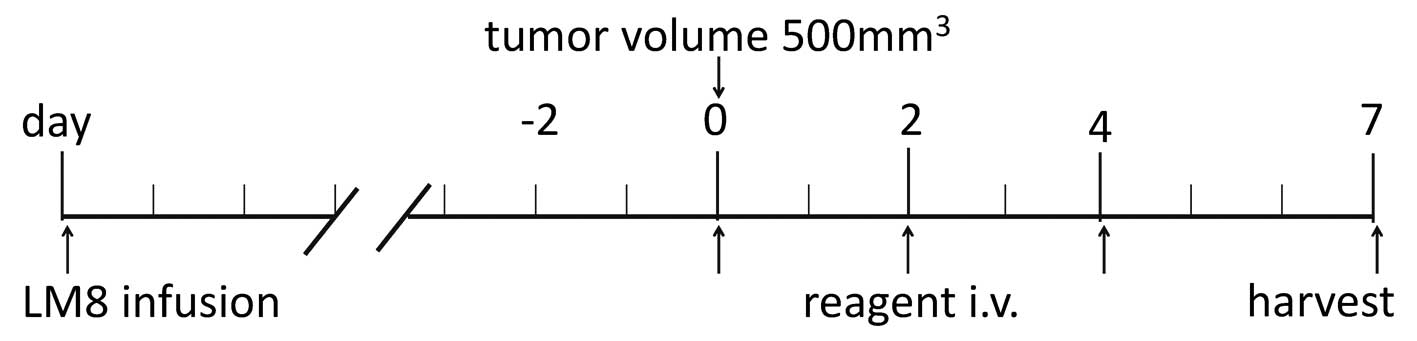
administration of the therapeutic agents was initiated. A scheme of
the administration protocol is presented in Fig. 1.
Combinations of the agents were administered as
follows: PBS (i.v., sham administration), CV (0.1 mg/g BW), IFO
(direct i.v. 0.1 mg/g BW), IV (i.v. 0.1 mg/g BW), IV+CSB, and
IV+CV. Five to eight animals were tested in each group. Agents were
given by tail vein injection (~50 μl) on days 0, 2 and 4, and then
the mice were sacrificed on day 7 under anesthesia. Tumor diameter
and body weight were evaluated every day. After the harvest, tumor
volumes and tumor weights were evaluated, and the entire organs
were processed for histopathological analyses. All the animal
experimental protocols were in accordance with the Guide for Animal
Experimentation, Ehime University, and approved by the Committee
for Animal Experimentation, Ehime University.
Histopathological analyses
Tumors and entire organs from the harvested mice
were formalin-fixed and paraffin-embedded. The viability of the
tumor tissue was evaluated as the area of viable tumor. In
addition, entire organ tissue sections stained with hematoxylin and
eosin, periodic acid-Schiff (PAS), and Elastica-Goldner were
screened by a skilled pathologist to determine the adverse
effects.
Fertility test
A fertility test was performed as follows. Three
male C3H/HeJ mice in each group that were administered IFO, IV or
IV+CV were cross-mated with 6-week-old female C3H/HeJ mice, and the
fertility of each animal was evaluated.
Statistical analyses
Body weight, tumor volume, and viability of the
tumor at the maximum sections were statistically compared among the
different groups with the Student’s t-test, using GraphPad
Prism® software (GraphPad Software Inc., La Jolla, CA,
USA).
Results
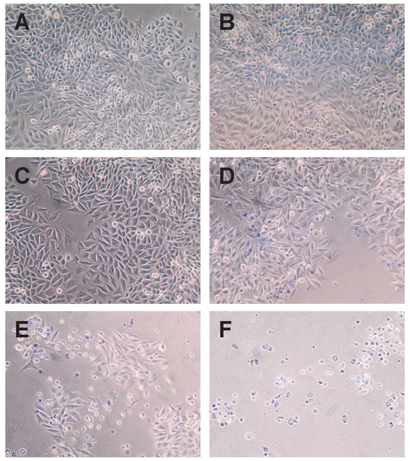
Antitumor effects in vitro
In vitro analyses showed that cultured LM8
mouse osteosarcoma cells treated with IV+CV manifested almost
complete cell death by the trypan blue stain assay, while PBS, CSB,
PV and CV showed almost no cell death. IFO resulted in 13%, IV
resulted in 28%, and IFO+CV resulted in 75% cell death (Fig. 2 and Table I).
 | Table INon-viable cell population in trypan
blue analysis. |
Table I
Non-viable cell population in trypan
blue analysis.
| Treatment | Population of
non-viable cells (%)(mean ± SD) |
|---|
| PBS | 1.5 ±0.9 |
| CSB | 2.1±1.2 |
| PV | 3.3±1.8 |
| CV | 3.1±1.9 |
| IFO | 13±3.4a |
| IV | 28±5.5a |
| IFO+CSB | 25±6.7b |
| IV+CSB | 40±9.2c |
| IFO+CV | 75±10.8d |
| IV+CV |
98±1.2e |
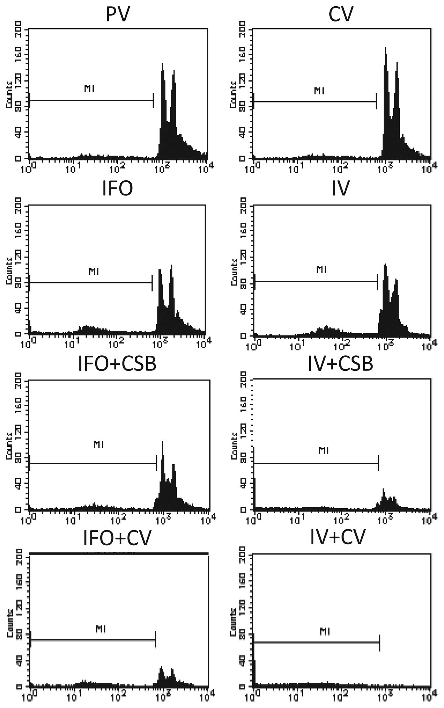
Apoptosis analyses in vitro
By PI analysis, almost all the cells treated with
IV+CV underwent cell death by apoptosis. In contrast, PBS, CSB, PN
and CV induced very limited cell death, whereas IFO and IV induced
apoptosis or necrosis in a small population (8.8–10.2%), and
IFO+CSB and IV+CSB increased the cell death effect to approximately
a quarter to one-third of the population (Fig. 3, Table
II).
| Table IIPopulation of non-viable cells (M1)
in flow cytometric analysis. |
Table II
Population of non-viable cells (M1)
in flow cytometric analysis.
| Treatment | Population of
non-viable cells (%) |
|---|
| PV | 1.6±1.1 |
| CV | 1.4±1.0 |
| IFO | 8.8±1.9a |
| IV | 10.2±2.9a |
| IFO+CSB | 16.5±3.9b |
| IV+CSB | 25.2±4.2b |
| IFO+CV | 32.8±5.9b |
| IV+CV | 97.1±1.9c |
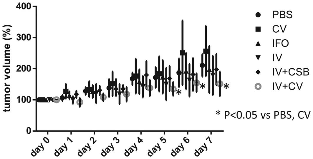
Antitumor effects in vivo
The body weights were not significantly different
among the different groups. The tumor volumes in the IV+CV group
were significantly reduced as compared to those of the control
groups (PBS and CV), and showed a tendency towards a decrease
compared with the PV and IFO direct i.v. groups on days 5–7
(Fig. 4).
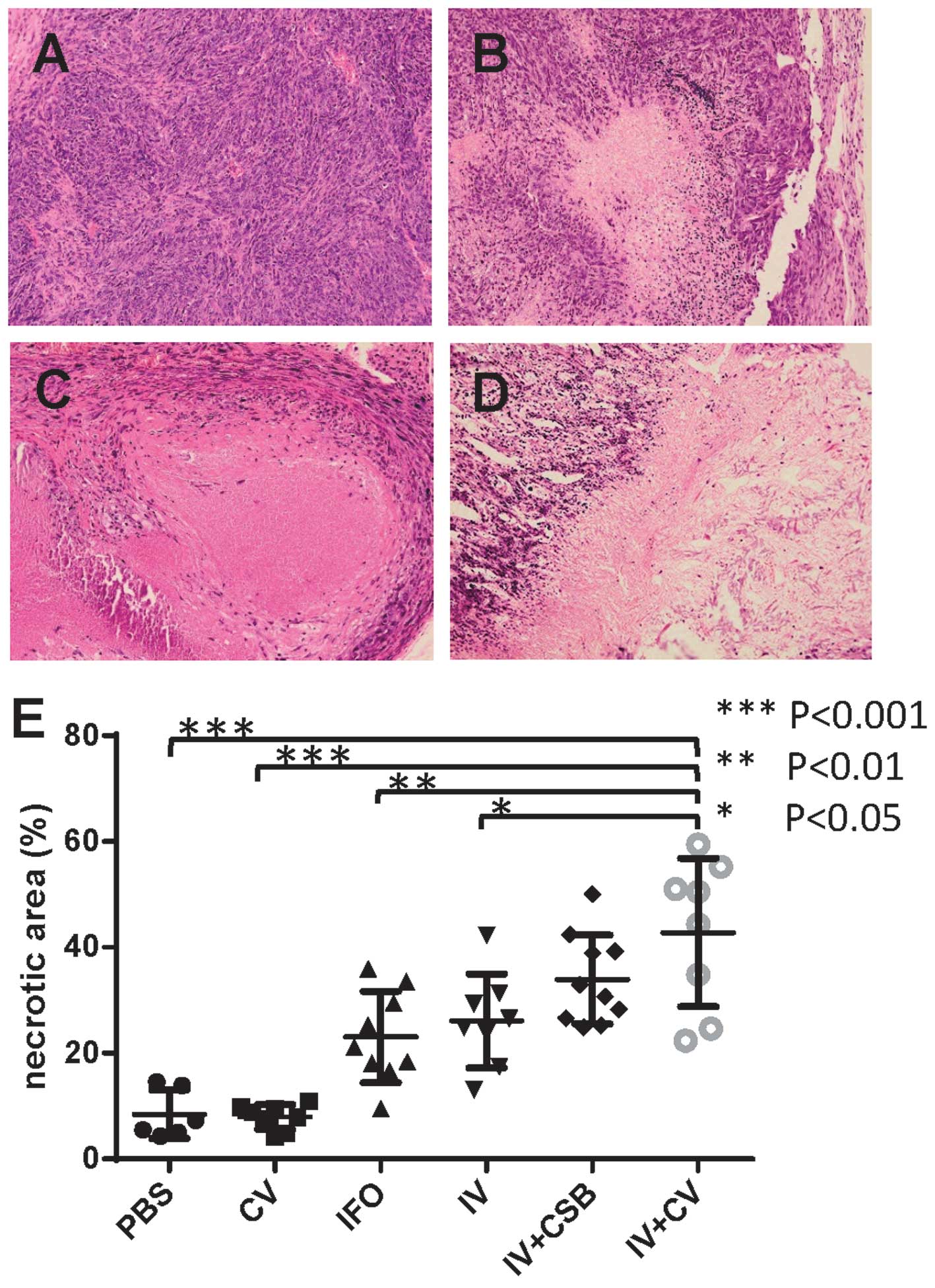
The histopathological analyses of the tumors
demonstrated that IFO, IV, IV+CSB, and IV+CV groups showed
significantly larger non-viable tumor areas as compared to the
controls. In addition, the IV+CV group manifested significantly
greater necrotic areas compared with the IFO and IV groups
(Fig. 5).
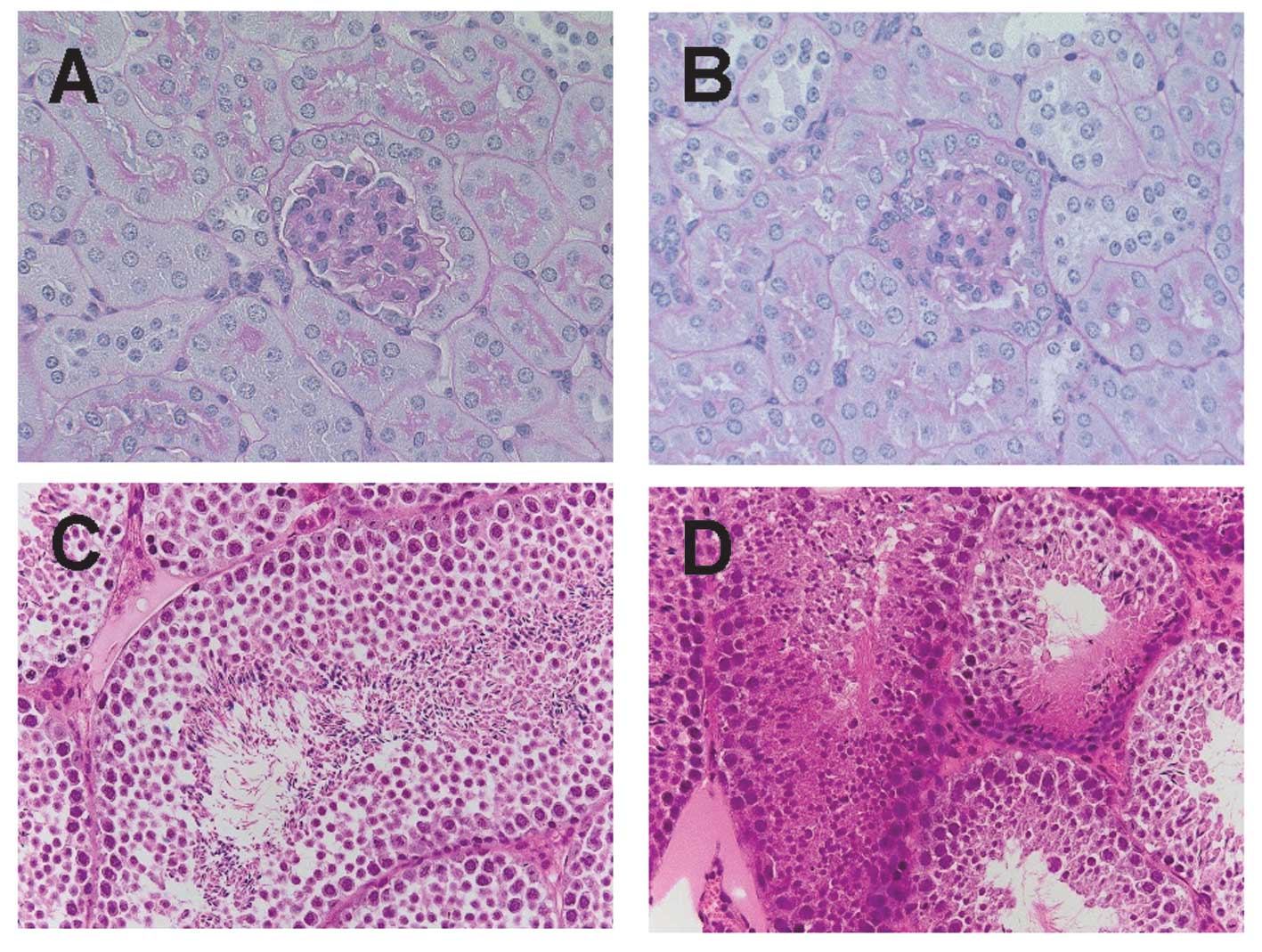
Histopathological analyses for adverse
effects in vivo
To clarify whether the Span 80 vesicles can prevent
adverse effects induced by chemotherapeutic agents,
histopathological examination of the entire organs was performed.
Marked differences were noted in the kidney and testis. In the
kidney, marked tubular injury, which was manifested as loss of
brush border, as well as glomerular changes such as expansion of
the mesangial matrix were observed in the IFO direct i.v. group as
compared to those in the IV and IV+CV groups (Fig. 6A and B). In addition, the IFO direct
i.v. group showed marked suppression of spermatogenesis with
necrosis of the germ cells, while the IV and IV+CV groups showed no
marked changes in spermatogenesis (Fig.
6C and D).
Fertility test
To clarify whether the DDS using Span 80
nano-vesicles could prevent infertility, a fertility test was
performed. The test revealed that the male mice after IV+CV
administration had normal fertility, and there were no
malformations in their progeny (data not shown).
Discussion
We present findings herein to support the evidence
that the DDS with Span 80 vesicles can enhance the antitumor
effects of IFO and caffeine-potentiated IFO chemotherapy, as well
as suppress the adverse effects induced by chemotherapy. In
vitro, IV+CV induced drastic cell death in a very early phase
in contrast to the ‘mild’ apoptosis induction by IFO alone, IV
alone or combinations of IFO+CSB, IV+CSB and IFO+CV. This suggests
that the immediate delivery of IFO and CSB into the cytosol leads
to extremely rapid apoptosis induction, although the detailed
mechanisms are still unknown.
The DDS with nano-vesicles has been developed with
many types of phospholipids and/or detergents (6–8,15).
Among them, the Span 80 nano-vesicle appears to be a promising
material due to its favorable physicochemical properties, such as
membrane fluidity and flexibility. Regarding membrane fluidity,
Span 80 vesicles were reported to have markedly high fluidity with
various cholesterol contents as compared to conventional
phospholipid liposomes, including
1,2-dipalmitoyl-sn-glycero-3-phosphocholine (DPPC) and
1-palmitoyl-2-oleoyl-sn-glycero-3-phosphocholine (POPC) liposomes.
In addition to the high fluidity, Span 80 vesicles also have
greater flexibility as compared to DPPC and POPC liposomes
(10).
Kato et al reported that while non-vesicular
aggregates are often observed in liposome suspensions, Span 80
vesicle suspensions can also include non-vesicular aggregates such
as tubulin structures. Meanwhile, it has been reported that Span 80
vesicles are kinetically-trapped aggregates and not thermodynamic
equilibrium structures, as in most vesicles formed from
phosphatidylcholines (liposomes) (9).
IFO has been reported to induce adverse effects in
the kidney (16–20), gonadal cells (21–23),
and bone marrow along with other organs (24,25).
In the present study, the mice in the IFO i.v. group also exhibited
a certain degree of tubular injury and glomerular changes as well
as severe suppression of spermatogenesis with gonadal cell
necrosis. In contrast, DDS with Span 80 vesicles manifested marked
suppression of the adverse effects in the kidney and testis. This
phenomenon could be due to tumor selectivity of the vesicles, which
may be partially derived from the avoidance of phagocytosis by
macrophages based on the pegylation of the vesicles, and could also
be from the lower permeability of vesicles at the blood-testis
barrier as compared to the direct injection of low molecular weight
molecules such as IFO (23,26).
Our findings suggest that the higher tumor
selectivity of Span 80 vesicles may be associated with their
pegylation as well as high fluidity and flexibility, which result
in higher vascular permeability and tendency to fuse with the
instable cell membrane of the tumors (27). Indeed, a cell fusion model of Span
80 vesicles has been recently reported (11). In addition, our data demonstrated
that CV produced significantly greater antitumor effects as
compared to the direct injection of CSB. This may also be due to
pegylation-associated tumor selectivity and the tendency for cell
fusion which may enable the immediate delivery of caffeine into the
cytoplasm. Regarding the selectivity of caffeine delivery, it may
result in the prevention of caffeine toxicity which may cause the
withdrawal of numerous patients from caffeine-potentiated
chemotherapy (5).
Currently, DDS using liposomal nano-vesicles
containing doxorubicin is applied with marked efficacy (6–8,28–30).
Next generation liposomes, which have targeting molecules on their
surface, have also been under development. A study of the
anti-carcinoma application of Span 80 vesicles containing
doxorubicin was recently reported (27). Span 80 not only has favorable
physicochemical properties, but is also safe since it is already
used as a stabilizer for injected drugs. In addition, Span 80
vesicles should cost drastically less than normal liposomes. Hence,
Span 80 vesicles could be a promising next generation material for
DDS.
In summary, the DDS with Span 80 vesicles may
enhance the antitumor effects of IFO and of caffeine-potentiated
IFO chemotherapy against osteosarcoma. Moreover, the usage of this
DDS may suppress the adverse effects of chemotherapy. Thus, this
DDS model has potential importance for clinical application in the
therapy of metastatic osteosarcoma.
Acknowledgements
We would like to thank Professor Keiichi Kato (Ehime
University Graduate School of Science and Engineering), Dr Yosuke
Omokawa (Nanocareer Inc. Ltd.), and Dr Keita Hayashi (Nara National
College of Technology) for their kind technical support and useful
advice. We also deeply thank Professor Peter Walde (ETH, Zürich,
Switzerland) for his helpful scientific discussion and advice. This
study was supported by a Grant-in-Aid of Scientific Research for
Basic Research (C) (nos. 23592191 and 25460496) from the Ministry
of Education, Culture, Sports, Science and Technology of Japan.
References
|
1
|
Wu PK, Chen WM, Chen CF, Lee OK, Haung CK
and Chen TH: Primary osteogenic sarcoma with pulmonary metastasis:
clinical results and prognostic factors in 91 patients. Jpn J Clin
Oncol. 39:514–522. 2009. View Article : Google Scholar : PubMed/NCBI
|
|
2
|
Clark JC, Dass CR and Choong PF: A review
of clinical and molecular prognostic factors in osteosarcoma. J
Cancer Res Clin Oncol. 134:281–297. 2008. View Article : Google Scholar
|
|
3
|
Kimura H, Tsuchiya H, Shirai T, et al:
Caffeine-potentiated chemotherapy for metastatic osteosarcoma. J
Orthop Sci. 14:556–565. 2009. View Article : Google Scholar : PubMed/NCBI
|
|
4
|
Tsuchiya H, Tomita K, Mori Y, et al:
Caffeine-assisted chemotherapy and minimized tumor excision for
nonmetastatic osteosarcoma. Anticancer Res. 18:657–666.
1998.PubMed/NCBI
|
|
5
|
Hayashi K, Tsuchiya H, Yamamoto N, et al:
Impact of serum caffeine monitoring on adverse effects and
chemotherapeutic responses to caffeine-potentiated chemotherapy for
osteosarcoma. J Orthop Sci. 14:253–258. 2009. View Article : Google Scholar : PubMed/NCBI
|
|
6
|
Yang C and Fu ZX: Liposomal delivery and
polyethylene glycol-liposomal oxaliplatin for the treatment of
colorectal cancer (Review). Biomed Rep. 2:335–339. 2014.PubMed/NCBI
|
|
7
|
Sultana S, Khan MR, Kumar M, Kumar S and
Ali M: Nanoparticles-mediated drug delivery approaches for cancer
targeting: a review. J Drug Target. 21:107–125. 2013. View Article : Google Scholar
|
|
8
|
Brochu H, Polidori A, Pucci B and Vermette
P: Drug delivery systems using immobilized intact liposomes: a
comparative and critical review. Curr Drug Deliv. 1:299–312. 2004.
View Article : Google Scholar
|
|
9
|
Kato K, Walde P, Koine N, et al:
Temperature-sensitive nonionic vesicles prepared from Span 80
(sorbitan monooleate). Langmuir. 24:10762–10770. 2008. View Article : Google Scholar : PubMed/NCBI
|
|
10
|
Hayashi K, Shimanouchi T, Kato K, Miyazaki
T, Nakamura A and Umakoshi H: Span 80 vesicles have a more fluid,
flexible and ‘wet’ surface than phospholipid liposomes. Colloids
Surf B Biointerfaces. 87:28–35. 2011. View Article : Google Scholar : PubMed/NCBI
|
|
11
|
Hayashi K, Tatsui T, Shimanouchi T and
Umakoshi H: Membrane interaction between Span 80 vesicle and
phospholipid vesicle (liposome): Span 80 vesicle can perturb and
hemifuse with liposomal membrane. Colloids Surf B Biointerfaces.
106:258–264. 2013. View Article : Google Scholar : PubMed/NCBI
|
|
12
|
Omokawa Y, Miyazaki T, Walde P, et al: In
vitro and in vivo anti-tumor effects of novel Span 80 vesicles
containing immobilized Eucheuma serra agglutinin. Int J Pharm.
389:157–167. 2010. View Article : Google Scholar : PubMed/NCBI
|
|
13
|
Hotz MA, Gong J, Traganos F and
Darzynkiewicz Z: Flow cytometric detection of apoptosis: comparison
of the assays of in situ DNA degradation and chromatin changes.
Cytometry. 15:237–244. 1994. View Article : Google Scholar : PubMed/NCBI
|
|
14
|
Asai T, Ueda T, Itoh K, et al:
Establishment and characterization of a murine osteosarcoma cell
line (LM8) with high metastatic potential to the lung. Int J
Cancer. 76:418–422. 1998. View Article : Google Scholar : PubMed/NCBI
|
|
15
|
Elbayoumi TA and Torchilin VP: Enhanced
cytotoxicity of monoclonal anticancer antibody 2C5-modified
doxorubicin-loaded PEGylated liposomes against various tumor cell
lines. Eur J Pharm Sci. 32:159–168. 2007. View Article : Google Scholar : PubMed/NCBI
|
|
16
|
Patterson WP and Khojasteh A:
Ifosfamide-induced renal tubular defects. Cancer. 63:649–651. 1989.
View Article : Google Scholar : PubMed/NCBI
|
|
17
|
Skinner R, Pearson AD, Coulthard MG, et
al: Assessment of chemotherapy-associated nephrotoxicity in
children with cancer. Cancer Chemother Pharmacol. 28:81–92. 1991.
View Article : Google Scholar : PubMed/NCBI
|
|
18
|
Skinner R, Sharkey IM, Pearson AD and
Craft AW: Ifosfamide, mesna, and nephrotoxicity in children. J Clin
Oncol. 11:173–190. 1993.PubMed/NCBI
|
|
19
|
McCune JS, Friedman DL, Schuetze S, Blough
D, Magbulos M and Hawkins DS: Influence of age upon
Ifosfamide-induced nephrotoxicity. Pediatr Blood Cancer.
42:427–432. 2004. View Article : Google Scholar : PubMed/NCBI
|
|
20
|
Oberlin O, Fawaz O, Rey A, et al:
Long-term evaluation of Ifosfamide-related nephrotoxicity in
children. J Clin Oncol. 27:5350–5355. 2009. View Article : Google Scholar : PubMed/NCBI
|
|
21
|
Yonemoto T, Ishii T, Takeuchi Y, Hagiwara
Y, Iwata S and Tatezaki S: Recently intensified chemotherapy for
high-grade osteosarcoma may affect fertility in long-term male
survivors. Anticancer Res. 29:763–767. 2009.PubMed/NCBI
|
|
22
|
Ridola V, Fawaz O, Aubier F, et al:
Testicular function of survivors of childhood cancer: a comparative
study between ifosfamide- and cyclophosphamide-based regimens. Eur
J Cancer. 45:814–818. 2009. View Article : Google Scholar : PubMed/NCBI
|
|
23
|
Longhi A, Macchiagodena M, Vitali G and
Bacci G: Fertility in male patients treated with neoadjuvant
chemotherapy for osteosarcoma. J Pediatr Hematol Oncol. 25:292–296.
2003. View Article : Google Scholar : PubMed/NCBI
|
|
24
|
Thomson B, Hawkins D, Felgenhauer J and
Radich J: RT-PCR evaluation of peripheral blood, bone marrow and
peripheral blood stem cells in children and adolescents undergoing
VACIME chemotherapy for Ewing’s sarcoma and alveolar
rhabdomyo-sarcoma. Bone Marrow Transplant. 24:527–533. 1999.
View Article : Google Scholar : PubMed/NCBI
|
|
25
|
Lotz JP, Andre T, Donsimoni R, et al: High
dose chemotherapy with ifosfamide, carboplatin, and etoposide
combined with autologous bone marrow transplantation for the
treatment of poor-prognosis germ cell tumors and metastatic
trophoblastic disease in adults. Cancer. 75:874–885. 1995.
View Article : Google Scholar : PubMed/NCBI
|
|
26
|
Williams D, Crofton PM and Levitt G: Does
ifosfamide affect gonadal function? Pediatr Blood Cancer.
50:347–351. 2008. View Article : Google Scholar
|
|
27
|
Hayashi K, Tatsui T, Shimanouchi T and
Umakoshi H: Enhanced cytotoxicity for colon 26 cells using
doxorubicin-loaded sorbitan monooleate (Span 80) vesicles. Int J
Biol Sci. 9:142–148. 2013. View Article : Google Scholar : PubMed/NCBI
|
|
28
|
Iwamoto T: Clinical application of drug
delivery systems in cancer chemotherapy: review of the efficacy and
side effects of approved drugs. Biol Pharm Bull. 36:715–718. 2013.
View Article : Google Scholar : PubMed/NCBI
|
|
29
|
Eckes J, Schmah O, Siebers JW, et al:
Kinetic targeting of pegylated liposomal doxorubicin: a new
approach to reduce toxicity during chemotherapy (CARL-trial). BMC
Cancer. 11:3372011. View Article : Google Scholar : PubMed/NCBI
|
|
30
|
Maeng JH, Lee DH, Jung KH, et al:
Multifunctional doxorubicin loaded superparamagnetic iron oxide
nanoparticles for chemotherapy and magnetic resonance imaging in
liver cancer. Biomaterials. 31:4995–5006. 2010. View Article : Google Scholar : PubMed/NCBI
|