Introduction
Gastric cancer (GC) is a leading disease in Eastern
Asia (including South Korea, Japan and China). The incidence and
mortality of GC in East Asian regions rank, respectively, second
and third among the most common types of cancer worldwide (1,2).
According to World Health Organization statistics, there were
988,000 new cases of GC worldwide and 736,000 deaths in 2008.
Approximately 60% of the cases were identified in East Asia (mainly
China). In China, approximately two-thirds of patients develop
advanced or metastatic disease, and >50% have recurrent disease
following curative surgery (3–5). For
these patients, chemotherapy remains the most effective treatment
of choice. However, the development of mutidrug resistance (MDR) to
cancer chemotherapy is a major obstacle to the effective treatment
of advanced GC (6). Moreover, the
mechanism of MDR remains obscure.
c-Myc gene is crucial in gastric carcinogenesis. The
c-Myc protein is a transcription factor that regulates a large
series of downstream genes. An association between c-Myc
deregulation and GC has been previously demonstrated (7). c-Myc overexpression has been described
in >40% of GC (7). It encodes a
helix-loop-helix leucine zipper transcription factor that dimerizes
with its partner protein, Max, to transactivate gene expression
(8,9). c-Myc regulates several large gene
families resulting in coordinated changes in cell proliferation and
metabolism. c-Myc stimulates genes involved in protein
biosynthesis, cancer metabolism, transcription factors, cell cycle
and some microRNAs, while inhibiting the expression of other
microRNAs and some tumor suppressor genes (8). The pleiotropic effects of c-Myc
expression occur at the molecular and cellular level and affect
almost every activity of cell life (10).
In vivo studies have demonstrated that a
single low-dose cisplatin treatment results in tumour growth
retardation and a 2-fold elevation in the level of c-Myc expression
(11,12). This reproducible elevation in the
expression of c-Myc is mirrored by reports of analysis conducted on
freshly isolated colon carcinoma tissues from patients with failed
cisplatin therapy (13). Although
these data demonstrate that relatively low doses of cisplatin can
evoke a significant increase in c-Myc expression, it is premature
to suggest there is a direct link between use of cisplatin and
c-Myc-modulated chemoresistance. Clearly, the mechanism for
chemoresistance remains to be investigated.
MicroRNAs (miRNAs) are a group of small RNAs, which
are single-stranded and consist of 19–25 nucleotides. They do not
code for any protein or peptide; however, they regulate gene
expression by various mechanisms. The aberrant miRNA expression and
its correlation with the development and progression of cancers is
an emerging field (14,15). Some miRNAs regulate the formation of
cancer stem cells and the acquisition of epithelial-mesenchymal
transition, which are critically associated with drug resistance
(16,17). Moreover, some miRNAs target genes
associated with drug sensitivity, resulting in the altered
sensitivity of cancer cells to anti-cancer drugs. Findings of
previous studies have also shown that the knockdown or
re-expression of specific miRNAs by synthetic antisense
oligonucleotides or pre-miRNAs induced drug sensitivity, leading to
increased inhibition of cancer cell growth, invasion, and
metastasis (18,19). Those results suggested that specific
targeting of miRNAs by different approaches potentially open new
avenues for cancer treatment by overcoming drug resistance, thereby
improving the outcome of cancer therapy.
The let-7 miRNAs are a family of 12
sequence-associated miRNAs that are distributed over eight genomic
clusters and are often downregulated in cancers (20). The let-7 miRNAs function as tumor
suppressors through the silencing of key oncogenes, such as
RAS and MYC (21,22).
Findings on ovarian cancer showed that let-7i expression was
significantly reduced in chemotherapy-resistant patients and in
vitro reduction of let-7i expression was associated with the
resistance of ovarian and breast cancer cells to cisplatin
(23). By investigating drug
resistance to cisplatin and 5-fluorouracil in 90 patients with GC
and comparing miRNA expression of patients before and after
chemotherapy, Kim et al (24) found that a high expression of let-7g
indicated sensitivity to chemotherapy.
We found that c-Myc may serve as a target gene of
let-7b through Targetscan and Pictar bioinformatics software. In
this study, we detected the expression of let-7b and c-Myc in
SGC7901 and drug-resistant SGC7901/VCR and SGC7901/ DDP GC cell
lines. The aim was to determine whether let-7b regulates the
sensitivity of chemotherapy to mutidrugs in GC by possibly
targeting c-Myc, and to confirm our hypothesis that inhibition of
c-Myc by let-7b reverses MDR in GC cells.
Materials and methods
Cell culture
The human SGC7901 gastric cancer cells were obtained
from the Cell Bank of the Chinese Academy of Sciences (Shanghai,
China). Its drug-resistant SGC7901/DDP and SGC7901/VCR cells were
purchased from the Keygen Biotech Development Co., Ltd. (Nanjing,
China). The cell lines were cultured in RPMI-1640 (Gibco, Grand
Island, NY, USA) medium with 10% fetal bovine serum (Sijiqing,
Hangzhou, China), and maintained in a humidified incubator at 37°C
with an atmosphere of 5% CO2.
Cell viability and drug sensitivity
assay
The SGC7901, SGC7901/VCR, SGC7901/DDP cells were
transfected with let-7b mimic or inhibitor and control mimic. The
transfected cells were seeded in 96-well plates for 24 h and then
treated with different concentrations of cisplatin (DDP) (Qilu
Pharmaceutical Co., Ltd., Shandong, China), 5-fluorouracil (5-FU)
(KingYork Group Co., Ltd., Tianjin, China) and vincristine (VCR)
(HuaLian Pharmaceutical Co., Ltd., Shanghai, China). After 48 h,
the cell viability was evaluated by the MTT assay according to the
manual. Absorbance at 490 nm was measured on an ELISA reader.
Dose-effect curves of anticancer drugs were drawn on semi-logarithm
coordinate paper and IC50 values were determined. Each
experiment was conducted in triplicate and repeated three
times.
Quantitative PCR
Total RNA was extracted using TRIzol reagent
(Invitrogen Inc., Carlsbad, CA, USA) and reverse-transcribed using
a high capacity RNA-cDNA kit (Applied Biosystems Inc., Foster City,
CA, USA). cDNA was quantified on an ABI Prism 7900 sequence
detection system (Applied Biosystems Inc.). PCR was performed using
Power SYBR-Green PCR master mix (Applied Biosystems Inc.). The U6
small nuclear RNA was used as an internal control for let-7b. The
GAPDH was used as an internal control for c-Myc. Primer sequences
used are listed in Table I.
 | Table IPrimer sequences for real-time
PCR. |
Table I
Primer sequences for real-time
PCR.
| Gene | Primer
sequence |
|---|
|
Pre-let-7 | F: TGA GGT AGT AGG
TTG TGT GGT
R: GGA AGG CAG TAG GTT GTA TAG |
| U6 | F: CTC GCT TCG GCA
GCA CA
R: AAC GCT TCA CGA ATT TGC GT |
| c-Myc | F: CCC AGC GAG ACA
TCT GGA AGA A
R: GAG AAG CCG CTC CAC ATG CAG TC |
| Actin | F: AAG GTG AAG GTC
GGA GTC AAC
R: GGG GTC ATT GAT GGC AAC AATA |
Western blot analysis
Cells were lysed in mammalian protein extraction
reagent (Pierce Inc., Rockford, IL, USA) with protease inhibitor
cocktail (Sigma, St. Louis, MO, USA). Following centrifugation at
5,000 × g for 15 min at 4°C, the protein concentration was measured
with a BCA protein assay kit (Pierce Inc., 23227). Total protein
(15 μg) was separated by 10% SDS-PAGE under denaturing conditions
and transferred to PVDF membranes (Millipore Inc., Billerica, MA,
USA). Membranes were blocked in 5% non-fat milk (Bio-Rad, Hercules,
CA, USA) and then incubated with the c-Myc antibody (Santa Cruz
Biotechnology, Inc., Santa Cruz, CA, USA). After incubation with a
secondary antibody conjugated with HRP (Amersham Biosciences Inc.,
Uppsala, Sweden) together with an HRP-conjugated primary antibody
for β-actin (Sigma), immunoreactive proteins were visualized using
the LumiGLO chemiluminescent substrate (Cell Signaling Technology
Inc., Danvers, MA, USA). Densitometric analyses were performed
using Scion Image software.
Luciferase reporter assay
The DNA encoding 3′UTR of c-Myc was PCR-amplified
from human genomic DNA and cloned downstream of firefly luciferase
reporter gene in pGL3-control plasmid (Promega Corp., Madison, WI,
USA). SGC7901 gastric cancer cells were plated in a 24-well plate
24 h prior to transfection at 50% confluence. Let-7b mimic (30 nM)
or control mimic (Ambion Inc., Austin, TX, USA) were transfected
using Lipofectamine RNAiMAX (Invitrogen Inc.). After 24 h
post-transfection, 0.125 μg of reporter vector or empty vector were
transfected using FuGENE6 transfection reagent (Roche Inc., Basel,
Switzerland). After 48-h reporter vector transfection, the cells
were collected, and reporter assays were performed using a dual
luciferase reporter assay system (Promega Corp.).
Transfection of let-7b mimic and
inhibitor oligonucleotides
Pre-miR miRNA precursor and control oligos were
purchased from Ambion Inc. miRCURY LNA miRNA inhibitors and control
oligos were purchased from Exiqon (Vedbaek, Denmark). Transfections
were performed using the Lipofectamine RNAiMAX transfection reagent
(Invitrogen Inc.), and then cells were incubated in the medium
containing the transfection mixture for 24–48 h.
Statistical analysis
Data were expressed as mean ± SD. One-way ANOVA
followed by Bonferroni correction was used to compare the data
among three or more groups, followed by the Student’s t-test.
Statistical analyses were performed using the SPSS 15.0 software
package for Windows (SPSS Inc.., Chicago, IL, USA). P<0.05 was
considered significant.
Results
IC50 of DDP, VCR and 5-FU in
SGC7901, SGC7901/DDP and SGC7901/VCR gastric cancer cells
SGC7901, SGC7901/VCR and SGC7901/DDP cells were
seeded in 96-well plates for 24 h and then treated with different
concentrations of DDP, VCR and 5-FU. After 48 h, the cell viability
was evaluated by the MTT assay according to the manual. Absorbance
at 490 nm was measured on an ELISA reader. Dose-effect curves of
anticancer drugs were drawn on semi-logarithm coordinate paper and
IC50 values were determined (Fig. 1A). IC50 of DDP in
SGC7901, SGC7901/DDP and SGC7901/VCR cells were 0.23, 2.98 and 2.14
μM/l, respectively. IC50 of VCR in SGC7901, SGC7901/DDP
and SGC7901/VCR cells were 0.35, 0.77 and 1.18 μM/l, respectively.
IC50 of 5-FU in SGC7901, SGC7901/DDP and SGC7901/VCR
cells were 0.32, 1.62 and 1.42 μM/l, respectively.
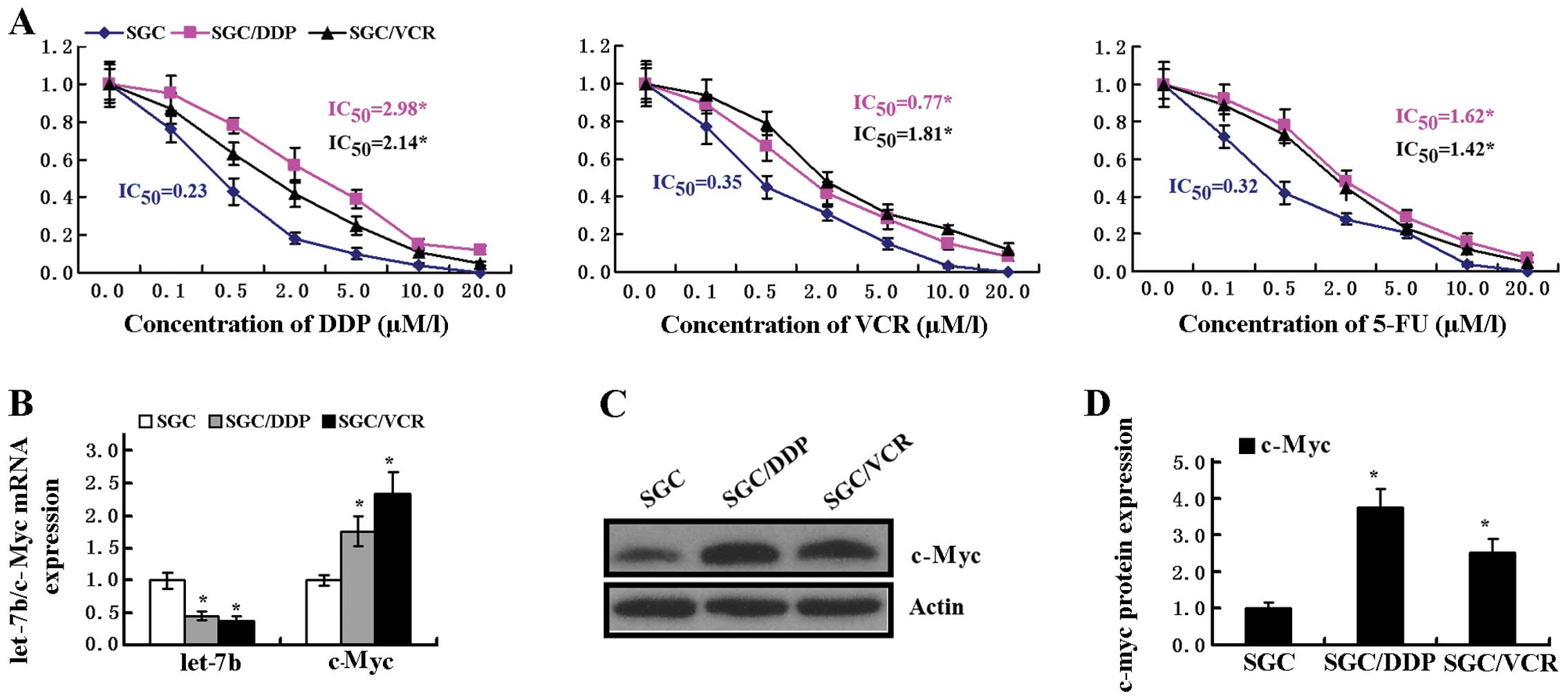 | Figure 1IC50 of DDP, VCR, 5-FU and
expression of let-7b and c-Myc in gastric cancer cells. (A)
SGC7901, SGC7901/VCR, SGC7901/DDP cells were seeded in 96-well
plates for 24 h and then treated with different concentrations of
DDP, VCR and 5-FU. After 48 h, the absorbance at 490 nm was
measured on an ELISA reader. Dose-effect curves of anticancer drugs
were drawn and IC50 values were determined; (B)
Expresion of let-7b and c-Myc mRNA in SGC7901, SGC7901/VCR,
SGC7901/DDP cells were analyzed by quantitive PCR
(*P<0.01, vs. SGC7901 cells); (C) Expresion of c-Myc
protein was evaluated by western blotting; (D) The relative
expression of c-Myc protein was higher in SGC7901/DDP and
SGC7901/VCR cells (*P<0.01, vs. SGC7901 cells). |
Expression of let-7b and c-Myc in
SGC7901, SGC7901/ DDP and SGC7901/VCR gastric cancer cells
To investigate the potential role of let-7b on MDR
in GC, the expression of let-7b in SGC7901, SGC7901/DDP and
SGC7901/VCR cells was evaluated by qPCR. A significant difference
was observed between SGC7901 and the drug-resistant SGC7901/DDP and
SGC7901/VCR cells (Fig. 1B). The
expression of c-Myc mRNA was increased in SGC7901/DDP and
SGC7901/VCR cells compared with that of SGC7901 cells (Fig. 1B). To confirm this phenomenon, we
also performed western blot analysis for c-Myc protein. The protein
levels of c-Myc were higher in SGC7901/DDP and SGC7901/VCR cells
than that in SGC7901 cells (Fig. 1C and
D). Taken together, we demonstrated that there is a potential
correlation between let-7b and c-Myc.
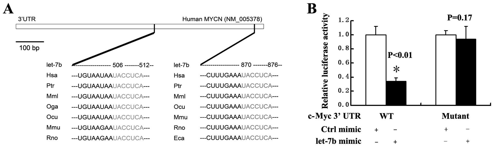
Let-7b directly targets the 3′UTR of
c-Myc
We predicted that there was a conserved let-7
binding site in the c-Myc 3′UTR by TargetScan (Fig. 2A). This hypothesis was confirmed
experimentally. To determine whether let-7b regulation to
c-Myc gene depends on its binding sites of 3′UTR sequences
on the target genes, we constructed reporter plasmids with
wild-type or mutant let-7b binding sites from 3′UTR of c-Myc gene
inserted in the downstream sequence of the luciferase gene. A
reporter assay was performed by co-transfecting SGC7901/DDP cells
with wild-type or mutant reporter plasmids and let-7b mimic or
control oligos. Let-7b potently decreased the luciferase activity
of wild-type reporter plasmid examined in this study, whereas it
had no effect on the mutant forms (Fig.
2B). We concluded that let-7b suppressed c-Myc gene
expression in a sequence-specific manner and the suppression
depends on let-7b binding sites within 3′UTR sequences of the
c-Myc genes.
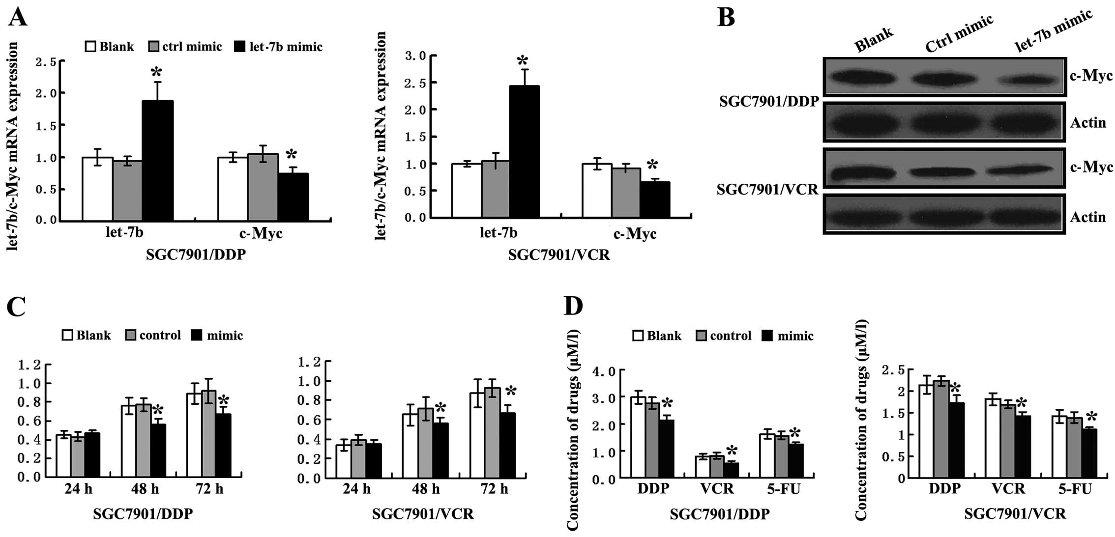
Transfection of let-7b mimic
downregulates the expression of c-Myc and increases the sensitivity
of chemotherapy in SGC7901/DDP and SGC7901/VCR cells
To investigate whether let-7b could regulate c-Myc
expression, let-7b mimic and control mimic, respectively, were
transfected in SGC7901/DDP and SGC7901/VCR cells. The expression of
let-7b and c-Myc in the two cell lines was detected by qPCR. As
shown in Fig. 3A, the expression of
let-7b was significantly higher in let-7b mimic-transfected cells
than that in control mimic-transfected cells. However, the
expression of c-Myc was significantly lower in let-7b
mimic-transfected cells than that in the control mimic-transfected
cells. From the results of western blotting, we found that
transfection of let-7b mimic significantly downregulated the
expression of c-Myc protein in the two cells (Fig. 3B). Therefore, we concluded that
let-7b suppresses c-Myc gene expression at the mRNA and
protein levels.
To determine whether transfection of let-7b mimic
affects cell proliferation in SGC7901/DDP and SGC7901/VCR cells,
metabolic activity at 24, 48 and 72 h after transfection was
determined by MTT assay. The cell viability was reduced
significantly in the two cells after transfection with the let-7b
mimic at 48 and 72 h as compared with the control mimic (Fig. 3C, P<0.05).
To explore the role of c-Myc on the sensitivity of
chemotherapy in GC cells, SGC7901/VCR and SGC7901/DDP cells were
transfected with let-7b or control mimic. The transfected cells
were seeded in 96-well plates for 24 h and then treated with
different concentrations of DDP, VCR and 5-FU. After 48 h, the cell
viability was evaluated by the MTT assay according to the manual,
dose-effect curves of anticancer drugs were drawn on semi-logarithm
coordinate paper and IC50 values were determined. As
shown in Fig. 3D, the
IC50 valus of DDP, VCR and 5-FU were 2.13±0.18,
0.54±0.08 and 1.23±0.09 μM/l in let-7b mimic-transfected
SGC7901/DDP cells, and were 2.76±0.22, 0.81±0.12 and 1.57±0.16 μM/l
in control mimic-transfected SGC7901/DDP cells. For SGC7901/ VCR
cells, the IC50 of DDP, VCR and 5-FU were 1.71±0.19,
1.41±0.11 and 1.11±0.05 μM/l in let-7b mimic-transfected cells, and
were 2.23±0.11, 1.69±0.09 and 1.39±0.12 μM/l in control mimic
transfected cells, respectively. Thus, the two drug-resistant GC
cells showed the sensitivity of chemotherapy to be significantly
increased in let-7b mimic-transfected cells at the 3rd day after
transfection.
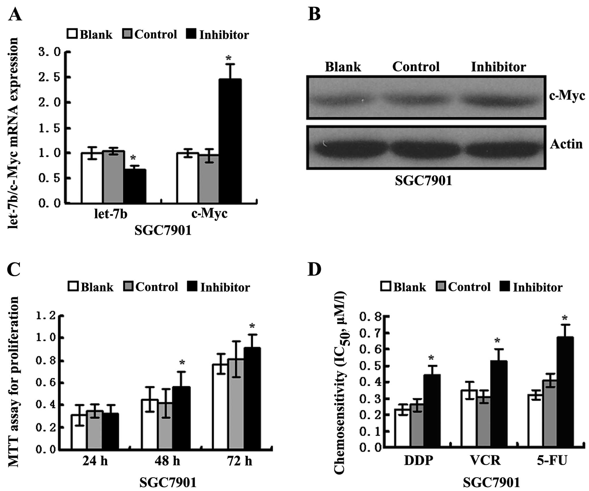
Transfection of let-7b inhibitor
upregulates the expression of c-Myc and decreases the sensitivity
of chemotherapy in SGC7901 cells
For loss-of-function experiments, let-7b inhibitor
was used to block endogenous let-7b expression in SGC7901 cell
lines. The expression of let-7b and c-Myc was detected by qPCR. As
shown in Fig. 4A, the expression of
let-7b was significantly lower in let-7b inhibitor-transfected
cells than that in control mimic-transfected cells. However, the
expression of c-Myc was significantly higher in let-7b
inhibitor-transfected cells than that in control mimic-transfected
cells. From the results of western blotting, we found that the
transfection of let-7b inhibitor significantly increased the
expression of c-Myc protein in SGC7901 cells (Fig. 4B). We also confirmed that
transfection of let-7b inhibitor enhances SGC7901 cell
proliferation at 48 and 72 h after transfection by MTT assay
(Fig. 4C, P<0.05).
The sensitivity of chemotherapy in SGC7901 cells was
also detected by MTT assay following transfection with let-7b
inhibitor or control mimic. As shown in Fig. 4D, the IC50 values of DDP,
VCR and 5-FU were 0.44±0.06, 0.53±0.07 and 0.67±0.08 μM/l in let-7b
inhibitor-transfected SGC7901 cells, and were 0.26±0.04, 0.31±0.04
and 0.41±0.04 μM/in control mimic-transfected cells, respectively.
We demonstrated that in drug-sensitive GC cells, the sensitivity of
chemotherapy was significantly decreased following let-7b inhibitor
transfection.
Discussion
Chemotherapy is an important therapeutic strategy
for advanced or recurrent gastric cancer (GC) treatment. However,
chemotherapy fails to eliminate all tumor cells because of
intrinsic or acquired MDR, which is the most common cause of tumor
recurrence (25,26), and the greatest obstacle for the
effective treatment of GC (6). The
underlying mechanisms of cellular resistance in cancer cells to
these DNA-damaging anticancer drugs have been broadly explored, but
have not been fully characterized (27,28).
P-gp was the first molecule identified as a modulator of MDR.
Subsequently, various other molecules were shown to be involved,
including transporters that eject anticancer drugs from cells, such
as MDR-associated protein (MRP) (29), genes regulating apoptosis, such as
p53 (30), telomere-binding
protein, such as TRF2(31), RhoE
GTPase (32) and transcription
factors, such as CDX2 (33) and
c-Myc (13). miRNAs have also beeen
considered to be involved in the field of cancer MDR
investigations. miRNAs are a class of small, non-coding RNA
molecules that repress protein expression through imperfect binding
to sequences in the 3′UTR of target mRNAs, Seven miRNAs were found
to be significantly and differentially expressed in tumors from
platinum-sensitive vs. platinum-resistant patients. These seven
miRNAs included miR-27a, miR-23a, miR-30c, let-7g, miR-199a-3p,
miR-378 and miR-625, which were overexpressed in platinum-resistant
patients (34). Results of a recent
study suggested that miRNA-200c regulates the sensitivity of
chemotherapy to cisplatin (DDP) in SGC7901/DDP gastric cancer cells
by directly targeting RhoE (32).
Chen et al (35) transfected
miRNA-200c into SGC7901/DDP gastric cancer cells, which increased
sensitivity to DDP, 5-fluorouracil, paclitaxel and doxorubicin.
In the present study, we found that the expression
of let-7b was lower in chemotherapy-resistant SGC7901/DDP and
SGC7901/ VCR gastric cancer cells than that in
chemotherapy-sensitive SGC7901 cells. By contrast, the expression
of c-Myc was higher in SGC7901/DDP and SGC7901/VCR cells than that
in SGC7901 cells. Using TargetScan and luciferase reporter assay,
we confirmed that let-7b suppresses c-Myc gene expression in
a sequence-specific manner and this suppression depends on let-7b
binding sites within 3′UTR sequences of the c-Myc genes.
Furthermore, we have demonstrated that transfection of let-7b mimic
increases drug sensitivity in chemotherapy-resistant SGC7901/DDP
and SGC7901/VCR cells by targeting the downregulation of c-Myc. For
loss-of-function experiments, the results suggest that in
drug-sensitive SGC7901 cells, the sensitivity of chemotherapy was
significantly decreased after let-7b inhibitor transfection.
An increasing number of reports have suggested that
let-7 is poorly expressed in a variety of human tumors and a
reduced let-7 level results in the overexpression of
let-7-responsive genes in tumors, including CyclinD, RAS, Myc and
Lin28/Lin28B (36–39). According to our previous report, a
double-negative feedback regulating loop of Lin28 and let-7 exists
in tumor cells and controls ALDH1+ cancer stem cells (40). let-7 interacts with two iPS genes,
Myc and Lin28, and these autoregulatory loops of
let-7/Myc (41) and let-7/Lin28
(40) may control stem cell
self-renewal and differentiation. High levels of Lin28 and Myc or a
low level of let-7 may promote the conversion of epithelial cells
to a more undifferentiated stage and maintain tumor cells in this
stem-like stage (40,42). A recent study found that Lin28B
enhances n-Myc levels and induces neuroblastoma, suggesting that
n-Myc is a key target of Lin28B (43). Moreover, the c-Myc and
n-Myc oncogenes are positive regulators of Lin28 and Lin28B,
respectively (44,45).
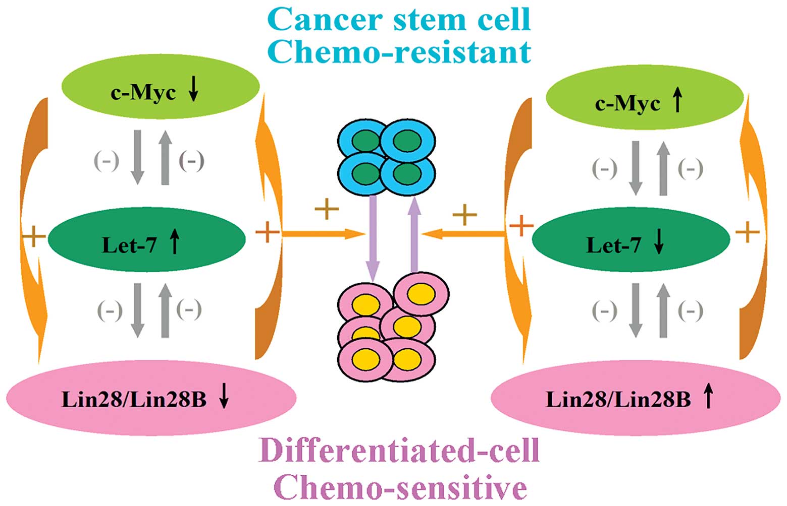
Emerging evidence suggests that cancer stem cells
(CSCs) may play critical roles in drug resistance and ultimately
recurrence (46–50). The present findings together with
those of previous reports (40,41,43–45)
support a model in which double-negative autoregulatory loops
(Lin28/let-7 and Myc/let-7) and a double-positive autoregulatory
loop (Lin28/Lin28B/Myc) existing in GC cells may control cancer
stem cell differentiation and regulate MDR (Fig. 5).
In conclusion, the present study results have
demonstrated that, let-7b increases drug sensitivity in
chemotherapy-resistant SGC7901/DDP and SGC7901/VCR gastric cancer
cells by targeting downregulation of c-Myc and that, let-7b mimic
reverses MDR by promoting cancer stem cell differentiation
controlled by double-negative autoregulatory loops (Lin28/ let-7
and Myc/let-7) and a double-positive autoregulatory loop (Lin28/
Lin28B/Myc) existing in GC cells, which remains to be confirmed.
These results suggest that let-7b plays key roles in MDR of GC. By
downregulating Lin28 and c-Myc and promoting cancer stem cell
differentiation, let-7b may function as a chemotherapy enhancer in
GC. This role in regulating GC cell MDR signifies an opportunity to
develop novel cancer therapies. Therefore, it is presumed that
enforced expression of let-7b may improve the chemotherapy
efficiency of patients with GC with a low let-7b expression.
Acknowledgements
This study was supported, in whole or in part, by
GWGL2013-47(HC), and Natural Science Foundation of Gansu Province
(145RJZA117, HC). We are greatly indebted to Dr Youcheng Zhang
(Lanzhou University Second Hospital) and Lin Zhang (University of
Pennsylvania School of Medicine) for the technical assistance.
References
|
1
|
Lee JH, Kim KM, Cheong JH and Noh SH:
Current management and future strategies of gastric cancer. Yonsei
Med J. 53:248–257. 2012. View Article : Google Scholar : PubMed/NCBI
|
|
2
|
Hudler P: Genetic aspects of gastric
cancer instability. Sci World J. 2012:7619092012. View Article : Google Scholar
|
|
3
|
Verdecchia A, Corazziari I, Gatta G, Lisi
D, Faivre J and Forman D; EUROCARE Working Group. Explaining
gastric cancer survival differences among European countries. Int J
Cancer. 109:737–741. 2004. View Article : Google Scholar : PubMed/NCBI
|
|
4
|
Bonenkamp JJ, Hermans J, Sasako M, et al:
Extended lymphnode dissection for gastric cancer. N Engl J Med.
340:908–914. 1999. View Article : Google Scholar : PubMed/NCBI
|
|
5
|
Kaneko S and Yoshimura T: Time trend
analysis of gastric cancer incidence in Japan by histological
types, 1975–1989. Br J Cancer. 84:400–405. 2001. View Article : Google Scholar : PubMed/NCBI
|
|
6
|
Fan D and Liu X: New progresses in
researches on multidrug resistance in gastric cancer. Chin J
Digest. 20:77–78. 2000.
|
|
7
|
Milne AN, Sitarz R, Carvalho R, Carneiro F
and Offerhaus GJ: Early onset gastric cancer: on the road to
unraveling gastric carcinogenesis. Curr Mol Med. 7:15–28. 2007.
View Article : Google Scholar : PubMed/NCBI
|
|
8
|
Eilers M and Eisenman RN: Myc’s broad
reach. Genes Dev. 22:2755–2766. 2008. View Article : Google Scholar : PubMed/NCBI
|
|
9
|
Herkert B and Eilers M: Transcriptional
repression: the dark side of myc. Genes Cancer. 1:580–586. 2010.
View Article : Google Scholar
|
|
10
|
Miller MD, Thomas SD, Islam A, Muench D
and Sedoris K: C-Myc and cancer metabolism. Clin Cancer Res.
18:5546–5553. 2012. View Article : Google Scholar : PubMed/NCBI
|
|
11
|
Walker TL, White JD, Esdale WJ, Burton MA
and DeCruz EE: Tumour cells surviving in vivo cisplatin
chemotherapy display elevated cmyc expression. Br J Cancer.
73:610–614. 1996. View Article : Google Scholar : PubMed/NCBI
|
|
12
|
Marazzi L, Parodi MT, Martino DD, Ferrari
S and Tonini GP: Coordinate change of c-myc, transferrin receptor
and H3 gene expression precedes induction of haemoglobin-producing
cells of the leukaemia K562 cell line treated with
cisdiamminedichlo-roplatinum II. Anticancer Res. 11:947–952.
1991.PubMed/NCBI
|
|
13
|
Kashani-Sabet M, Lu Y, Leong L, Haedicke K
and Scanlon KJ: Differential oncogene amplification in tumour cells
from a patient treated with cisplatin and 5-FU. Eur J Cancer.
26:383–390. 1990. View Article : Google Scholar : PubMed/NCBI
|
|
14
|
Vandenboom TG II, Li Y, Philip PA and
Sarkar FH: MicroRNA and cancer: tiny molecules with major
implications. Curr Genomics. 9:97–109. 2008. View Article : Google Scholar
|
|
15
|
Iorio MV and Croce CM: MicroRNAs in
cancer: small molecules with a huge impact. J Clin Oncol.
27:5848–5856. 2009. View Article : Google Scholar : PubMed/NCBI
|
|
16
|
Peter ME: Regulating cancer stem cells the
miR way. Cell Stem Cell. 6:4–6. 2010. View Article : Google Scholar : PubMed/NCBI
|
|
17
|
Adam L, Zhong M, Choi W, et al: miR-200
expression regulates epithelial-to-mesenchymal transition in
bladder cancer cells and reverses resistance to epidermal growth
factor receptor therapy. Clin Cancer Res. 15:5060–5072. 2009.
View Article : Google Scholar : PubMed/NCBI
|
|
18
|
Bourguignon LY, Spevak CC, Wong G, Xia W
and Gilad E: Hyaluronan-CD44 interaction with protein kinase
C(epsilon) promotes oncogenic signaling by the stem cell marker
Nanog and the Production of microRNA-21, leading to down-regulation
of the tumor suppressor protein PDCD4, anti-apoptosis, and
chemotherapy resistance in breast tumor cells. J Biol Chem.
284:26533–26546. 2009. View Article : Google Scholar : PubMed/NCBI
|
|
19
|
Blower PE, Chung JH, Verducci JS, et al:
MicroRNAs modulate the chemosensitivity of tumor cells. Mol Cancer
Ther. 7:1–9. 2008. View Article : Google Scholar : PubMed/NCBI
|
|
20
|
Roush S and Slack FJ: The let-7 family of
microRNAs. Trends Cell Biol. 18:505–516. 2008. View Article : Google Scholar : PubMed/NCBI
|
|
21
|
Lee YS and Dutta A: The tumor suppressor
microRNA let-7 represses the HMGA2 oncogene. Genes Dev.
21:1025–1030. 2007. View Article : Google Scholar : PubMed/NCBI
|
|
22
|
Kumar MS, Lu J, Mercer KL, Golub TR and
Jacks T: Impaired microRNA processing enhances cellular
transformation and tumorigenesis. Nat Genet. 39:673–677. 2007.
View Article : Google Scholar : PubMed/NCBI
|
|
23
|
Yang N, Kaur S, Volinia S, et al: MicroRNA
microarray identifies Let-7i as a novel biomarker and therapeutic
target in human epithelial ovarian cancer. Cancer Res.
68:10307–10314. 2008. View Article : Google Scholar : PubMed/NCBI
|
|
24
|
Kim CH, Kim HK, Rettig RL, et al: miRNA
signature associated with outcome of gastric cancer patients
following chemotherapy. BMC Med Genomics. 4:792001. View Article : Google Scholar
|
|
25
|
Broxterman HJ, Gotink KJ and Verheul HM:
Understanding the causes of multidrug resistance in cancer: a
comparison of doxorubicin and sunitinib. Drug Resist Updat.
12:114–126. 2009. View Article : Google Scholar : PubMed/NCBI
|
|
26
|
Fojo T: Multiple paths to a drug
resistance phenotype: mutations, translocations, deletions and
amplification of coding genes or promoter regions, epigenetic
changes and microRNAs. Drug Resist Updat. 10:59–67. 2007.
View Article : Google Scholar : PubMed/NCBI
|
|
27
|
Gatti L and Zunino F: Overview of tumor
cell chemoresistance mechanisms. Methods Mol Med. 111:127–148.
2005.PubMed/NCBI
|
|
28
|
Fan D, Zhang X, Chen X, et al: Bird’s-eye
view on gastric cancer research of the past 25 years. J
Gastroenterol Hepatol. 20:360–365. 2005. View Article : Google Scholar : PubMed/NCBI
|
|
29
|
Chuman Y, Sumizawa T, Takebayashi Y, et
al: Expression of the multidrug resistance associated protein (MRP)
gene in human colorectal, gastric and non-small-cell lung
carcinomas. Int J Cancer. 66:274–279. 1996. View Article : Google Scholar : PubMed/NCBI
|
|
30
|
Matsuhashi N, Saio M, Matsuo A, Sugiyama Y
and Saji S: The evaluation of gastric cancer sensitivity to
5-FU/CDDP in terms of induction of apoptosis: Time- and p53
expression-dependency of anti-cancer drugs. Oncol Rep. 14:609–615.
2005.PubMed/NCBI
|
|
31
|
Ning H, Li T, Zhao L, et al: TRF2 promotes
multidrug resistance in gastric cancer cells. Cancer Biol Ther.
5:950–956. 2006. View Article : Google Scholar : PubMed/NCBI
|
|
32
|
Chang L, Guo F, Wang Y, et al:
MicroRNA-200c regulates the sensitivity of chemotherapy of gastric
cancer SGC7901/DDP cells by directly targeting RhoE. Pathol Oncol
Res. 20:93–98. 2014. View Article : Google Scholar
|
|
33
|
Yan LH, Wang XT, Yang J, et al: Reversal
of multidrug resistance in gastric cancer cells by CDX2
downregulation. World J Gastroenterol. 19:4155–4165. 2013.
View Article : Google Scholar : PubMed/NCBI
|
|
34
|
Eitan R, Kushnir M, Lithwick-Yanai G, et
al: Tumor microRNA expression patterns associated with resistance
to platinum based chemotherapy and survival in ovarian cancer
patients. Gynecol Oncol. 114:253–259. 2009. View Article : Google Scholar : PubMed/NCBI
|
|
35
|
Chen Y, Zuo J, Liu Y, Gao H and Liu W:
Inhibitory effects of miRNA-200c on chemotherapy-resistance and
cell proliferation of gastric cancer SGC7901/DDP cells. Chin J
Cancer. 29:1006–1011. 2010. View Article : Google Scholar : PubMed/NCBI
|
|
36
|
Schultz J, Lorenz P, Gross G, Ibrahim S
and Kunz M: MicroRNA let-7b targets important cell cycle molecules
in malignant melanoma cells and interferes with
anchorage-independent growth. Cell Res. 18:549–557. 2008.
View Article : Google Scholar : PubMed/NCBI
|
|
37
|
Sampson VB, Rong NH, Han J, et al:
MicroRNA let-7a down-regulates MYC and reverts MYC-induced growth
in Burkitt lymphoma cells. Cancer Res. 67:9762–9770. 2007.
View Article : Google Scholar : PubMed/NCBI
|
|
38
|
Johnson SM, Grosshans H, Shingara J, et
al: RAS is regulated by the let-7 microRNA family. Cell.
120:635–647. 2005. View Article : Google Scholar : PubMed/NCBI
|
|
39
|
Rybak A, Fuchs H, Smirnova L, et al: A
feedback loop comprising lin-28 and let-7 controls pre-let-7
maturation during neural stem-cell commitment. Nat Cell Biol.
10:987–993. 2008. View Article : Google Scholar : PubMed/NCBI
|
|
40
|
Yang XJ, Lin XJ, Zhong XM, et al: Double
negative feedback loop between reprogramming factor LIN28 and
microRNA let-7 regulates aldehyde dehydrogenase 1-positive cancer
stem cells. Cancer Res. 70:9463–9472. 2010. View Article : Google Scholar : PubMed/NCBI
|
|
41
|
Chang TC, Zeitels LR, Hwang HW, et al:
Lin-28B transactivation is necessary for Myc-mediated let-7
repression and proliferation. Proc Natl Acad Sci USA.
106:3384–3389. 2009. View Article : Google Scholar : PubMed/NCBI
|
|
42
|
Cotterman R and Knoepfler PS: N-Myc
regulates expression of pluripotency genes in neuroblastoma
including lif, klf2, klf4, and lin28b. PLoS One. 4:e57992009.
View Article : Google Scholar : PubMed/NCBI
|
|
43
|
Molenaar JJ, Domingo-Fernández R, Ebus ME,
et al: LIN28B induces neuroblastoma and enhances MYCN levels via
let-7 suppression. Nat Genet. 44:1199–1206. 2012. View Article : Google Scholar : PubMed/NCBI
|
|
44
|
Laurenti E, Varnum-Finney B, Wilson A, et
al: Hematopoietic stem cell function and survival depend on c-Myc
and N-Myc activity. Cell Stem Cell. 3:611–624. 2008. View Article : Google Scholar : PubMed/NCBI
|
|
45
|
Stanton BR, Perkins AS, Tessarollo L,
Sassoon DA and Parada LF: Loss of Nmyc function results in
embryonic lethality and failure of the epithelial component of the
embryo to develop. Gene Dev. 6:2235–2247. 1992. View Article : Google Scholar
|
|
46
|
Konopleva M, Tabe Y, Zeng Z and Andreeff
M: Therapeutic targeting of micro environmental interactions in
leukemia: mechanisms and approaches. Drug Resist Updat. 12:103–113.
2009. View Article : Google Scholar : PubMed/NCBI
|
|
47
|
Voulgari A and Pintzas A:
Epithelial-mesenchymal transition in cancer metastasis: mechanisms,
markers and strategies to overcome drug resistance in the clinic.
Biochim Biophys Acta. 1796:75–90. 2009.PubMed/NCBI
|
|
48
|
Wang Z, Li Y, Banerjee S and Sarkar FH:
Emerging role of Notch in stem cells and cancer. Cancer Lett.
279:8–12. 2009. View Article : Google Scholar :
|
|
49
|
Wang ZW, Li YW, Ahmad A, et al: Targeting
miRNAs involved in cancer stem cell and EMT regulation: an emerging
concept in overcoming drug resistance. Drug Resist Updat.
13:109–118. 2010. View Article : Google Scholar : PubMed/NCBI
|
|
50
|
Ahmed N, Abubaker K, Findlay J and Quinn
M: Cancerous ovarian stem cells: obscure targets for therapy but
relevant to chemoresistance. J Cell Biochem. 114:21–34. 2013.
View Article : Google Scholar
|