Introduction
Pancreatic ductal adenocarcinoma (PDAC) has a high
mortality rate due to its aggressive growth and high rates of
metastasis (1) and chemoresistance
(2). Increasing evidence supports
the roles of cancer stem cells (CSCs) in the initiation,
maintenance, proliferation, metastasis and resistance of malignant
tumors to therapy (3). Numerous
studies have demonstrated the existence of CSC fractions in PDAC
(4), which have been detected by
CSC-specific markers, including CD24 (5–7), CD44
(5,6,8,9), CD133
(10–13), CXCR4 (10,14),
epithelial cell adhesion molecule (EpCAM; epithelial-specific
antigen, ESA) (5), nestin (15) and combinations of these markers
(5,10). There is an urgent need for the
discovery of novel regulatory molecules of CSCs and for effective
approaches that target this specific cancer cell population.
As the only known mammalian carboxypeptidase
inhibitor, latexin (Lxn) shares 30% sequence similarity with
tazarotene-induced gene 1 (TIG1), which is downregulated or absent
in many types of tumors (16).
Several studies have reported that Lxn has important roles in stem
cell proliferation and tumor suppression. Liang et al
revealed that Lxn functions in the negative regulation of the
hematopoietic stem cell (HSC) population in mice by causing a
decrease in cell replication, and an increase in apoptosis
(17). Lxn-deficient HSCs have been
shown to possess enhanced colony-forming ability (18). Muthusamy et al reported that
the increased expression of Lxn in melanoma cell lines is
correlated with a reduction in expression of the stem cell
transcription factors, such as octamer-binding transcription factor
4 (OCT4), sex determining region Y-box 2 (SOX2), kruppel-like
factor 4 (KLF4), NANOG and MYCN, indicating that Lxn may exert its
tumor-suppressive function by altering the stem cell-like
properties of melanoma cells (19).
Our previous study also found that Lxn induced apoptosis and
inhibited the proliferation of CD133+ MIA PaCa-2
pancreatic cancer stem-like cells (20). Lxn may thus be a negative regulator
of CSCs.
CD133 is one of the most important cancer-initiating
(stem) cell markers (21–23), and its expression has been confirmed
in solid cancers, such as colon (24), brain (25,26)
and pancreatic cancers (27–29),
and cholangiocarcinoma (30).
Studies of CD133 expression in PDAC have been conducted in Korea
(27) and China (28,29),
and the results indicate that CD133 expression tends to be
associated with a higher incidence of metastasis. Moreover, CD133
is independently related to worse prognosis in PDAC patients
(27). However, an investigation of
the relationship between Lxn and CD133 expression in PDAC has not
yet been conducted, and it would be interesting to determine
whether an association exists. Therefore, we performed
immunohistochemical analysis to investigate the possible roles of
Lxn in the pathology and prognosis of 43 PDAC patients. We then
analyzed the possible relationship between Lxn and CD133
expression. Furthermore, we studied the effect of Lxn gene
overexpression on the proliferation and apoptosis of CD133-positive
pancreatic cancer cells, which were sorted by magnetic-activated
cell sorting (MACS). We hope that the present study provides
information on the targeting of CSCs for development of an
effective therapy for pancreatic cancer.
Materials and methods
Patients and tissue samples
Forty-three formalin-fixed paraffin-embedded
pancreatic tumor samples and 32 corresponding adjacent
non-cancerous samples were obtained from the Department of
Pathology of the Second Affiliated Hospital of Wenzhou Medical
University, China. None of the patients who provided samples
received radiotherapy or chemotherapy prior to surgery. Each tissue
specimen was histologically evaluated by at least two experienced
pathologists. Tumor grading and staging were based on the American
Joint Committee on Cancer/International Union Against Cancer
staging manual (2009). The carcinoma patients included 18 men and
25 women who ranged in age from 43 to 78 years (mean age of 59.6
years). The primary clinicopathological features of the patients
are shown in Table I. All PDAC
tissues samples were collected using protocols approved by the
Ethics Committee of the Second Affiliated Hospital of Wenzhou
Medical University, and informed consent was obtained from every
patient.
 | Table IRelationship between latexin or CD133
expression and clinicopathological features of the pancreatic
cancer samples. |
Table I
Relationship between latexin or CD133
expression and clinicopathological features of the pancreatic
cancer samples.
| Clinicopathological
features | Parameter | CD133
| P-value | Latexin
| P-value |
|---|
| Low | High | Low | High |
|---|
| Age (years) | ≥60 | 10 | 12 | 0.864 | 13 | 9 | 0.451 |
| <60 | 9 | 12 | | 10 | 11 | |
| Gender | Male | 8 | 10 | 0.977 | 11 | 7 | 0.395 |
| Female | 11 | 14 | | 12 | 13 | |
| Tumor site | Head | 12 | 13 | 0.553 | 14 | 11 | 0.697 |
| body, tail | 7 | 11 | | 9 | 9 | |
| Size | >3 cm | 7 | 17 | 0.026 | 18 | 6 | 0.002 |
| ≤3 cm | 12 | 7 | | 5 | 14 | |
| Histologic
grade | High to
moderate | 15 | 9 | 0.016 | 6 | 18 | 0.000 |
| Low | 4 | 15 | | 17 | 2 | |
| Metastasis | Yes | 5 | 14 | 0.036 | 15 | 4 | 0.007 |
| No | 14 | 10 | | 8 | 16 | |
| TNM stage | 1–2 | 16 | 8 | 0.003 | 9 | 15 | 0.018 |
| 3–4 | 3 | 16 | | 14 | 5 | |
| Survival | Mean (months) | 27.4 | 16.2 | 0.000 | 14.7 | 28.5 | 0.000 |
Immunohistochemical analysis
Tissue sections (4-μm thick) of the
paraffin-embedded pancreatic cancer samples were cut and placed
onto slides coated with polylysine. The Lxn and CD133 expression
levels were examined in the pancreatic cancer samples following a
standard immunohistochemistry protocol. Briefly, after
deparaffinization and rehydration, the slides were immersed in
boiling ethylenediamine tetraacetic acid (EDTA) buffer (pH 8.0) for
antigen retrieval. Then, the sections were incubated in 3% hydrogen
peroxide for 10 min at room temperature to quench endogenous
peroxidase activity. This step was followed by incubation in 5%
normal bovine serum albumin for 20 min to block non-specific
conjugation. Subsequently, the slides were incubated with an
anti-Lxn rabbit polyclonal antibody (ab103485, diluted 1:100;
Abcam, London, UK) or with an anti-CD133 rabbit polyclonal antibody
(orb18124, diluted 1:200; Biorbyt, San Francisco, CA, USA) at 4°C
overnight. All of the sections were treated with a biotinylated
secondary antibody for 2 h and were then incubated with a
streptavidin-horseradish peroxidase (HRP) complex. After washes in
phosphate-buffered saline (PbS), the sections were stained with
3,3′-diaminobenzidine (DAb). Finally, they were rinsed with
distilled water, counterstained with Mayer's hematoxylin,
dehydrated and mounted. The Lxn and CD133 expression levels in the
pancreatic tumor samples were evaluated by two independent,
experienced experts in clinical pathology. For immunohistochemical
evaluation of Lxn and CD133 protein expression, both the extent and
intensity of immunopositivity were considered, according to the
method of Hao et al (31).
The degree of intensity was scored as follows: 0, negative; 1,
weak; 2, moderate; and 3, strong. The extent of positivity was
scored as follows: 0, <5%; 1, >5–25%; 2, >25–50%; 3,
>50–75%; and 4, >75% of the cells in the respective lesions.
The final scores were determined by multiplying the scores for
intensity by those for the extent of positivity, which yielded
values ranging from 0 to 12. High expression was defined by a score
of ≥4, and low expression was defined by a score of <4.
Cell sorting and culturing
The human pancreatic cancer cell line MIA PaCa-2 was
purchased from the American Type Culture Collection (ATCC;
Manassas, VA, USA) and was maintained in Dulbecco's modified
Eagle's medium (DMEM) supplemented with 10% fetal bovine serum
(FbS), 100 U/ml penicillin G and 10 μg/ml streptomycin (both
from Gibco, Grand Island, NY, USA). CD133+ cells of the
human pancreatic carcinoma cell line MIA PaCa-2 were isolated by
MACS. A total of 5×10⁷ MIA PaCa-2 cells were
trypsinized, washed and resuspended in 300 μl PbS. After an
addition of 100 μl FcR blocking reagent, the cells were
incubated for 30 min at 4°C in 100 μl of a monoclonal CD133
antibody labeled with Microbeads (Miltenyi Biotec Ltd., Bergisch
Gladbach, Germany: CD133/1; clone AC133, order no. 130-050-801).
CD133+ cells were enriched with a MiniMACS magnet and MS
columns (Miltenyi Biotech Ltd.) and were flushed out via
application of a plunger that was supplied with the column.
CD133+ cells that were isolated by MACS were seeded at a
density of 1,000 cells/ml and cultured in serum-free medium (SFM)
containing DMEM/F12 (Gibco), 20 ng/ml epidermal growth factor (EGF;
PeproTech Asia, Rehovot, Israel), 1% B27 (Gibco), 0.4% FbS, 5
μg/ml bovine insulin (both from Sigma-Aldrich, St. Louis,
MO, USA), and 100 U/ml penicillin-streptomycin at 37°C in a
humidified atmosphere with 5% CO2.
Cell preparation and transfection
CD133+ MIA PaCa-2 cells in the
logarithmic growth phase were collected and combined with 1 ml
trypsin-EDTA. The cell suspension was then aspirated for 2 min, and
2 ml complete culture medium was added. After vigorous mixing, the
cells were centrifuged at 1,200 × g, and the supernatant was
discarded. Next, the cells were resuspended in 2 ml SFM. A volume
of 200 μl of cell suspension (~1×10⁵ cells) was
seeded in a 10-cm culture dish with 8 ml SFM. Forty-eight hours
later, ~5×10⁵ cells were prepared for transfection.
Transfection was performed with Lipofectamine 2000
(Invitrogen, Carlsbad, CA, USA) according to the manufacturer's
instructions. Briefly, 250 μl SFM was added to each 1.5 ml
extreme pressure (EP) tube, followed by the addition of different
doses of pCMV6-AC-GFP-LXN (1, 3 and 5 μg) [Lxn (NM_020169)
Human cDNA ORF Clone; OriGene, Rockville, MD, USA] or pCMV6-AC-GFP
empty vector. The resulting solution (A solution) was mixed for 5
min. Next, 250 μl SFM and 10 μl Lipofectamine 2000
were added to each 1.5 ml EP tube and mixed for 5 min (B solution).
The B solution was added to the A solution and mixed for 20 min at
room temperature. After the transfection complex formed, it was
added to the prepared cells, which were cultured for 24 h.
qRT-PCR
Total RNA was isolated with TRIzol (Invitrogen)
according to the manufacturer's instructions. Reverse transcription
was performed with a RevertAid™ First Strand cDNA Synthesis kit
(Fermentas, Burlington, ON, Canada) using 2 μg of total RNA
in a final reaction volume of 20 μl. qRT-PCR was performed
with a SYbR® Premix Ex Taq™ (Perfect Real-Time) PCR kit
(Takara, Dalian, China) and a LightCycler 480 (Roche Biochemicals,
Indianapolis, IN, USA). The primer sequences were as follows: Lxn
sense, 5′-GAAGGTCAAACAAGCCAGCA-3′ and antisense,
5′-AACCCAGGCTAAATGTAGAACG-3′; β-actin sense,
5′-CGTGGACATCCGCAAAGAC-3′ and antisense,
5′-AAGAAAGGGTGTAACGCAACTAAG-3′. The qRT-PCR conditions were 30 sec
at 95°C, followed by 45 cycles at 95°C for 10 sec and 60°C for 20
sec. Melting curve analysis was performed to verify the purity of
the products and to exclude undesired primer dimers. All analyses
were performed in triplicate in three independent experiments. The
relative amount of target gene mRNA was normalized to that of the
control (β-actin).
Western blot analysis
Harvested cells were washed with cold PbS and then
lysed with radioimmunoprecipitation assay (RIPA) lysis buffer
(Beyotime Bio, Haimen, China) containing 50 mM Tris (pH 7.4), 15 mM
NaCl, 1% Triton X-100, 1% sodium deoxycholate, 0.1% sodium dodecyl
sulfate (SDS) and protease inhibitors. Cell lysates were
centrifuged at 12,000 × g at 4°C for 20 min, and the total protein
level in the supernatants was measured with a Beyotime BCA protein
assay kit (Beyotime Bio) according to the manufacturer's protocol.
Equal amounts (30 μg) of protein were electrophoresed on a
10% SDS-PAGE gel and transferred to a polyvinylidene fluoride
(PVDF) membrane (Beyotime Bio), which was incubated overnight at
4°C with the following primary antibodies: anti-Lxn antibody
(ab103485, diluted 1:500; Abcam) and anti-β-actin antibody (aa128,
diluted 1:1,000; Beyotime Bio). Subsequently, the membrane was
incubated at room temperature for 2 h with the following
HRP-labeled secondary antibodies: HRP-labeled goat anti-mouse IgG
(a0216, diluted 1:1,000) and HRP-labeled goat anti-rabbit IgG
(aa208, diluted 1:1,000) (both from Beyotime Bio). Protein bands
were detected using enhanced chemiluminescence detection reagents
(Applygen Technologies, Beijing, China) and were documented with
AlphaEaseFC 4.0 software (Alpha Innotech Co., San Leandro, CA,
USA). All analyses were performed in triplicate in three
independent experiments. The relative amount of target protein was
normalized to that of the control (β-actin).
Detection of apoptosis
CD133+ MIA PaCa-2 cells were prepared and
transfected (see the above paragraph entitled 'Cell preparation and
transfection'). The experimental cells were divided into four
groups according to the different doses of pCMV6-AC-GFP-LXN (0.1,
0.2 and 0.4 μg) or pCMV6-AC-GFP empty vector. Forty-eight
hours after transfection, cells were collected from the treated and
control groups and digested with EDTA trypsinase. The collected
cells were washed twice with ice-cold PbS and were then
precipitated by centrifugation at 500 × g for 10 min. Cell pellets
were resuspended in 1X Annexin V binding buffer. Then, 5 μl
Annexin V (20 μg/ml) and 5 μl propidium iodide (PI;
50 μg/ml) (both from Beyotime, Jiangsu, China) were added to
a 100-μl aliquot of cell suspension, and the cells were
incubated in the dark for 15 min at room temperature (25°C). Flow
cytometry was performed with a FACSCalibur system
(Becton-Dickinson, San Jose, CA, USA). Data from a total of 10,000
events were analyzed with CellQuest software (Becton-Dickinson
Immunocytometry Systems, San Jose, CA, USA). Finally, the
percentage of Annexin V-positive or PI-positive cells was
calculated.
Cell proliferation assay
CD133+ MIA PaCa-2 cells were prepared and
transfected (see the above paragraph entitled 'Cell preparation and
transfection'). The experimental cells were divided into five
groups according to the different doses of pCMV6-AC-GFP-LXN (0.1,
0.2, 0.3 and 0.4 μg) or pCMV6-AC-GFP empty vector. A Cell
Counting Kit-8 (CCK-8; Dojindo, Kumamoto, Japan) was used to assay
the antiproliferative activity of Lxn. Briefly, cells were plated
at a density of 5,000 cells/well into 96-well plates containing
SFM. Then, the transfection complex, which included different doses
of pCMV6-AC-GFP-LXN (0.1, 0.2, 0.3 and 0.4 μg) or the
control (pCMV6-AC-GFP empty vector), was added to the wells at a
final volume of 100 μl/well. The cells were then incubated
for 24, 48 and 72 h. Subsequently, 10 μl CCK-8 reagent was
added to each well, and the cells were incubated for another 4 h.
Optical density (OD) was measured at a wavelength of 450 nm using a
microplate reader (ELx800; Bio-Tek, Shoreline, WA, USA). All assays
were performed in triplicate in three independent experiments.
Statistical analysis
Statistical analysis was performed with IbM SPSS
statistics 22.0 software program (IbM Corp., New York, NY, USA).
Differences between the treated groups were analyzed by one-way
analysis of variance (ANOVA). Fisher's exact, Pearson's Chi-square
tests or Spearman's correlation coefficient test (for trends in
proportions), and the Kaplan-Meier method with the log-rank test or
Cox regression method (for univariate or multivariate overall
survival analysis) were used to assess the associations between Lxn
or CD133 expression and pathological indices. A P<0.05 was
considered to indicate a statistically significant result.
Results
Lxn and CD133 expression in pancreatic
carcinoma and adjacent non-cancerous pancreatic tissues
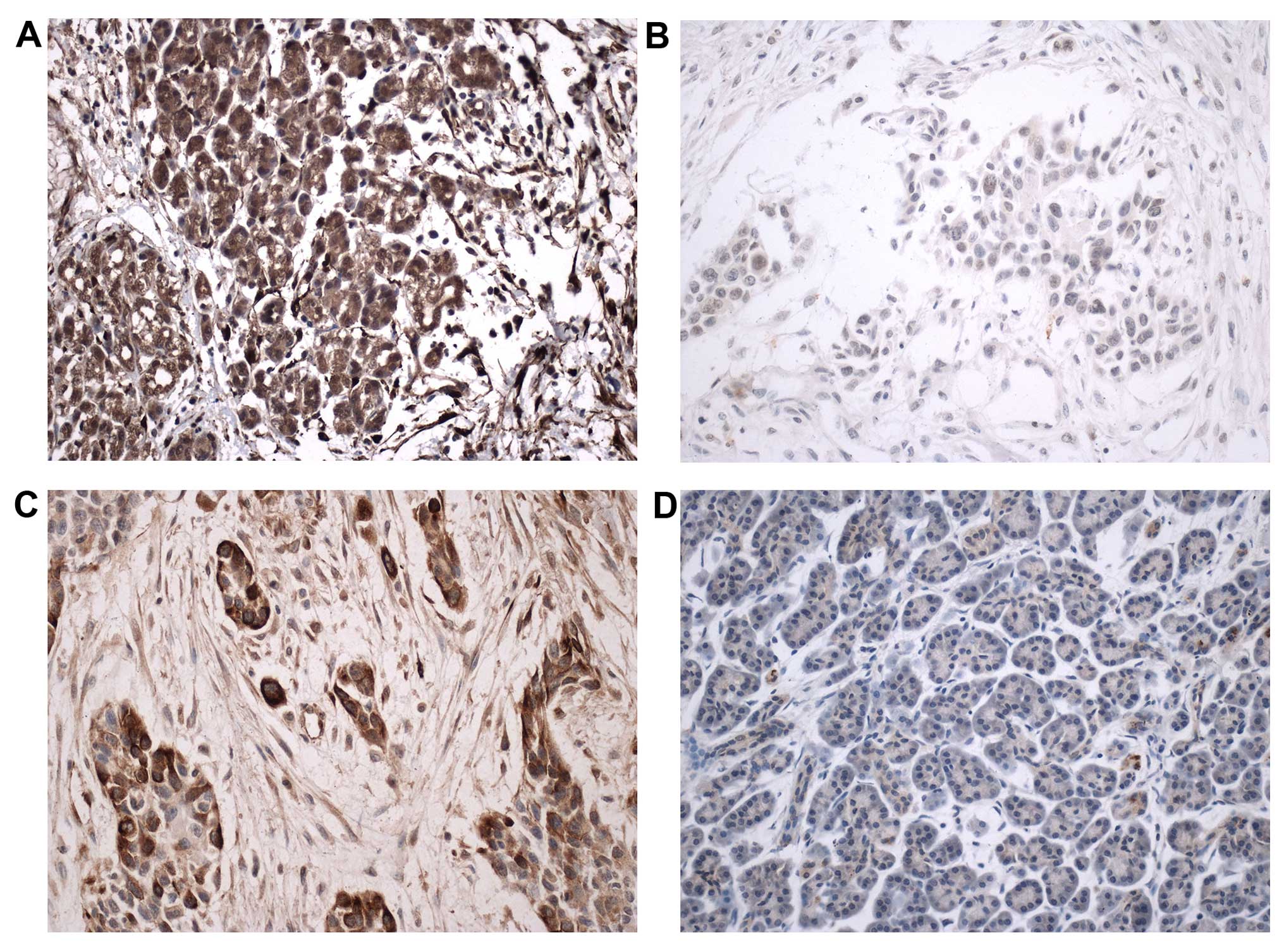
The Lxn protein was highly expressed or was
diffusely positive in the cytoplasm and membrane of the pancreatic
cells in 29 (29/32, 90.6%) adjacent non-cancerous pancreatic
tissues (Fig. 1A). Lxn showed
diffuse expression in the cytoplasm and membrane of the cancer
cells in the carcinoma samples (20, 46.5%). Its expression was low
or negative in 23 (53.5%) tumor samples. Most of the poorly
differentiated cancer cells were negative for the Lxn protein
(Fig. 1B). A significant difference
in Lxn expression was observed between the pancreatic carcinoma and
adjacent non-cancerous pancreatic tissues (P=0.000).
High CD133 expression was observed in 24 (55.8%)
carcinoma samples, including diffuse and strong expression in the
membrane and cytoplasm of the cancer cells. Most cancer cells in
the pancreatic carcinoma tissues demonstrated diffuse and strong
positive staining for the CD133 protein in the membrane and
cytoplasm (Fig. 1C). CD133 protein
expression was low or negative in all 32 adjacent non-cancerous
tissues (Fig. 1D). A significant
difference in CD133 expression was observed between the pancreatic
carcinoma and adjacent non-cancerous pancreatic tissues
(P=0.000).
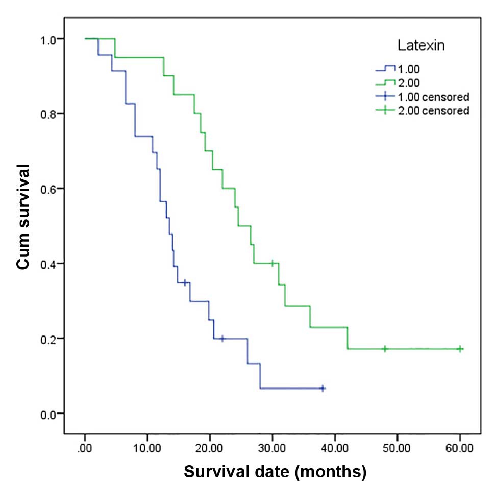
Relationship between Lxn expression and
age, gender, tumor site and size, histological grade, clinical
stage and prognosis
Lxn expression was correlated with tumor size,
histological grade, metastasis, TNM stage and survival but not with
age, gender or tumor site in the 43 pancreatic tumor samples
(Table I). The overall mean
survival time between the individuals with tumors with low Lxn
expression (14.7 months) and those with tumors with high Lxn
expression (28.5 months) was significantly different (P=0.000)
(Fig. 2). These results revealed
that low Lxn expression was significantly related to a shorter mean
survival time (P=0.000) of the cancer patients. According to
multivariate analysis performed using Cox regression, Lxn
expression was an independent prognostic factor for PDAC
(P=0.000).
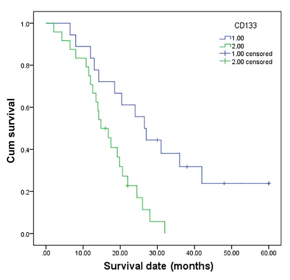
Relationship between CD133 expression and
age, gender, tumor site, tumor size, histological grade, clinical
stage and prognosis
CD133 expression was correlated with histological
grade, metastasis, TNM stage and survival but not with age, gender
or tumor site in the group of 43 pancreatic tumor samples (Table I). The follow-up data revealed a
significant difference in the overall mean survival time between
the patients with a tumor with CD133 high expression (16.2 months)
and those with a tumor with low CD133 expression (27.4 months)
(P=0.000) (Fig. 3). These findings
revealed that high CD133 expression was significantly related to a
shorter mean survival time of the cancer patients. Multivariate
analysis conducted using Cox regression revealed that CD133
expression was also an independent prognostic factor for PDAC
(P=0.000).
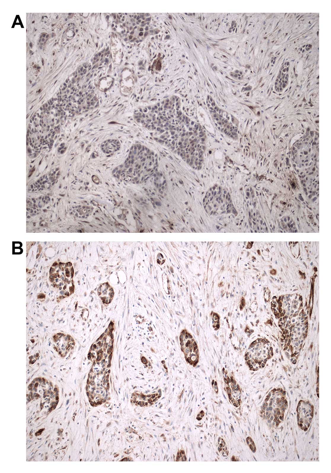
Relationship between Lxn and CD133
expression
When Lxn was not expressed or was expressed at a low
level (Fig. 4A), CD133 was often
found to be highly expressed in cancer cells from the pancreatic
tumors (Fig. 4B). Lxn expression
was detected in only 25% (6/24) of the pancreatic tumors with high
CD133 expression, whereas it was highly expressed in up to 73.7%
(14/19) of the tumors with low CD133 expression. A significant
inverse relationship was found between the expression of Lxn and
CD133 (r=−0.485, P=0.001; Fig.
5).
Effect of pCMV6-AC-GFP-LXN transfection
on Lxn gene expression in the CD133+ MIA PaCa-2
pancreatic cancer cells
As shown in Fig. 6,
the western blotting and qRT-PCR results indicated that Lxn
expression in transfected CD133+ MIA PaCa-2 pancreatic
cancer cells was significantly higher than that in the control
cells and that its expression increased with the pCMV6-AC-GFP-LXN
dose in a concentration-dependent manner (P<0.05). The Lxn gene
was successfully transfected into the CD133+ MIA PaCa-2
pancreatic cancer cells.
Effects of pCMV6-AC-GFP-LXN transfection
on apoptosis and cell growth in the CD133+ MIA PaCa-2
pancreatic cancer cells
We determined the effects of pCMV6-AC-GFP-LXN
transfection on the rate of apoptosis of the CD133+ MIA
PaCa-2 pancreatic cancer cells relative to that of the control
cells. As shown in Fig. 7B–D,
transfection of the cells with pCMV6-AC-GFP-LXN for 48 h increased
the percentage of early and late apoptotic cells as well as
necrotic cells in a dose-dependent manner.
We also determined the cell death-promoting effects
of pCMV6-AC-GFP-LXN transfection on the CD133+ cells. As
shown in Fig. 8, with increasing
doses of transfected pCMV6-AC-GFP-LXN, the inhibitory effects on
CD133+ cells was also increased. Treatment with 0.1 and
0.4 μg pCMV6-AC-GFP-LXN for 48 h resulted in cell growth
inhibition rates of 4.7 and 26.4%, respectively. Similarly, the
inhibitory effects on CD133+ cells increased as a
function of the incubation time. For example, treatment with 0.4
μg pCMV6-AC-GFP-LXN for 24 and 72 h resulted in cell
inhibition rates of 15 and 36.2%, respectively. Therefore, Lxn
suppressed the proliferation of CD133+ cells in a time-
and concentration-dependent manner (Table II).
| Table IIEffect of Lxn gene overexpression on
the proliferation of CD133-positive MIA PaCa-2 cells (OD450, mean ±
SD). |
Table II
Effect of Lxn gene overexpression on
the proliferation of CD133-positive MIA PaCa-2 cells (OD450, mean ±
SD).
| Lxn
μmol/l | OD at 24 h | Inhibition ratio
(%) | OD at 48 h | Inhibition ratio
(%) | OD at 72 h | Inhibition ratio
(%) |
|---|
| Con | 0.92±0.013 | 0.2±0.00 | 0.46±0.012 | 0.3±0.01 | 0.30±0.011 | 0.4±0.01 |
| 0.1 | 0.86±0.019 | 6.5±0.14a | 0.40±0.019 | 4.7±0.22a | 0.27±0.011 | 6±0.22a |
| 0.2 | 0.81±0.142 | 8.7±1.53a | 0.38±0.0208 | 8.5±0.47a | 0.25±0.012 | 14.1±0.68a,b |
| 0.3 | 0.76±0.0318 | 12±0.5a | 0.33±0.022 | 13.2±0.88a | 0.19±0.002 | 32±0.34a,b |
| 0.4 | 0.69±0.0218 | 15±0.47a | 0.27±0.022 | 26.4±2.2a,b | 0.17±0.008 | 36.2±1.7a,b |
Discussion
Lxn has primarily been studied in the nervous system
since it is involved in the specification of cortical brain regions
during development (32), as well
as in the speed of nerve transmission in the adult peripheral
nervous system (33). Lxn has ~30%
sequence homology, but has much greater structural homology, with
TIG1 or retinoic acid receptor responder 1 (RARRES1), the
expression of which is downregulated in a variety of tumor types in
humans (16,34–37).
Lxn and TIG1 are closely linked genetically and may represent
members of a family of functionally related genes. Liang et
al identified the Lxn locus as the primary determinant of HSC
frequency variation between two inbred mouse strains. Exogenous Lxn
expression in the hematopoietic compartment has been demonstrated
to negatively regulate the number of HSCs. Muthusamy et al
also demonstrated that Lxn was downregulated in >50% of
melanomas by promoter hypermethylation. Furthermore, they showed
that Lxn has a tumor-suppressive role and that it negatively
regulates the expression of tumor-sustaining stem cell factors
(19). Recently, Lxn has been
demonstrated to be a tumor-suppressor gene in gastric cancer
(38), hepatocellular carcinoma
(39), melanoma (19), leukemia (40), prostate (41) and breast cancer (42). Our previous study revealed that Lxn
induced the apoptosis and inhibited the proliferation of
CD133+ MIA PaCa-2 pancreatic cancer cells (20). However, to the best of our
knowledge, little is known concerning the role of Lxn in PDAC. To
investigate the relationship between Lxn and the
clinicopathological indices in PDAC patients, we analyzed the
relationship between Lxn and CD133 expression in PDAC tissues by
immunohistochemistry. For the PDAC patients with follow-up data,
our results indicated that low Lxn expression was correlated with
tumor size (P=0.002), histological grade (P=0.000), metastasis
(P=0.007) and clinical stage (P=0.018). Furthermore, our data
showed that low Lxn protein expression was highly associated with
overall survival (P=0.000), suggesting that its expression may be a
potential prognostic factor for PDAC. Thus, Lxn may have a
regulatory role in PDAC as a tumor suppressor.
Currently, CD133 is one of the most frequently
studied markers in pancreatic tumor stem cell research (43–45).
However, some inconsistent results have been obtained with regard
to the relationship between CD133 expression and the
clinicopathological features of PDAC (23,27,43–45).
Nomura et al reported that CD133 expression contributes to
'stemness,' tumorigenicity, epithelial-mesenchymal transition (EMT)
induction, invasion and metastasis in pancreatic cancer (23). Chen et al reported that
co-expression of CD133, CD44v6 and human tissue factor is
associated with metastasis and poor prognosis in pancreatic
carcinoma patients (28). A study
conducted in Korea revealed that upregulation of CD133 expression
in PDAC is related to poor prognosis (27), while Immervoll et al reported
no correlation between CD133 expression and patient survival
(43). One study showed that CD133
expression is positively associated with venous invasion but not
with tumor grade (44), while
another study demonstrated that CD133 expression is significantly
inversely correlated with tumor grade, but not with disease stage
(45). The present study
investigated CD133 protein expression in 43 PDAC patients with
follow-up data, and the results indicated that its expression was
correlated with histological grade (P=0.016) and clinical stage
(P=0.003). Furthermore, our data showed that CD133 protein
expression was highly associated with overall survival (P=0.000),
suggesting that its expression may be a potential prognostic factor
for PDAC. We found that 55.8% of the PDAC samples were
CD133-positive. Our results are consistent with those of a previous
study of PDAC conducted in Korea (27) and with other studies performed by
two research groups in Chongqing (28) and Taiwan (29), China, reporting rates of CD133
positivity of 66.7, 59.6 and 43.7%, respectively; in addition,
these studies considered CD133 expression to be a potential
prognostic indicator.
Currently, our knowledge of the relationship between
Lxn and CD133 is still lacking. We reported that Lxn expression in
CD133+ MIA PaCa-2 pancreatic tumor cells was
significantly lower than that in CD133− MIA PaCa-2
cells. Moreover, exogenous Lxn inhibits the proliferation of
CD133+ MIA PaCa-2 pancreatic cancer stem-like cells
(20). Our findings suggest that
expression of Lxn is inversely correlated with that of CD133
(P=0.001), implying that the loss of Lxn function may occur in
cancer cells with high CD133 expression. Furthermore, we found that
CD133-positive MIA PaCa-2 cells overexpressing Lxn exhibited
significantly increased apoptosis and low proliferative activity,
indicating that Lxn may act as a tumor suppressor that targets
CD133-positive pancreatic cancer cells.
Acknowledgments
The present study received research funding from the
Zhejiang Provincial Natural Science Foundation of China (Y2100546),
the Wenzhou Municipal Science and Technology Bureau of Zhejiang
Province, China (Y20090086). The statistical methods used in the
present study were reviewed by Zuo-Kai Xie from the General
Statistics Office of The Second Affiliated Hospital and Yuying
Children's Hospital of Wenzhou Medical University.
References
|
1
|
Jemal A, Siegel R, Ward E, Hao Y, Xu J,
Murray T and Thun MJ: Cancer statistics, 2008. CA Cancer J Clin.
58:71–96. 2008.
|
|
2
|
Sultana A, Smith CT, Cunningham D,
Starling N, Neoptolemos JP and Ghaneh P: Meta-analyses of
chemotherapy for locally advanced and metastatic pancreatic cancer.
J Clin Oncol. 25:2607–2615. 2007.
|
|
3
|
Tan BT, Park CY, Ailles LE and Weissman
IL: The cancer stem cell hypothesis: A work in progress. Lab
Invest. 86:1203–1207. 2006.
|
|
4
|
Hruban RH, Maitra A and Goggins M: Update
on pancreatic intraepithelial neoplasia. Int J Clin Exp Pathol.
1:306–316. 2008.
|
|
5
|
Li C, Heidt DG, Dalerba P, Burant CF,
Zhang L, Adsay V, Wicha M, Clarke MF and Simeone DM: Identification
of pancreatic cancer stem cells. Cancer Res. 67:1030–1037.
2007.
|
|
6
|
Huang P, Wang CY, Gou SM, Wu HS, Liu T and
Xiong JX: Isolation and biological analysis of tumor stem cells
from pancreatic adenocarcinoma. World J Gastroenterol.
14:3903–3907. 2008.
|
|
7
|
Ikenaga N, Ohuchida K, Mizumoto K, Yu J,
Kayashima T, Hayashi A, Nakata K and Tanaka M: Characterization of
CD24 expression in intraductal papillary mucinous neoplasms and
ductal carcinoma of the pancreas. Hum Pathol. 41:1466–1474.
2010.
|
|
8
|
Hong SP, Wen J, Bang S, Park S and Song
SY: CD44-positive cells are responsible for gemcitabine resistance
in pancreatic cancer cells. Int J Cancer. 125:2323–2331. 2009.
|
|
9
|
Li C, Wu JJ, Hynes M, Dosch J, Sarkar B,
Welling TH, Pasca di Magliano M and Simeone DM: c-Met is a marker
of pancreatic cancer stem cells and therapeutic target.
Gastroenterology. 141:2218–2227.e5. 2011.
|
|
10
|
Hermann PC, Huber SL, Herrler T, Aicher A,
Ellwart JW, Guba M, Bruns CJ and Heeschen C: Distinct populations
of cancer stem cells determine tumor growth and metastatic activity
in human pancreatic cancer. Cell Stem Cell. 1:313–323. 2007.
|
|
11
|
Olempska M, Eisenach PA, Ammerpohl O,
Ungefroren H, Fandrich F and Kalthoff H: Detection of tumor stem
cell markers in pancreatic carcinoma cell lines. Hepatobiliary
Pancreat Dis Int. 6:92–97. 2007.
|
|
12
|
Kim MP, Fleming JB, Wang H, Abbruzzese JL,
Choi W, Kopetz S, McConkey DJ, Evans DB and Gallick GE: ALDH
activity selectively defines an enhanced tumor-initiating cell
population relative to CD133 expression in human pancreatic
adenocarcinoma. PLoS One. 6:e206362011.
|
|
13
|
Maeda S, Shinchi H, Kurahara H, Mataki Y,
Maemura K, Sato M, Natsugoe S, Aikou T and Takao S: CD133
expression is correlated with lymph node metastasis and vascular
endothelial growth factor-C expression in pancreatic cancer. Br J
Cancer. 98:1389–1397. 2008.
|
|
14
|
Maréchal R, Demetter P, Nagy N, Berton A,
Decaestecker C, Polus M, Closset J, Devière J, Salmon I and Van
Laethem JL: High expression of CXCR4 may predict poor survival in
resected pancreatic adenocarcinoma. Br J Cancer. 100:1444–1451.
2009.
|
|
15
|
Kawamoto M, Ishiwata T, Cho K, Uchida E,
Korc M, Naito Z and Tajiri T: Nestin expression correlates with
nerve and retro-peritoneal tissue invasion in pancreatic cancer.
Hum Pathol. 40:189–198. 2009.
|
|
16
|
Youssef EM, Chen XQ, Higuchi E, Kondo Y,
Garcia-Manero G, Lotan R and Issa JP: Hypermethylation and
silencing of the putative tumor suppressor Tazarotene-induced
gene 1 in human cancers. Cancer Res. 64:2411–2417. 2004.
|
|
17
|
Liang Y, Jansen M, Aronow B, Geiger H and
Van Zant G: The quantitative trait gene latexin influences the size
of the hematopoietic stem cell population in mice. Nat Genet.
39:178–188. 2007.
|
|
18
|
Mitsunaga K, Kikuchi J, Wada T and
Furukawa Y: Latexin regulates the abundance of multiple cellular
proteins in hematopoietic stem cells. J Cell Physiol.
227:1138–1147. 2012.
|
|
19
|
Muthusamy V, Premi S, Soper C, Platt J and
Bosenberg M: The hematopoietic stem cell regulatory gene latexin
has tumor-suppressive properties in malignant melanoma. J Invest
Dermatol. 133:1827–1833. 2013.
|
|
20
|
Xue ZX, Zheng JH, Zheng ZQ, Cai JL, Ye XH,
Wang C, Sun WJ, Zhou X, Lu MD, Li PH, et al: Latexin inhibits the
proliferation of CD133+ miapaca-2 pancreatic cancer
stem-like cells. World J Surg Oncol. 12:4042014.
|
|
21
|
Shmelkov SV, St Clair R, Lyden D and Rafii
S: AC133/CD133/Prominin-1. Int J Biochem Cell Biol. 37:715–719.
2005.
|
|
22
|
Banerjee S, Nomura A, Sangwan V, Chugh R,
Dudeja V, Vickers SM and Saluja A: CD133+ tumor
initiating cells in a syngenic murine model of pancreatic cancer
respond to Minnelide. Clin Cancer Res. 20:2388–2399. 2014.
|
|
23
|
Nomura A, Banerjee S, Chugh R, Dudeja V,
Yamamoto M, Vickers SM and Saluja AK: CD133 initiates tumors,
induces epithelial-mesenchymal transition and increases metastasis
in pancreatic cancer. Oncotarget. 6:8313–8322. 2015.
|
|
24
|
Ricci-Vitiani L, Lombardi DG, Pilozzi E,
Biffoni M, Todaro M, Peschle C and De Maria R: Identification and
expansion of human colon-cancer-initiating cells. Nature.
445:111–115. 2007.
|
|
25
|
Singh SK, Clarke ID, Terasaki M, Bonn VE,
Hawkins C, Squire J and Dirks PB: Identification of a cancer stem
cell in human brain tumors. Cancer Res. 63:5821–5828. 2003.
|
|
26
|
Singh SK, Hawkins C, Clarke ID, Squire JA,
Bayani J, Hide T, Henkelman RM, Cusimano MD and Dirks PB:
Identification of human brain tumour initiating cells. Nature.
432:396–401. 2004.
|
|
27
|
Kim HS, Yoo SY, Kim KT, Park JT, Kim HJ
and Kim JC: Expression of the stem cell markers CD133 and nestin in
pancreatic ductal adenocarcinoma and clinical relevance. Int J Clin
Exp Pathol. 5:754–761. 2012.
|
|
28
|
Chen K, Li Z, Jiang P, Zhang X, Zhang Y,
Jiang Y, He Y and Li X: Co-expression of CD133, CD44v6 and human
tissue factor is associated with metastasis and poor prognosis in
pancreatic carcinoma. Oncol Rep. 32:755–763. 2014.
|
|
29
|
Hou YC, Chao YJ, Tung HL, Wang HC and Shan
YS: Coexpression of CD44-positive/CD133-positive cancer stem cells
and CD204-positive tumor-associated macrophages is a predictor of
survival in pancreatic ductal adenocarcinoma. Cancer.
120:2766–2777. 2014.
|
|
30
|
Chu X, Zhao P, Lv Y and Liu L: Decreased
expression of TFPI-2 correlated with increased expression of CD133
in cholangiocarcinoma. Int J Clin Exp Pathol. 8:328–336. 2015.
|
|
31
|
Hao XP, Willis JE, Pretlow TG, Rao JS,
MacLennan GT, Talbot IC and Pretlow TP: Loss of fragile histidine
triad expression in colorectal carcinomas and premalignant lesions.
Cancer Res. 60:18–21. 2000.
|
|
32
|
Arimatsu Y: Latexin: A molecular marker
for regional specification in the neocortex. Neurosci Res.
20:131–135. 1994.
|
|
33
|
Jin M, Ishida M, Katoh-Fukui Y, Tsuchiya
R, Higashinakagawa T, Ikegami S and Arimatsu Y: Reduced pain
sensitivity in mice lacking latexin, an inhibitor of
metallocarboxypeptidases. Brain Res. 1075:117–121. 2006.
|
|
34
|
Aagaard A, Listwan P, Cowieson N, Huber T,
Ravasi T, Wells CA, Flanagan JU, Kellie S, Hume DA, Kobe B, et al:
An inflammatory role for the mammalian carboxypeptidase inhibitor
latexin: Relationship to cystatins and the tumor suppressor TIG1.
Structure. 13:309–317. 2005.
|
|
35
|
Jing C, El-Ghany MA, Beesley C, Foster CS,
Rudland PS, Smith P and Ke Y: Tazarotene-induced gene 1 (TIG1)
expression in prostate carcinomas and its relationship to
tumorigenicity. J Natl Cancer Inst. 94:482–490. 2002.
|
|
36
|
Kwong J, Lo KW, Chow LS, Chan FL, To KF
and Huang DP: Silencing of the retinoid response gene TIG1
by promoter hyper-methylation in nasopharyngeal carcinoma. Int J
Cancer. 113:386–392. 2005.
|
|
37
|
Zhang J, Liu L and Pfeifer GP: Methylation
of the retinoid response gene TIG1 in prostate cancer
correlates with methylation of the retinoic acid receptor beta
gene. Oncogene. 23:2241–2249. 2004.
|
|
38
|
Li Y, Basang Z, Ding H, Lu Z, Ning T, Wei
H, Cai H and Ke Y: Latexin expression is downregulated in human
gastric carcinomas and exhibits tumor suppressor potential. BMC
Cancer. 11:1212011.
|
|
39
|
Ni QF, Tian Y, Kong LL, Lu YT, Ding WZ and
Kong LB: Latexin exhibits tumor suppressor potential in
hepatocellular carcinoma. Oncol Rep. 31:1364–1372. 2014.
|
|
40
|
Liu Y, Howard D, Rector K, Swiderski C,
Brandon J, Schook L, Mehta J, Bryson JS, Bondada S and Liang Y:
Latexin is downregulated in hematopoietic malignancies and
restoration of expression inhibits lymphoma growth. PLoS One.
7:e449792012.
|
|
41
|
Oldridge EE, Walker HF, Stower MJ, Simms
MS, Mann VM, Collins AT, Pellacani D and Maitland NJ: Retinoic acid
represses invasion and stem cell phenotype by induction of the
metastasis suppressors RARRES1 and LXN. Oncogenesis. 2:e452013.
|
|
42
|
Zhang H, Ren Y, Pang D and Liu C: Clinical
implications of AGbL2 expression and its inhibitor latexin in
breast cancer. World J Surg Oncol. 12:1422014.
|
|
43
|
Immervoll H, Hoem D, Sakariassen PØ,
Steffensen OJ and Molven A: Expression of the 'stem cell marker'
CD133 in pancreas and pancreatic ductal adenocarcinomas. BMC
Cancer. 8:482008.
|
|
44
|
Kure S, Matsuda Y, Hagio M, Ueda J, Naito
Z and Ishiwata T: Expression of cancer stem cell markers in
pancreatic intraepithelial neoplasias and pancreatic ductal
adenocarcinomas. Int J Oncol. 41:1314–1324. 2012.
|
|
45
|
Vizio B, Mauri FA, Prati A, Trivedi P,
Giacobino A, Novarino A, Satolli MA, Ciuffreda L, Camandona M,
Gasparri G, et al: Comparative evaluation of cancer stem cell
markers in normal pancreas and pancreatic ductal adenocarcinoma.
Oncol Rep. 27:69–76. 2012.
|