Introduction
Colon cancer, the second major cause of
cancer-related death in the US, has also become a common malignancy
in Asia due to recent changes in diet. Individuals with colorectal
cancer that has spread from the colon or rectum to other parts of
the body often undergo chemotherapy or radiation using a
combination of targeted drugs to alleviate symptoms of the disease
and reduce local failure and distant metastasis or prolong survival
time, thereby improving the clinical outcome of patients (1,2).
Vascular endothelial growth factor (VEGF) plays an
essential role in the progression of cancer by stimulating the new
growth of blood vessels (3).
Essentially, VEGF stimulates the body to provide a blood supply for
a newly developing cancer (4).
SU5416 inhibits the effects of VEGF in the body and is now being
evaluated in patients with metastatic colorectal cancer (5). The combination of radiotherapy and
targeted drugs with anti-angiogenic or anti-vascular effects may be
the most prospective approach to improving therapeutic anticancer
results (3,6,7).
In the present study, we investigated the
enhancement of radiation-induced antitumor and antimetastatic
effects in colorectal cancer cells induced by combination treatment
with SU5416. We found that the addition of SU5416 markedly enhanced
the therapeutic efficacy of radiation in colon cancer cells through
an increase in apoptosis and inhibition of metastatic potential.
Taken together, our results show that SU5416 can be successfully
combined with a radiation regimen to potentiate its antitumor and
antimetastatic activities. The present study provides a scientific
rationale for further exploration of the efficacy of the
combination of radiation and VEGF-targeted therapy in controlling
the growth of colon cancer cells for future clinical trials.
Materials and methods
Antibodies and chemicals
Antibody against β-actin was purchased from Santa
Cruz Biotechnology (Santa Cruz, CA, USA). Antibodies against
phosphorylated extracellular signal-regulated kinase (p-ERK), ERK,
p-p38 and p38 were purchased from Cell Signaling Technology
(Danver, MA, USA). Anti-γ H2A histone family, member X (γH2AX)
antibody was purchased from Millipore (Billerica, MA, USA). The
angiogenesis inhibitor SU5416 was synthesized at Sugen Inc. as
previously described (8). For in
vitro experiments, SU5416 was dissolved in DMSO as a 2 mmol/l
stock solution that was stored at −20°C.
Cell culture
The human colorectal cancer cell lines HT29 and
HCT116 were grown in Roswell Park Memorial Institute (RPMI)-1640
medium supplemented with 10% fetal bovine serum (FBS), glutamine,
HEPES and antibiotics at 37°C in a 5% CO2 humidified
incubator.
Irradiation
Cells were plated in 60-mm dishes and incubated at
37°C in a 5% CO2 humidified incubator, and grown to
70–80% confluency. Cells were irradiated with a 137Cs
γ-ray source (Atomic Energy of Canada, Ltd., Ontario, Canada) at a
dose rate of 3.81 Gy/min.
Colony forming assay
SU5416 (2 μmol/l) was preincubated for 24 h
before radiation exposure and then incubated for a total of 72 h.
After 14–20 days, colonies were stained with 0.4% crystal violet
(Sigma-Aldrich, St. Louis, MO, USA). The plating efficiency (PE)
represents the percentage of seeded cells that grew into colonies
under specific culture conditions for a given cell line. The
survival fraction, expressed as a function of irradiation, was
calculated as follows: survival fraction = colonies counted/(cells
seeded × PE/100). To evaluate the radiosensitizing effects of
SU5416, the enhancement ratio was calculated as the dose (Gy) for
radiation alone divided by the dose for radiation plus SU5416 that
yielded a surviving fraction of 50% (D50).
Detection of apoptotic cells by Annexin V
staining
After SU5416 exposure, the cells were treated with
radiation for 48 h. Cells were washed with ice-cold
phosphate-buffered saline (PBS), trypsinized and resuspended in 1X
binding buffer [10 mm HEPES/NaOH (pH 7.4), 140 mm NaCl and 2.5 mm
CaCl2] at 1×10⁶ cells/ml. Aliquots (100
μl) of the cell solution were mixed with 5 μl Annexin
V-fluorescein isothiocyanate (FITC) (BD Pharmingen) and 10
μl propidium iodide (PI) stock solution (50 μg/ml in
PBS) by gentle vortexing, followed by a 15-min incubation at room
temperature in the dark. Cells were resuspended in 1X binding
buffer (400 μl) and analyzed on a FACScan flow cytometer (BD
Biosciences, Franklin Lakes, NJ, USA). At least 10,000 cells were
counted for each sample and data analysis was performed using
CellQuest software (BD Biosciences).
Measurement of intracellular reactive
oxygen species (ROS)
The levels of intracellular ROS were assessed using
the fluorescent probe 2′,7′-dichlorofluorescein diacetate
(H2DCF-DA). The cells were then incubated with 10
μM H2DCF-DA for 30 min at 37°C, and the green
fluorescence corresponding to the levels of intracellular ROS was
detected through a 520-nm long-pass filter on an Olympus FV-1000
laser fluorescence microscope.
Senescence-associated β-galactosidase
(SA-β-gal) assay
Colon cancer cells were washed twice in PBS, fixed
at room temperature for 6–7 min in 2% formaldehyde/0.2%
glutaraldehyde, then washed three times in PBS and incubated at
37°C with SA-β-gal staining solution [1 mg/m;
5-bromo-4-chloro-3-indolyl β-D-galactoside (Sigma-Aldrich)] in
buffer containing 100 mM citric acid, 200 mM sodium phosphate, 5 mM
potassium ferrocyanide, 5 mM potassium ferrocyanide, 150 mM NaCl
and 2 mM MgCl2 at pH 6.0. Staining was evident after 4–6
h. The cells were washed with PBS and then with distilled water
before microscopic examination. Cells at passage 14 were used as a
control.
Analysis of cell cycle progression
Cells were seeded in 60-mm dishes with 60%
confluency. After 24 h, cells were trypsinized, harvested, and
fixed in 1 ml of 70% cold ethanol in test tubes and incubated at 4
℃ for overnight. After incubation, cells were centrifuged at 2,000
rpm for 3 min, and the cell pellets were resuspended in 500
μl propidium iodine (10 μg/ml) containing 300
μg/ml RNase (Sigma, MO, USA). Cell cycle distribution was
calculated from 10,000 cells with CellQuest software using
FACScaliber (both from Becton Dickinson, CA, USA).
Immunohistochemistry
Immunohistochemistry was performed to determine the
nuclear distribution of γ-H2AX in individual cells. Cells were
grown on chamber slides for 1 day before irradiation or SU5416
treatment. For combination treatment, SU5416 was added to the cells
before the radiation treatment. All treatments were performed while
cells were attached to the slides. After treatment, the cells were
fixed in 4% paraformaldehyde and permeabilized with 0.7% Triton
X-100 in PBS. After blocking with 10% FBS/1% bovine serum albumin
for 1 h, detection was performed using a 1:1,000 dilution of a
FITC-labeled mouse monoclonal γ-H2AX antibody (Millipore).
Western blotting
Colon cancer cells were exposed to SU5416 and then
treated with radiation for 1 and 24 h. The cells were lysed using
radioimmunoprecipitation assay buffer, and equal amounts of
proteins were separated by SDS-polyacrylamide gel electrophoresis
and transferred to nitrocellulose membranes. The membranes were
blocked with 1% (v/v) non-fat dry milk in Tris-buffered saline with
0.05% Tween-20 and then incubated with the indicated antibodies.
Blots were incubated with primary antibodies at 1:1,000 dilutions
and with secondary antibodies at 1:5,000 dilutions. Immunoreactive
protein bands were visualized by enhanced chemiluminescence
(Amersham Biosciences, Piscataway, NJ, USA) and scanned.
Wound healing scratch assay
Human colon cancer cells were seeded onto 6-well
plates (Corning, Corning, NY, USA) at 2.5×10⁴/well in 3
ml RPMI-1640 medium supplemented with 10% FBS. At 2 days, the
monolayers were disrupted mechanically using a sterile 200
μl tip. The assay was performed in duplicate. The wells were
photographed every 24 h. Cells were then stained with 0.2% crystal
violet. Cell migration was monitored using a Nikon Eclipse Ti
microscope with a DS-Fi1 camera. The cells were counted using
ImageJ software (United States National Institutes of Health,
Bethesda, MD, USA).
Invasion assay
The invasive ability of the cells in vitro
was measured using a Transwell invasion assay kit (Chemicon,
Millipore, Norcross, GA, USA) according to the manufacturer's
protocol. Briefly, cells were seeded onto the membrane of the upper
chamber of the Transwell at a concentration of 4×10⁵/ml
in 150 μl serum-free RPMI medium and were left untreated or
were treated with the indicated doses of SU5416, radiation, or a
combination of the two. The medium in the lower chamber contained
10% FBS as a source of chemoattractants. After incubation for 24 h,
the cells that passed through the Matrigel-coated membrane were
stained with a cell stain solution containing crystal violet and
photographed.
Statistical analysis
Statistical significance was determined by the
Student's t-test. Differences were considered significant if the
p-value was <0.05 or 0.001.
Results
Radiosensitizing effect of SU5416 on
colon cancer cell proliferation
To investigate whether SU5416 treatment sensitizes
colon cancer cells to ionizing radiation (IR)-induced cell killing,
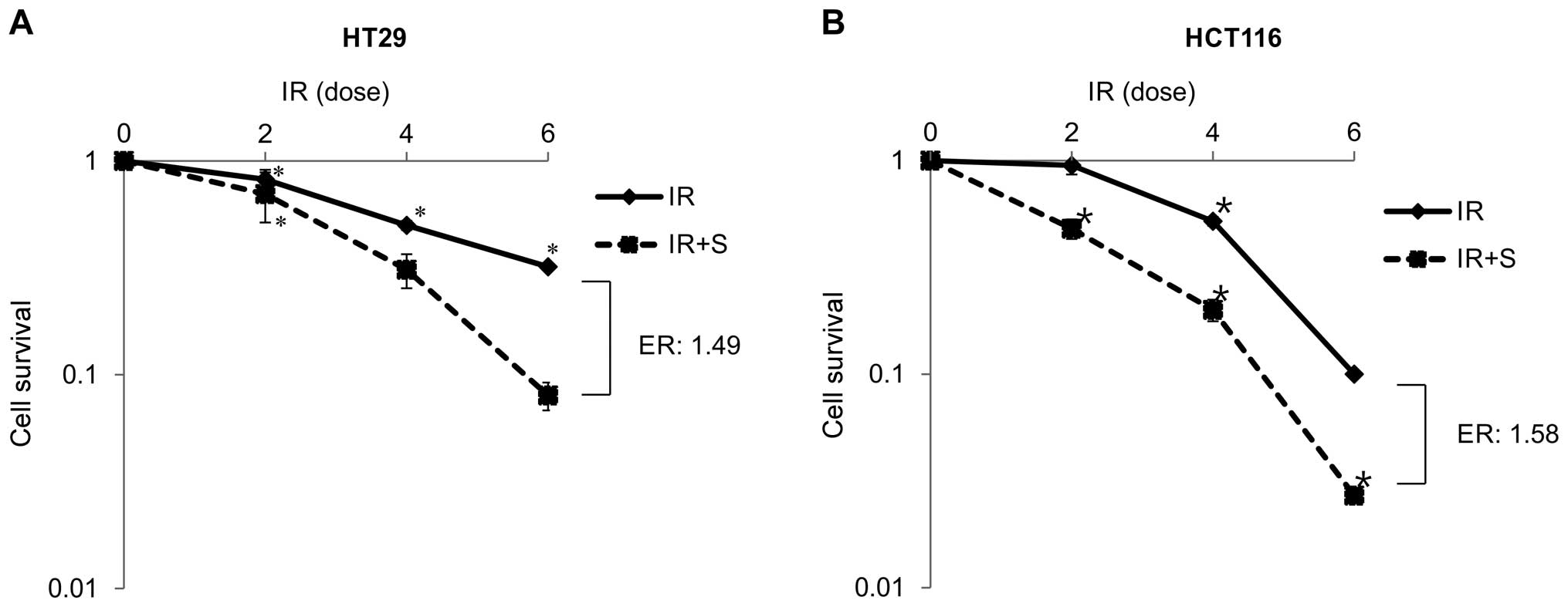
we conducted a clonogenic survival assay. Fig. 1A and B shows the radiation
dose-response curves of radiosensitive HCT116 cells and
radioresistant HT29 cells irradiated with γ-rays in the presence of
SU5416. The survival rate decreased to a greater extent for cells
treated with SU5416 before irradiation compared with cells
irradiated without SU5416, suggesting that SU5416 acts as a
potential radiosensitizer to increase the sensitivity of colon
cancer cells to IR-induced cell killing. The SU5416 effect,
expressed as the enhancement ratio compared with radiation alone
was calculated at D50.
SU5416 enhances radiation-induced
senescence and apoptosis
To further explore the mechanisms by which SU5416
increases the radiation sensitivity of colon cancer cells, we
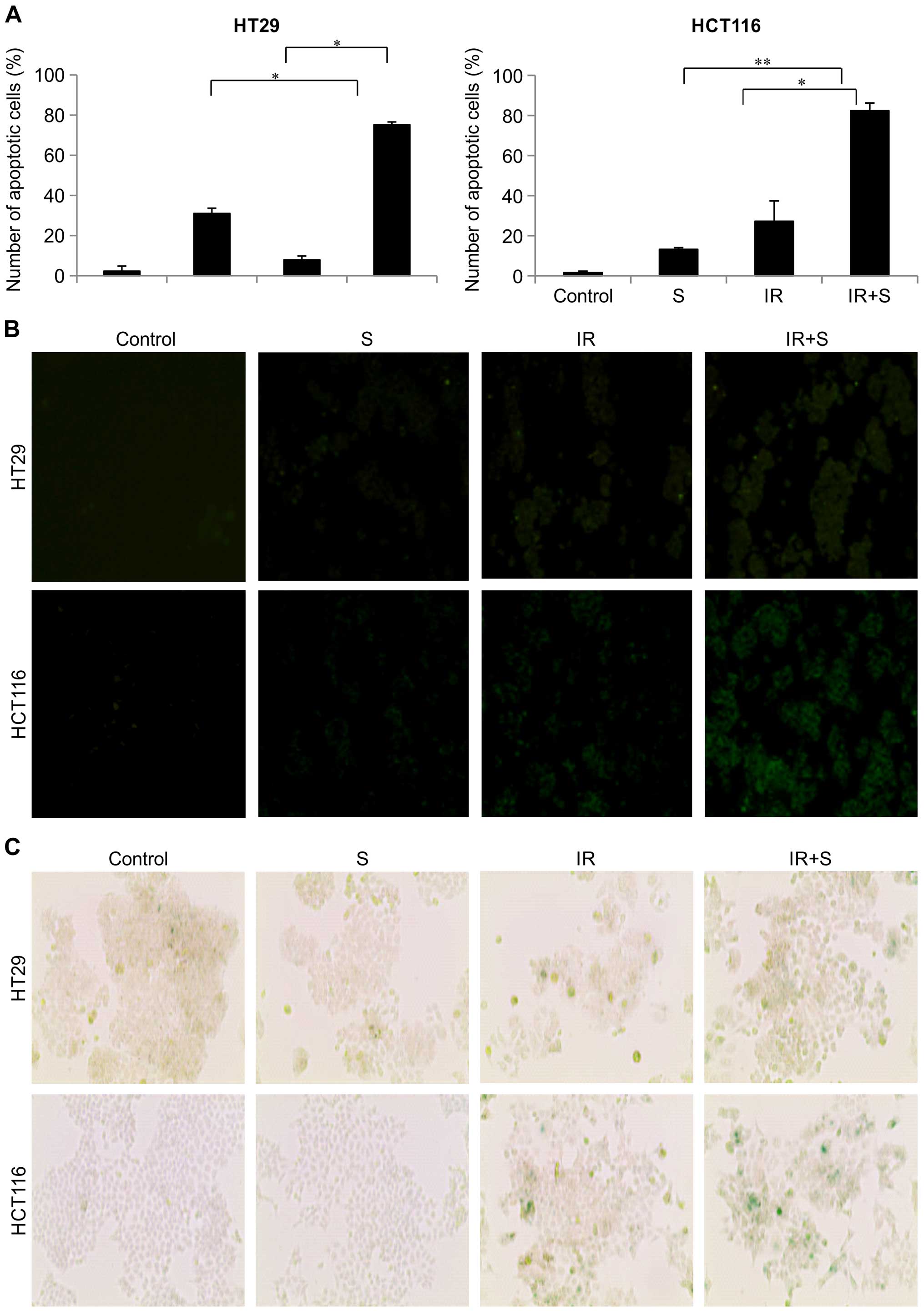
investigated whether SU5416 promotes IR-induced senescence and
apoptosis. We performed staining of the early apoptotic markers
Annexin V and PI in colon cancer cell lines; radiation and SU5416
combination treatment for 72 h significantly increased the
percentage of early apoptotic cells (Fig. 2A). To investigate the relationship
between ROS production and enhancement of radiation-induced
apoptosis by SU5416, we examined the effects of SU5416 on ROS
production in colon cancer cells. ROS production was induced to a
greater extent by the combination of SU5416 and radiation than by
SU5416 or radiation alone (Fig.
2B). Recent studies have shown that IR induces premature
senescence in cancer cells in a dose-dependent manner, suggesting
that the induction of senescence plays an important role in
IR-induced tumor suppression (9).
We stained cells for SA-β-gal and found that the expression of
SA-β-gal correlated with the loss of proliferation and was observed
after approximately 5 days of exposure to various doses (Fig. 2C). The results showed that combined
treatment with SU5416 and IR induced more SA-β-gal-positive
senescent colon cancer cells than did either SU5416 or IR treatment
alone (Fig. 2C). These results
suggest that SU5416 may radiosensitize colon cancer cells by
enhancing IR-induced premature senescence and apoptosis.
Effects of SU5416 and radiation on cell
cycle phase distribution
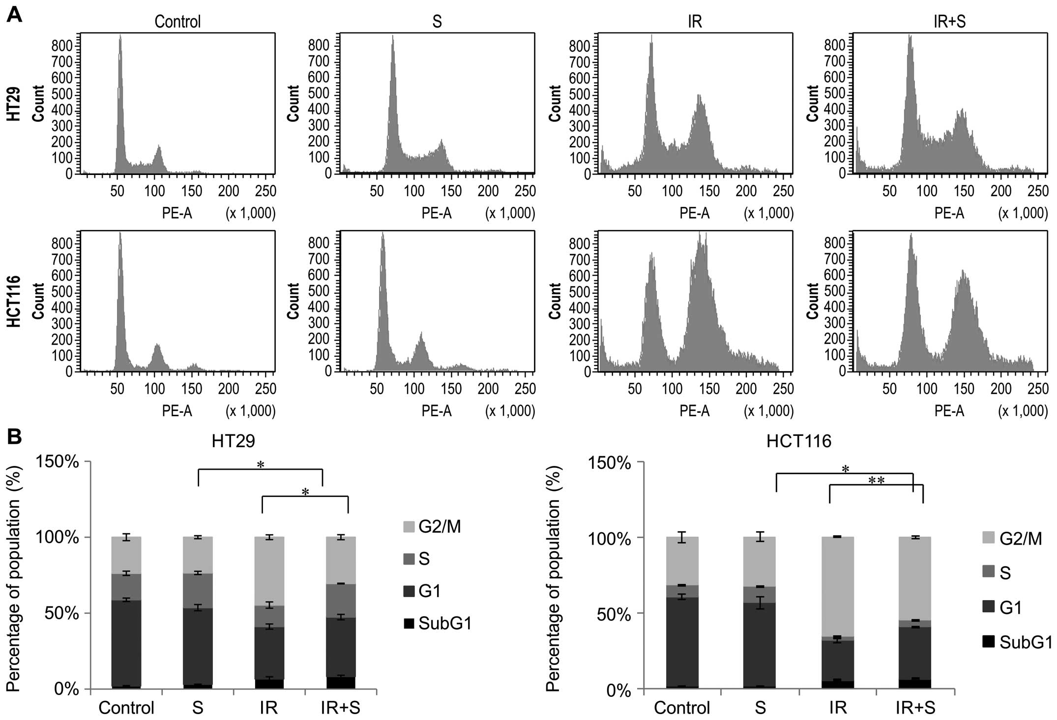
To further investigate the mechanisms underlying the
cell cycle after treatment with SU5416, we performed flow
cytometric analysis of PI-stained cells in the two colon cancer
cell lines (Fig. 3A). As shown in
Fig. 3B, the sub-G1 phase
indicating the apoptotic population was not markedly altered after
treatment with SU5416 alone; however, combined treatment caused
slight accumulation of cells in the sub-G1 phase. Combination
therapy caused a decrease in cells in the G2/M phase, suggesting
efficient induction of cell cycle arrest at the G2/M phase in both
colon cancer cell lines.
Effect of SU5416 on radiation-induced DNA
damage and mitogen-activated protein kinase (MAPK) expression
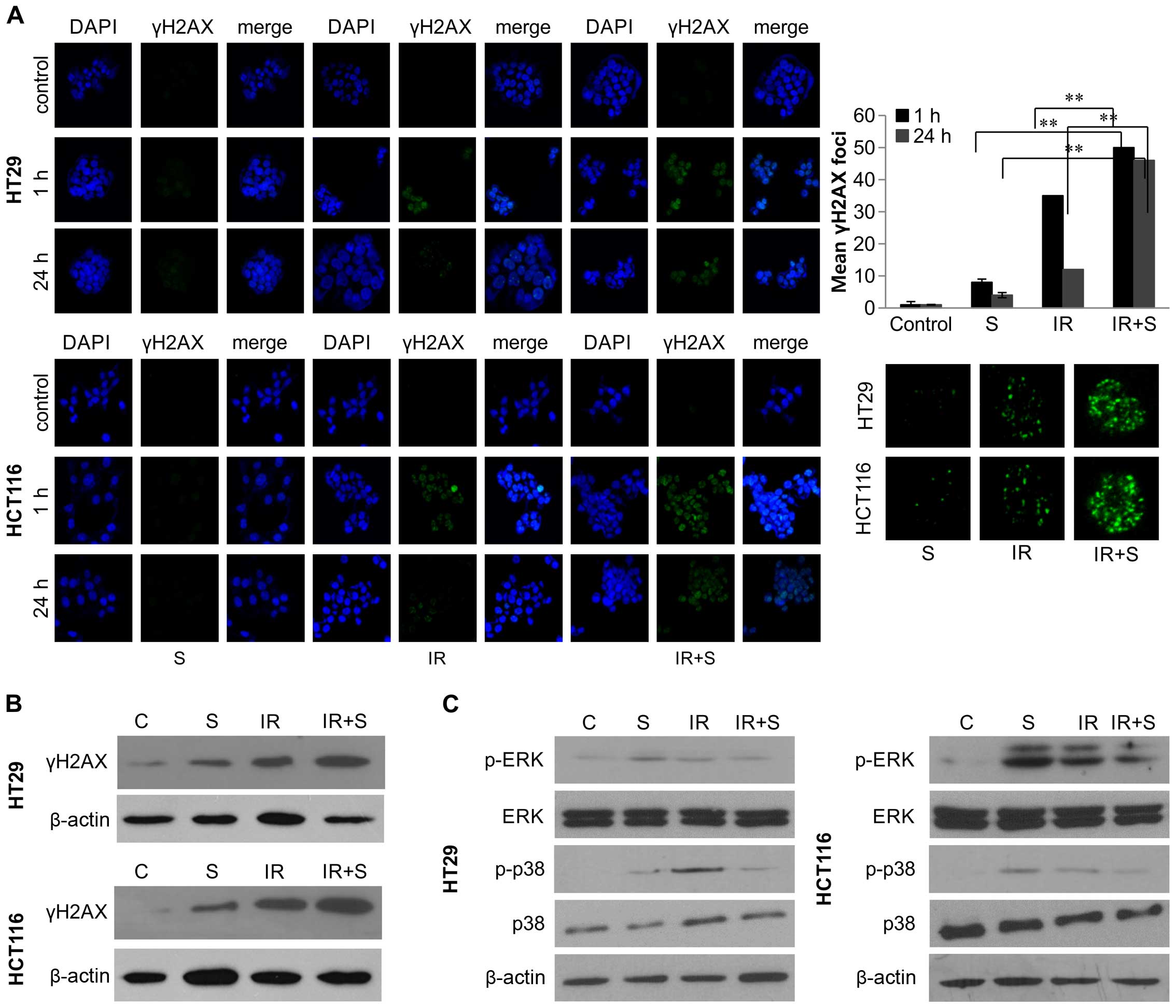
To analyze the effect of SU5416 on DNA double-strand
break (DSB) processing, the level of phosphorylated H2AX (γ-H2AX),
a marker of DSBs, was examined by immunohistochemistry and western
blotting at 0, 1 and 24 h after treatment. As shown in Fig. 4, prolonged expression of γ-H2AX was
observed 24 h after radiation exposure in the presence of SU5416
(Fig. 4A and B). Two MAPK members,
ERK and p38, are involved in DNA damage responses (10). However, it has yet to be determined
if SU5416 treatment affects the activities of ERK and p38 in colon
cancer cells. To address this issue, western blot analyses were
performed to assess the expression levels of p-ERK, ERK, p-p38 and
p38 in the colon cancer cells. The results showed that SU5416
treatment significantly inhibited the expression levels of p-ERK
and p-p38 in the irradiated colon cancer cells (Fig. 4C).
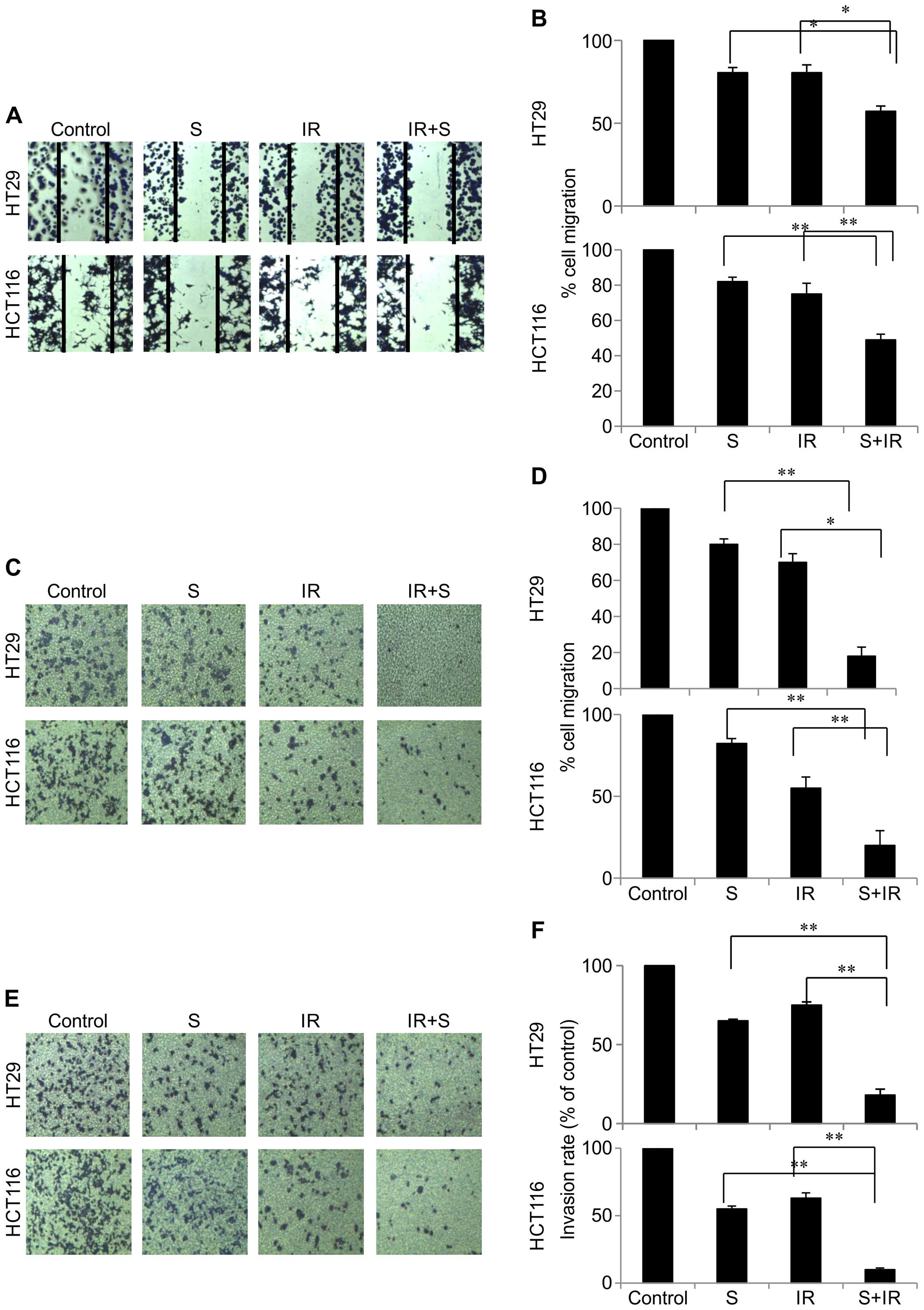
Combination therapy significantly
inhibits tumor cell motility and tumor cell invasion
Cell migration and invasion play crucial roles in
tumor metastasis (11). To further
investigate the antimetastatic effect of SU5416, the ability of
SU5416-treated colon cancer cell migration was assessed by
scratch-wound and Transwell assays. The results from the
scratch-wound assay showed that the wound healing of the
combination treatment was gradually reduced compared with the wound
healing ability of the cells treated with SU5416 or radiation alone
(Fig. 5A and B). We next
investigated the anti-invasive and anti-migratory activities of
SU5416 on colon cancer cells using a Transwell system. As shown in
Fig. 5, SU5416 in combination with
radiation significantly inhibited the migration and invasion of the
two colon cancer cell lines in the chamber Transwell assays
(Fig. 5C–F). These results suggest
that the combination of SU5416 and radiation affected the abilities
of cell migration and invasion.
Discussion
Numerous patients who experience radiotherapy
develop local failure. To improve the efficacy of treatment and
reduce side-effects, there is an increasing interest in various
combinations of radiotherapy using novel targeted therapies
(12). Among them, inhibition of
tumor angiogenesis is one such targeted therapy. The importance of
angiogenesis in the progression of human cancers has been
highlighted by recent studies that have associated angiogenic
phenotypes with patient survival (13). Growing tumors induce angiogenesis to
ensure an adequate delivery of oxygen and nutrients, and several
angiostatic drugs have been approved for the treatment of cancer
patients. Preclinical studies suggest that anti-angiogenic agents
enhance tumor control in response to radiation (14), and large numbers of such compounds
are already in clinical studies or have been approved for the
treatment of certain malignancies (15). Both pre-clinical and clinical
studies have shown that radiotherapy can affect tumor angiogenesis
and that inhibition of angiogenesis can potentiate the effect of
radiotherapy. Thus, inhibition of angiogenesis combined with
radiotherapy is a promising strategy for cancer therapy. In the
present study, we showed that the combination of VEGF signaling
inhibition and radiation significantly enhanced the antitumor
effects of each monotherapy in colon cancer cells.
For these studies, we used SU5416 as an inhibitor of
VEGF signaling in colon cancer cells. Recent clinical studies have
shown that SU5416 is active against metastatic colorectal cancer
when combined with fluorouracil/leucovorin (16). Recent clinical studies have also
shown that the combination of SU5416 and radiation may provide
clinical benefits in patients with certain cancer cell types
(17). However, it is largely
unknown how SU5416 treatment may affect IR-induced sensitivity of
colon cancer cells. Therefore, the aim of the present study was to
explore the molecular mechanisms of potential SU5416
treatment-induced sensitization of colon cancer cells to
radiotherapy. In the present study, we provide a scientific
rationale for the clinical application of SU5416 as a
radiosensitizer in colorectal cancer by evaluating the effect of
single and combined treatments on tumor cell survival, apoptosis,
senescence, cell cycle regulation, DNA repair activity and tumor
cell invasiveness. Our investigations demonstrated that SU5416
combined with radiation significantly decreased clonogenic survival
and that SU5416 enhanced the radiosensitivity of colon cancer cells
by promoting apoptosis through ROS (Figs. 1 and 2). Senescence is a critical mechanism
underlying chemotherapy and radiotherapy-induced tumor suppression.
Therefore, we demonstrated that SU5416 treatment markedly increased
SA-β-gal staining in the irradiated colon cancer cells, suggesting
that SU5416 may sensitize colon cancer cells to radiotherapy via
enhancing IR-induced premature senescence (Fig. 2C). In addition, when SU5416 was
administered before radiation, the colon cancer cells failed to
undergo mitosis. This SU5416-mediated inhibition of cell cycle
progression appeared to be caused by failure of the cells to
undergo transition from the G2 to M phase (Fig. 3). The critical mechanism of tumor
cell killing by IR is DNA damage and, most significantly, DSBs
(18). The rationale for using
cytotoxic chemotherapy as a radiosensitizer is based on the
rationale that additional DNA damage may lower the threshold of
cell death induced by IR. Our results showed that the combination
treatment delayed the clearance of γ-H2AX, suggesting that SU5416
maintains DNA damage and thus, increases the radiosensitivity of
cells (Fig. 4).
Migration is a main event in the metastatic cascade
of cancers, and expression of VEGF-2 plays an important role in
cell migration by activating a number of intracellular signaling
pathways (19–21). Therefore, we examined the effect of
our combination therapy on the migratory characteristics of colon
cancer cells. Our data showed that the combination treatment
effectively inhibited tumor cell invasion and migration mediated by
a Transwell chamber assay (Fig. 5).
VEGF mediates angiogenesis predominantly via the activation of
PI3K/Akt and MAPK signaling cascades (22). ERK and p38 are key downstream
components of the RAF/MEK/ERK signaling pathway, and aberrant
signaling can promote cell immortalization, proliferation and
resistance to radiation (22).
Fig. 4 shows that
radiation-activated p-ERK and p-p38 expression were suppressed by
pre-irradiation treatment with SU5416.
Taken together, our results demonstrated for the
first time that SU5416-induced radiosensitization is associated
with inhibition of tumor cell survival, marked increases in ROS
production, senescence induction, DNA DSBs and tumor cell
invasiveness in irradiated colon cancer cells, suggesting that
increased IR-induced premature senescence could be exploited as a
novel strategy to sensitize colon cancer cells to radiotherapy. Our
data provide a molecular biological basis for the use of
chemoradiation to enhance the therapeutic efficacy against colon
cancer cells. For future research, in vivo mouse models and
evaluation of toxicity to normal cells should be conducted to
determine the clinical applications of SU5416 and minimize
complications.
Acknowledgments
The present study was supported by the National
Research Foundation of Korea (NRF) grants (NRF-2014R1A1A3053958 and
NRF-2014029534) funded by the Korean government (MSIP).
References
|
1
|
Krook JE, Moertel CG, Gunderson LL, Wieand
HS, Collins RT, Beart RW, Kubista TP, Poon MA, Meyers WC, Mailliard
JA, et al: Effective surgical adjuvant therapy for high-risk rectal
carcinoma. N Engl J Med. 324:709–715. 1991. View Article : Google Scholar : PubMed/NCBI
|
|
2
|
Seiwert TY, Salama JK and Vokes EE: The
concurrent chemoradiation paradigm - general principles. Nat Clin
Pract Oncol. 4:86–100. 2007. View Article : Google Scholar : PubMed/NCBI
|
|
3
|
Siemann DW, Bibby MC, Dark GG, Dicker AP,
Eskens FA, Horsman MR, Marmé D and Lorusso PM: Differentiation and
definition of vascular-targeted therapies. Clin Cancer Res.
11:416–420. 2005.PubMed/NCBI
|
|
4
|
Jubb AM, Pham TQ, Hanby AM, Frantz GD,
Peale FV, Wu TD, Koeppen HW and Hillan KJ: Expression of vascular
endothelial growth factor, hypoxia inducible factor 1α, and
carbonic anhydrase IX in human tumours. J Clin Pathol. 57:504–512.
2004. View Article : Google Scholar : PubMed/NCBI
|
|
5
|
Fong TA, Shawver LK, Sun L, Tang C, App H,
Powell TJ, Kim YH, Schreck R, Wang X, Risau W, et al: SU5416 is a
potent and selective inhibitor of the vascular endothelial growth
factor receptor (Flk-1/KDR) that inhibits tyrosine kinase
catalysis, tumor vascularization, and growth of multiple tumor
types. Cancer Res. 59:99–106. 1999.PubMed/NCBI
|
|
6
|
Huber PE, Bischof M, Jenne J, Heiland S,
Peschke P, Saffrich R, Gröne HJ, Debus J, Lipson KE and Abdollahi
A: Trimodal cancer treatment: Beneficial effects of combined
antiangiogenesis, radiation, and chemotherapy. Cancer Res.
65:3643–3655. 2005. View Article : Google Scholar : PubMed/NCBI
|
|
7
|
Bischof M, Abdollahi A, Gong P, Stoffregen
C, Lipson KE, Debus JU, Weber KJ and Huber PE: Triple combination
of irradiation, chemotherapy (pemetrexed), and VEGFR inhibition
(SU5416) in human endothelial and tumor cells. Int J Radiat Oncol
Biol Phys. 60:1220–1232. 2004. View Article : Google Scholar : PubMed/NCBI
|
|
8
|
Krystal GW, Honsawek S, Kiewlich D, Liang
C, Vasile S, Sun L, McMahon G and Lipson KE: Indolinone tyrosine
kinase inhibitors block Kit activation and growth of small cell
lung cancer cells. Cancer Res. 61:3660–3668. 2001.PubMed/NCBI
|
|
9
|
Lu B, Geng L, Musiek A, Tan J, Cao C,
Donnelly E, McMahon G, Choy H and Hallahan DE: Broad spectrum
receptor tyrosine kinase inhibitor, SU6668, sensitizes radiation
via targeting survival pathway of vascular endothelium. Int J
Radiat Oncol Biol Phys. 58:844–850. 2004. View Article : Google Scholar : PubMed/NCBI
|
|
10
|
Kharbanda S, Saleem A, Shafman T, Emoto Y,
Taneja N, Rubin E, Weichselbaum R, Woodgett J, Avruch J, Kyriakis
J, et al: Ionizing radiation stimulates a Grb2-mediated association
of the stress-activated protein kinase with phosphatidylinositol
3-kinase. J Biol Chem. 270:18871–18874. 1995. View Article : Google Scholar : PubMed/NCBI
|
|
11
|
Leber MF and Efferth T: Molecular
principles of cancer invasion and metastasis (Review). Int J Oncol.
34:881–895. 2009.PubMed/NCBI
|
|
12
|
Begg AC, Stewart FA and Vens C: Strategies
to improve radiotherapy with targeted drugs. Nat Rev Cancer.
11:239–253. 2011. View
Article : Google Scholar : PubMed/NCBI
|
|
13
|
Cherrington JM, Strawn LM and Shawver LK:
New paradigms for the treatment of cancer: The role of
anti-angiogenesis agents. Adv Cancer Res. 79:1–38. 2000. View Article : Google Scholar : PubMed/NCBI
|
|
14
|
Wachsberger P, Burd R and Dicker AP: Tumor
response to ionizing radiation combined with antiangiogenesis or
vascular targeting agents: Exploring mechanisms of interaction.
Clin Cancer Res. 9:1957–1971. 2003.PubMed/NCBI
|
|
15
|
Jain RK, Duda DG, Clark JW and Loeffler
JS: Lessons from phase III clinical trials on anti-VEGF therapy for
cancer. Nat Clin Pract Oncol. 3:24–40. 2006. View Article : Google Scholar : PubMed/NCBI
|
|
16
|
Stadler WM, Heimann R, Karczmar GS,
Gajewski T, Kindler H, MacEaneny P, Zamora M, Medved M and Vokes E:
Clinical evaluation of tumor angiogenesis markers in metastatic
cancer. Proc Am Soc Clin Oncol. 20:3822001.
|
|
17
|
Timke C, Zieher H, Roth A, Hauser K,
Lipson KE, Weber KJ, Debus J, Abdollahi A and Huber PE: Combination
of vascular endothelial growth factor receptor/platelet-derived
growth factor receptor inhibition markedly improves radiation tumor
therapy. Clin Cancer Res. 14:2210–2219. 2008. View Article : Google Scholar : PubMed/NCBI
|
|
18
|
Bernier J, Hall EJ and Giaccia A:
Radiation oncology: A century of achievements. Nat Rev Cancer.
4:737–747. 2004. View
Article : Google Scholar : PubMed/NCBI
|
|
19
|
Brooks PC: Cell adhesion molecules in
angiogenesis. Cancer Metastasis Rev. 15:187–194. 1996. View Article : Google Scholar : PubMed/NCBI
|
|
20
|
Basset P, Okada A, Chenard MP, Kannan R,
Stoll I, Anglard P, Bellocq JP and Rio MC: Matrix
metalloproteinases as stromal effectors of human carcinoma
progression: Therapeutic implications. Matrix Biol. 15:535–541.
1997. View Article : Google Scholar : PubMed/NCBI
|
|
21
|
Johnsen M, Lund LR, Rømer J, Almholt K and
Danø K: Cancer invasion and tissue remodeling: Common themes in
proteolytic matrix degradation. Curr Opin Cell Biol. 10:667–671.
1998. View Article : Google Scholar : PubMed/NCBI
|
|
22
|
Roberts PJ and Der CJ: Targeting the
Raf-MEK-ERK mitogen-activated protein kinase cascade for the
treatment of cancer. Oncogene. 26:3291–3310. 2007. View Article : Google Scholar : PubMed/NCBI
|