Introduction
Rhus verniciflua Stokes (RVS) renamed as
Toxicodendron vernicifluum belongs to the Anacardiaceae
family, commonly known as the lacquer tree. RVS has been
traditionally used as an herbal medicine in East Asian countries
for the treatment of gastritis, stomach cancer and atherosclerosis
(1,2). RVS contains compounds such as gallic
and protocatechuic acids, quercetin, fustin, fisetin, sulfuretin
and butein. There is increasing evidence that RVS extract and its
components have antioxidant, anti-inflammatory and/or anticancerous
activities (3–6). Numerous in vitro and in
vivo studies have further shown the antitumor effects of the
RVS extract and/or its components on various human cancer types or
cell lines, including stomach, breast and liver cancer,
osteosarcoma and lymphoma (7–10).
Possible mechanisms underlying the antitumor effects of the RVS
extract and/or its components have also been previously reported,
including induction of apoptosis and inhibition of the
phosphoinositol-3-kinase (PI3K)-Akt/protein kinase B (PKB) pathway
(7), activation of the
AMP-activated protein kinase (AMPK) (11), cell cycle arrest (12) and reduction of manganese
superoxidase (MnSOD) activity or glutathione (GSH) content
(13).
However, herbal medicinal/pharmacological use of RVS
or its extract has been limited due to the presence of an
allergenic substance, urushiol (a mixture of several derivatives of
catechol) which causes severe contact dermatitis in sensitive
individuals (8,14). It is therefore proposed that removal
of urushiol from RVS or its extract is required for its
pharmacological or medicinal uses. Accordingly, it has been shown
that a standardized extract of the allergen-free RVS is efficacious
for the treatment of advanced or metastatic cancers (10). Although numerous studies have been
published concerning the antitumor effect of the allergen-free RVS
extract, molecular and cellular mechanisms by which the
allergen-free RVS extract exerts its anti-growth and/or
apoptosis-inducing effects on human cancer cells are still not
fully understood at present.
In the present study, we prepared an extract of the
allergen-removed RVS (aRVS) based on a traditional method and
investigated its effect on the growth of various human cancer
cells, including lung (A549), breast (MCF-7) and prostate (DU-145)
cancer cell lines. In the present study, we report for the first
time that aRSV extract has strong antiproliferative, anti-survival
and pro-apoptotic effects on A549 human lung cancer cells and the
effects are mediated through modulation of the expression levels
and/or activities of caspase-9/-3, Bcl-2, Mcl-1, Bax, p53 and
S6.
Materials and methods
Reagents and chemicals
Fetal bovine serum (FBS),
4-(2-hydroxyethyl)-1-piperazineethanesulfonic (HEPES), L-glutamate,
1% (w/v) penicillin-streptomycin and phosphate-buffered saline
(PBS) were obtained from Gibco (Paisley, Scotland, UK). Dulbecco's
modified Eagle's medium (DMEM), Roswell Park Memorial Institute
(RPMI)-1640 medium and anti-actin mouse monoclonal antibody were
purchased from Sigma-Aldrich (St. Louis, MO, USA). Cell culture
dishes were purchased from Nunc (Roskilde, Denmark). An Annexin
V-FLUOS staining kit was provided by Roche Diagnostics GmbH
(Mannheim, Germany). Enzyme-linked chemiluminescence (ECL) western
detection reagents were purchased from Thermo Scientific (Waltham,
MA, USA). Bradford reagent was purchased from Bio-Rad (Hercules,
CA, USA). N-benzyloxycarbonyl-Val-Ala-Asp-fluoromethylketone
(z-VAD-fmk) and a protease inhibitor cocktail (100×) were purchased
from Calbiochem (Madison, WI, USA). Antibodies for procaspase-9 and
-3 were purchased from Stressgen (Ann Arbor, MI, USA). Antibodies
for B-cell lymphoma-2 (Bcl-2) and myeloid cell leukemia-1 (Mcl-1),
Bcl-2-associated X protein (Bax), phosphorylated (p)-p53, total
(T)-p53, p-S6, T-S6, HuR and cytochrome c were purchased
from Santa Cruz Biotechnology (Santa Cruz, CA, USA). An antibody
for poly(ADP-ribose) polymerase (PARP) was purchased from Roche
(Basel, Switzerland). An antibody for extracellular signal
regulated kinase-1/2 (ERK-1/2) was purchased from Epitomics
(Burlingame, CA, USA).
Preparation of the aRVS extract
R. verniciflua Stokes (RVS) was purchased
from Chamotdeul Co., Ltd. (Okcheon, Chungcheongbuk-do, Korea). In
the commercial preparation, 8-year-old Rhus trees were
collected and dried during winter. The barks containing the toxic
substance urushiol were extracted from the wood by treatment with
ceramic water. The wood was then naturally dried in a field
setting. After pulverizing the chopped wood into small pieces and
converting it to powder, the powder samples were added to a
heat-treated drying hopper. The preparation procedure was conducted
according to the protocol of the Agricultural Corporation OTSAM
(Okcheon, Chungcheongbuk-do, Korea). After pretreatment, the
non-woven, packed Rhus powder was extracted with purified water in
a 1:2 (w/v) ratio in a 50 l pilot extractor at 121°C and 1.2
kg/cm2 for 24 h. The aRVS-containing flavonoids obtained
from 75 g of Rhus extract powder were extracted in 1,500 l
of water at 121°C and 1.2 kg/cm2 for 2 h. The extract
was filtered using a 1 mm filtration device and concentrated to 15
degrees Brix (°Bx) at 60°C for 4 h at a pressure of 760 mmHg. The
purified aRVS was kept refrigerated at 10°C prior to
experimentation.
Cell culture
A549 human lung carcinoma, MCF-7 human breast cancer
and DU-145 human prostate cancer cells, and normal human diploid
fibroblasts (HDF) were grown in DMEM supplemented with 10% (v/v)
FBS and 1% (w/v) penicillin-streptomycin with 5% (v/v)
Co2 in a 37°C humidified incubator. Respective cells
were allowed to adhere and grown for 24 h prior to treatment with
the aRVS extract.
Cell viability assay
Cell viability assay was performed as previously
described (12). Briefly,
respective human cancer cells and normal HDF were seeded at a
density of 5×10³ cells/well in a 96-well plate. After 24
h of incubation, tester cells were treated with the aRVS extract at
a given concentration. The optimal dose not affecting cytotoxicity
was determined and evaluated using the Cell Counting Kit-8 solution
(Dojindo, Gaithersburg, MD, USA). The aRVS powder was applied for
pretreatment at concentrations of 0, 0.125, 0.25, 0.5 or 1 mg/ml
for 24 h. Relative absorbance of cell viability was measured at 450
nm using a Sunrise microplate absorbance reader (Tecan, Männedorf,
Switzerland) and evaluated in triplicate experiments compared with
the untreated control.
Cell count assay and cell morphological
analysis
A549 cells were seeded in 24-well plates and treated
without or with different concentrations of aRVS extract for 24 h.
The number of surviving cells, which cannot be stained with trypan
blue dye, were counted using standard light microscopy.
Approximately, <100 cells were counted for the analysis. The
cell count assay was carried out in triplicate. Data are expressed
as the mean ± standard error (SE) of three independent experiments.
Survival is expressed as a percentage of the control. Phase
contrast images of the conditioned cells were also captured by an
Olympus phase contrast microscope equipped with a digital camera
(Nikon).
Propidium iodide (PI)-Annexin V
staining
Apoptosis was detected by PI-Annexin V staining
using an Annexin V-FLUOS Staining kit according to the
manufacturer's instructions. The detailed procedure has been
previously described (13).
Preparation of whole cell lysates
Untreated or aRVS-treated A549 cells were washed
with PBS and exposed to cell lysis buffer [50 mM Tris-Cl (pH 7.4),
150 mM NaCl, 0.1% sodium dodecyl sulfate, 0.25% sodium
deoxycholate, 1% Triton X-100, 1% Nonidet P-40, 1 mM EDTA, 1 mM
EGTA and protease inhibitor cocktail (1X)]. The cell lysates were
collected in a 1.5 ml tube and centrifuged for 20 min at 4°C at
12,000 rpm. The supernatant was saved and the protein
concentrations were determined using Bradford reagent.
Western blot analysis
Proteins (50 µg) were separated by SDS-PAGE
(10%) and transferred onto nitrocellulose membranes (Millipore,
Billerica, MA, USA). The membranes were washed with TBS (10 mM
Tris, 150 mM NaCl) supplemented with 0.05% (vol/vol) Tween-20
(TBST) followed by blocking with TBST containing 5% (w/v) non-fat
dried milk. The membranes were incubated overnight with antibodies
specific for the protein of interest at 4°C. The membranes were
exposed to secondary antibodies coupled to horseradish peroxidase
at room temperature for 2 h. The membranes were washed, and
immunoreactivities were detected by enzyme-linked chemiluminescence
(ECL) reagents. The relative expression levels of target proteins
to actin as the loading control protein were quantitatively
analyzed using a densitometer.
Subcellular fractionation
To observe the treatment effect of the aRVS extract
on the mobilization of cytochrome c from the mitochondria to
the cytosol, A549 cells (0.5×10⁶/2 ml/well) were seeded
in 6-well plates the day before the aRVS extract treatment. The
cells were treated without or with aRVS extract (0.5 mg/ml) for 24
h. The control or the aRVS extract-treated cells were then washed
in PBS and successively extracted as described. First, the cells
were extracted with buffer A [10 mM Tris-Cl (pH 7.5), 10 mM NaCl, 3
mM MgCl2, 0.5% (v/v) Nonidet P-40, 1 mM DTT]. The sample
was then centrifuged and the resultant supernatant was saved as
cytosolic proteins. The remaining pellet was washed in the same
buffer A and then extracted in buffer B (10 mM Tris-HCl (pH 7.5),
0.5 M NaCl, 3 mM MgCl2, 0.5% (v/v) Nonidet P-40, 1 mM
DTT) to obtain nuclear proteins.
Measurement of DNA fragmentation
A549 cells were seeded in a 60-mm dish and treated
with or without the aRVS extract in the absence or presence of
z-VAD-fmk for 24 h. The conditioned cells were harvested, washed
and lysed in a buffer [50 mM Tris (pH 8.0), 0.5% sarkosyl, 0.5
mg/ml proteinase K and 1 mM EDTA] at 55°C for 3 h, followed by the
addition of RNase A (0.5 µg/ml) and incubation at 55°C for
18 h. The lysates were centrifuged at 10,000 × g for 20 min.
Genomic DNA was extracted with equal volumes of a neutral
phenol-chloroform-isoamyl alcohol mixture (25:24:1), and analyzed
by electrophoresis on a 1.8% agarose gel. The DNA was visualized
and photographed under UV illumination after staining with ethidium
bromide.
Statistical analyses
GraphPad Prism (GraphPad, San Diego, CA, USA) was
used for the statistical analysis of the cell proliferation assay.
The assay was processed for the assessment of differences between
the aRVS extract-treated and control groups by Student's t-test.
The values are expressed as the mean ± standard deviation (SD).
Statistical significance was evaluated by p-values at 0.001, 0.01
and 0.05. Cell count analysis was carried out in triplicate. Data
are expressed as the mean ± standard error (SE). Significance
(p<0.05) was determined by one-way ANOVA.
Results
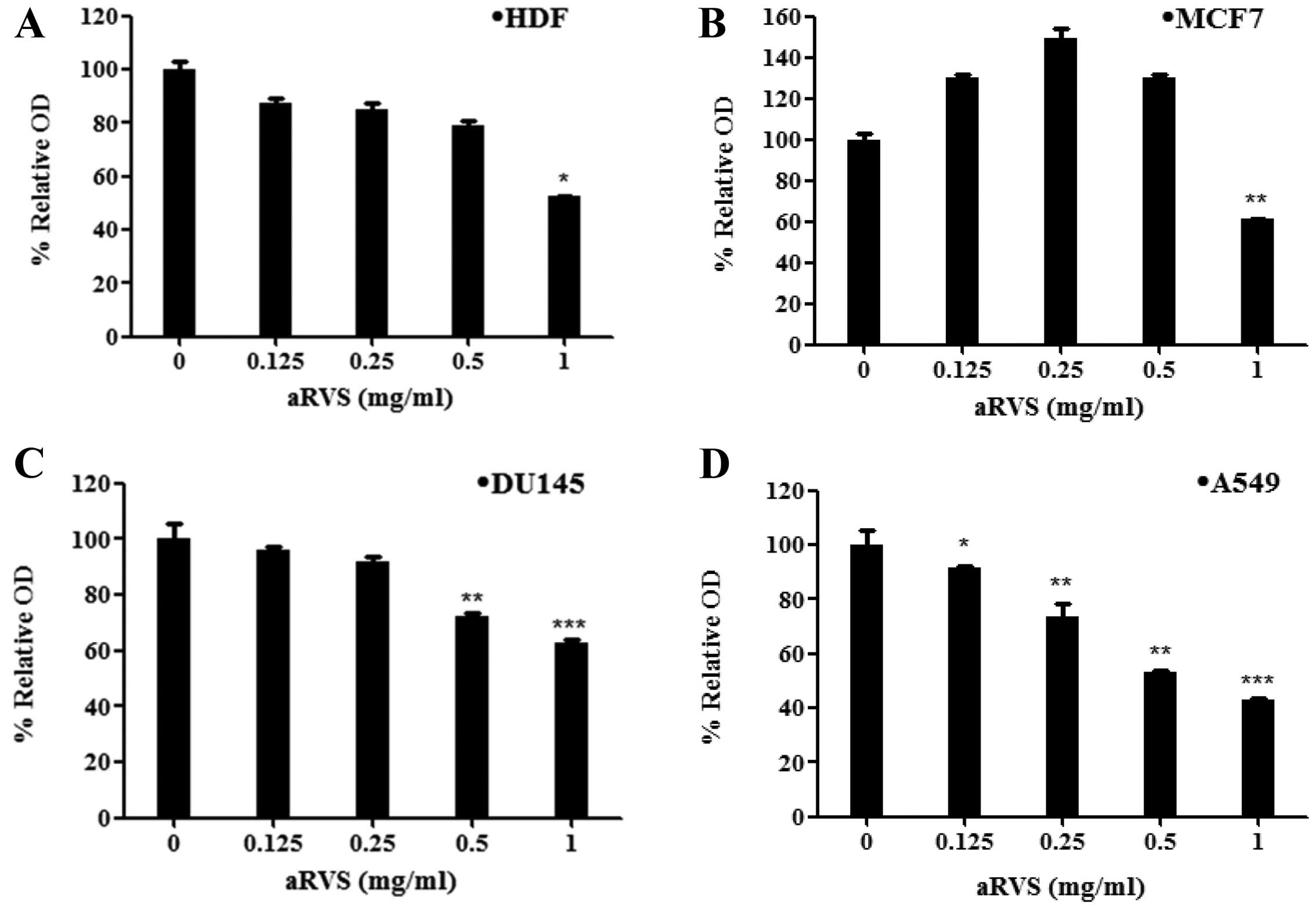
The aRVS extract has strong
antiproliferative activity in the A549 and DU145 cancer cells
Initially, we investigated the treatment effect of
the aRVS extract at different concentrations for 24 h on the
proliferation of various human cancer cell lines, including A549
(lung cancer), MCF-7 (breast cancer) or DU-145 (prostate cancer),
by MTS-based cell proliferation analysis. In the present study,
normal HDFs were also included to assess the effects of the aRVS
extract on normal cell proliferation (Fig. 1A). Treatment with the aRVS extract
dose-dependently inhibited proliferation of the DU-145 and A549
cells as shown in Fig. 1C and D.
Distinctly, the aRVS extract treatment at 0.125, 0.25 or 0.5 mg/ml
enhanced proliferation of the MCF-7 cells (Fig. 1B), but treatment with the aRVS
extract at 1.0 mg/ml led to a large decrease in the cell
proliferation. Although treatment with the aRVS extract at 0.125,
0.25 or 0.5 mg/ml had little effect on proliferation of the HDFs,
treatment with the aRVS extract at 1.0 mg/ml also largely decreased
HDF proliferation. Due to the strong antiproliferative effect on
the A549 cells and no cytotoxicity to normal HDFs, we chose the
A549 cells and the 0.5 mg/ml concentration of the aRVS extract for
further studies.
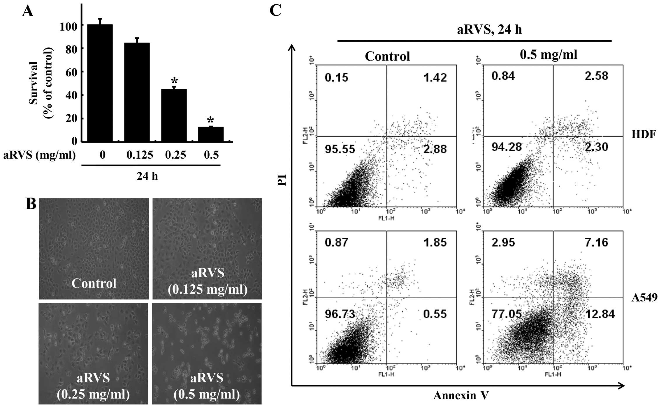
The aRVS extract has strong anti-survival
and pro-apoptotic effects on the A549 cells
We next studied the treatment effect of the aRVS
extract at different concentrations for 24 h on the survival of the
A549 cells by cell count analysis. As shown in Fig. 2A, treatment with the aRVS extract
led to a concentration-dependent decrease in survival of the A549
cells. Data from microscopic observation, as shown in Fig. 2B, also revealed that the aRVS
extract concentration-dependently reduced the number of A549 cells.
Seemingly, when exposed to aRVS extract, A549 cells began to detach
from the surface of the culture plate and appeared buoyant and were
altered from a round shape to a sharp form at the cell poles. We
next determined the effect of the aRVS extract on the apoptosis of
A549 cells by flow cytometry. For the evaluation of apoptosis
herein, the relative proportion of non-viable cells was
quantitatively measured as cells undergoing the early stage of
apoptosis (Annexin stained, non-disrupted cells) or as cells
entering the late stage of apoptosis (disrupted or lysed cells) in
response to the aRVS extract treatment. As shown in Fig. 2C, results of flow cytometry
demonstrated that upon treatment with the aRVS extract at 0.5 mg/ml
for 24 h, many Annexin V-stained viable A549 cells were shifted to
the early apoptotic stage (0.55–12.84%), whereas apoptotic change
of HDF was not observed (2.88–2.30%).
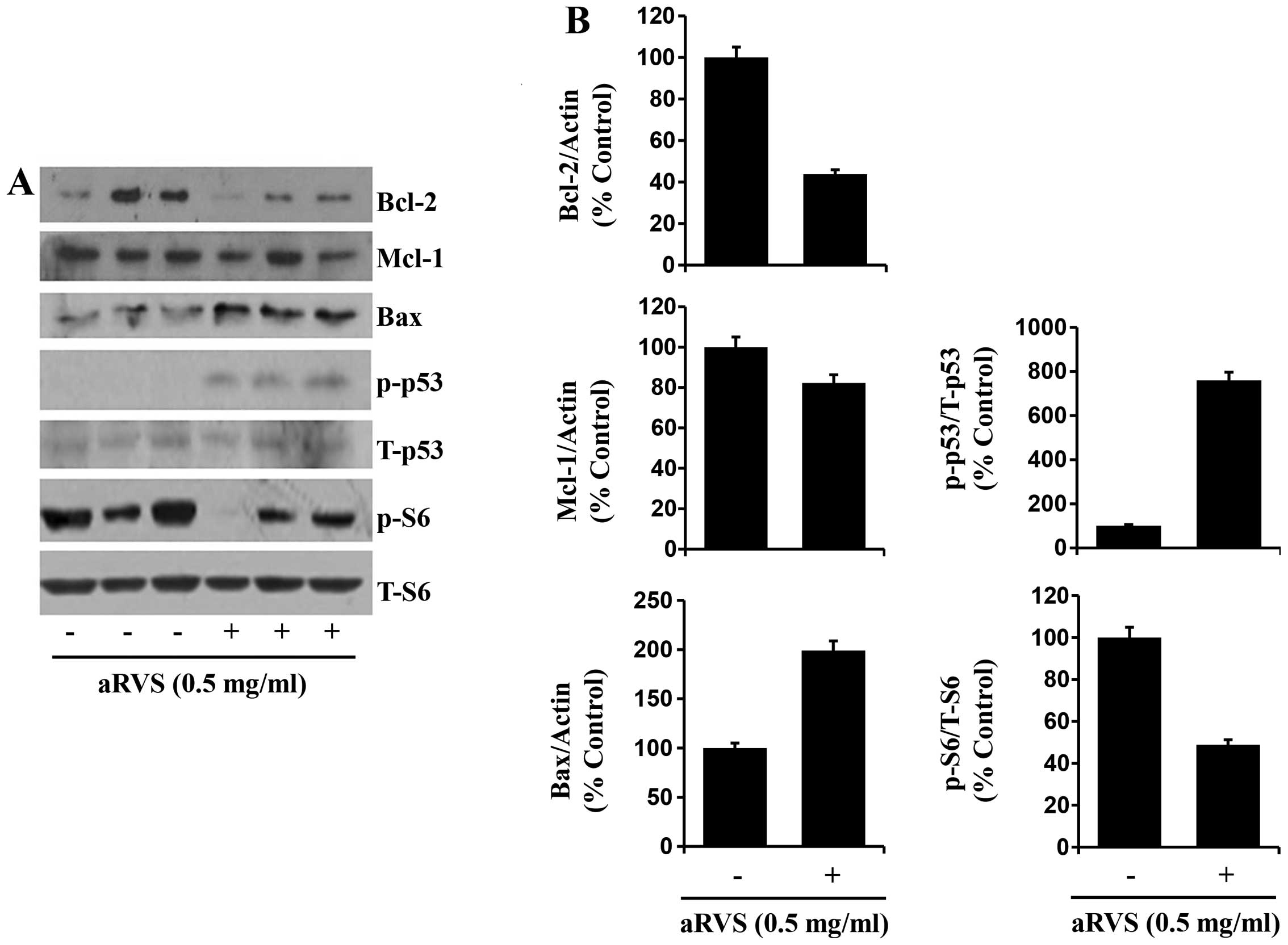
The aRVS extract alters the expression
levels and/or activities of proteins in the A549 cells
We next determined the treatment effect of the aRVS
extract on the expression levels and/or activities of cancer cell
growth- or apoptosis-related proteins in the A549 cells. As shown
in Fig. 3A, treatment with the aRVS
extract at 0.5 g/ml led to downregulation of Bcl-2 and Mcl-1
proteins but upregulation of Bax protein in the A549 cells.
Furthermore, there was an increase in the levels of the
phosphorylated p53 protein but a decrease in the levels of
phosphorylated S6 protein in the A549 cells treated with the aRVS
extract. Fig. 3B are the
densitometry data of Fig. 3A that
show Bcl-2, Mcl-1 and Bax protein levels normalized to actin
protein levels, and p-p53 and p-S6 protein levels normalized to
total p53 and S6 protein levels, respectively.
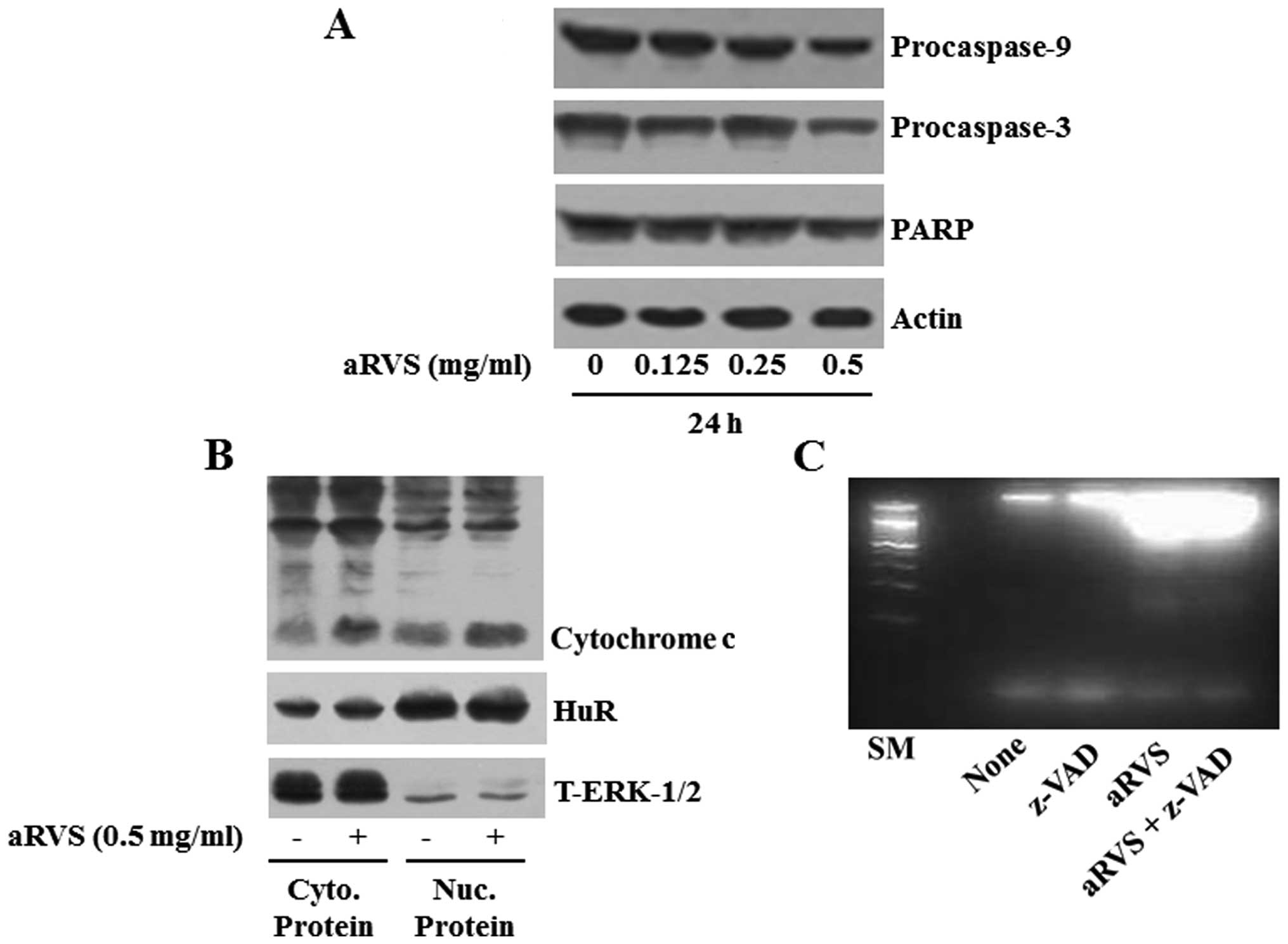
The aRVS extract activates caspase-9/-3
in A549 cells, which is important for aRVS extract-induced
apoptosis
We next investigated the treatment effect of the
aRVS extract on the activity of caspase-9 and -3, other
apoptosis-related proteins, in the A549 cells. In the present
study, the degree of caspase activation by the aRVS extract in the
A549 cells was assessed by decreased expression levels of
procaspase-9 or -3 (inactive form). As shown in Fig. 4A, treatment with the aRVS extract
dose-dependently reduced expression levels of procaspase-9 and -3
in the A549 cells. PARP is a known downstream substrate of
caspases. There was a dose-dependent decrease in the levels of PARP
in the A549 cells treated with the aRVS extract for 24 h,
supporting the activation of caspases. We next carried out
biochemical fractionation experiments (preparation of cytosolic and
subsequent nuclear proteins) to observe the levels of cytosolic
cytochrome c in the A549 cells treated with the aRVS extract
for 24 h. As shown in Fig. 4B,
treatment with the aRVS extract led to an increase in cytosolic
cytochrome c levels in the A549 cells. HuR is a nuclear
protein but shuttles both the cytosolic and nuclear compartment in
cells (15). ERK-1/2 is reported to
be abundantly expressed in the cytosolic compartment (16). Low/high levels of HuR and high/low
levels of ERK-1/2 in the cytosolic and nuclear fractions,
respectively, suggest the fractionation efficiency. Using
z-VAD-fmk, a pan-caspase inhibitor, we next determined the role of
the activation of caspases in the aRVS extract-induced apoptosis of
the A549 cells. As shown in Fig.
4C, aRVS extract-induced apoptosis of the A549 cells was
strongly blocked in the presence of z-VAD-fmk.
Discussion
RVS extract or a standardized extract of the
allergen-free RVS is reported to have anticancer effects on several
human cancers and/or cells, including stomach, breast and liver
cancer, osteosarcoma and lymphoma (7,9–12). The
efficacy and safety of a standardized allergen-free RVS extract as
maintenance therapy in patients with advanced non-small cell lung
cancer or pulmonary adenocarcinoma has also been previously
reported (17). Regulation of human
lung cancer cell growth and apoptosis by aRVS has not been
previously reported. In the present study, we demonstrate for the
first time that an extract of the allergen-removed Rhus
verniciflua Stokes (aRVS) prepared using a traditional method
has strong antiproliferative, anti-survival and pro-apoptotic
effects on A549 human lung cancer cells. Our data also suggest that
the anticancer effects of the aRVS extract on A549 cells are
mediated through modulation of the expression levels and/or
activities of caspase-9/-3, Bcl-2, Mcl-1, Bax, p53, and S6
proteins.
In initial experiments, we demonstrated that the
aRVS extract strongly inhibits proliferation and survival and also
induces apoptosis of A549 cells (Figs.
1D, 2A–C and 4C), as evidenced by results from
microscopic observation, PI-Annexin V staining and/or DNA
fragmentation experiments. Induction of apoptosis is closely
related to two pathways; the intrinsic (mitochondrial) pathway and
the extrinsic (death receptor) pathway. Among the proteins involved
in both these pathways mediating apoptosis, central to both
apoptosis pathways are the caspases, a group of essential proteases
required for the execution of cell death by apoptotic stimuli
(18). It has also been
demonstrated that in resting cells, caspases are synthesized as
zymogens (inactive precursors), but upon exposure to apoptotic
stimuli, they become processed via partial proteolytic cleavage and
activated in cells (19) and that
activated caspases participate in the cleavage of many target
proteins, including PARP or PKC-δ, and other vital proteins
(20). Thus, the present findings
that the aRVS extract induces activation of caspase-9/-3 (Fig. 4A) and increases cytosolic cytochrome
c levels (Fig. 4B) and that
z-VAD-fmk, a pan-caspase inhibitor blocks aRVS extract-induced
apoptosis (Fig. 4C) in the A549
cells strongly suggest that the activation of caspase-9/-3 through
the intrinsic pathway is critical for the aRVS extract-induced
apoptosis of the A549 cells.
An interesting finding of the present study is that
the aRVS extract differentially regulated the expression of the
family of Bcl-2 proteins, including Bcl-2, Mcl-1 and Bax in the
A549 cells (Fig. 3A and B). Bcl-2
and Mcl-1 are pro-survival and anti-apoptotic proteins that are
involved in apoptosis initiation and caspase activation by
regulating the mitochondrial membrane integrity (21–23).
In contrast, Bax is a pro-apoptotic protein that forms a
heterodimer with Bcl-2, and functions as an apoptotic activator
(23,24). It has also been shown that Bax
interacts with, and increases the opening of, the mitochondrial
voltage-dependent anion channel, which leads to the loss in
membrane potential and the release of cytochrome c (24–26).
Thus, considering an RVS extract-mediated decrease in expression of
Bcl-2 and Mcl-1, but an increase in the expression of Bax in the
A549 cells (Fig. 3A and B), the
present study suggests that both the loss of Bcl-2 and Mcl-1 and
Bax upregulation may contribute to activation of the intrinsic
caspase pathway, growth inhibition and/or apoptosis of A549 cells
exposed to the aRVS extract.
Another notable finding in the present study is the
ability of the aRVS extract to increase levels of phosphorylated
p53 protein without affecting its total protein expression levels
in the A549 cells (Fig. 3A and B).
p53 is a tumor-suppressor and has been shown to mediate a variety
of antiproliferative and/or pro-apoptotic processes in response to
diverse forms of cellular stress (27,28).
Studies have recently demonstrated that apoptosis of the A549 cells
is accompanied by p53 upregulation (27,29)
and siRNA-mediated p53 knockdown markedly reduces apoptosis of A549
cells (27), suggesting a role of
p53 upregulation in cell apoptosis. It is thus speculative that an
increase in phosphorylated p53 may further become a part of the
aRVS extract-mediated anticancer effects on A549 cells. S6 is a
ribosomal protein involved in protein synthesis (30,31).
Compelling evidence strongly suggests that hyperphosphorylation of
the S6 protein is associated with increased growth or survival of
cancer cells and is a therapeutic target in lung tumors (31,32).
In the present study, we demonstrated that the aRVS extract largely
reduced levels of phosphorylated S6 protein in the A549 cells
(Fig. 3A and B), which may indicate
that S6 hypophosphorylation may also mediate or facilitate the
anticancer effects of the aRVS extract on the A549 cells. Moreover,
we demonstrated that in addition to the A549 lung cancer cells, the
aRVS extract at a 0.5 mg/ml concentration also inhibited the growth
of DU-145 prostate cancer cells (Fig.
1C). These results may have importance to state that the cancer
cell growth inhibitory effect of the aRVS extract at 0.5 mg/ml is
not restricted to A549 cells.
In summary, we firstly demonstrated that aRSV has
strong anti-growth and pro-apoptotic effects on A549 human lung
cancer cells and the effects are mediated through the activation of
caspases, downregulation of Bcl-2 and Mcl-1, Bax upregulation, p53
hyperphosphorylation and S6 hypophosphorylation. Our findings
presented here, may shed light on the possibility of applying an
aRVS extract to the treatment of lung cancer, as a single and/or
combinatorial regimen with other known anti-lung cancer
therapies.
Acknowledgments
The present study was supported by the National
Research Foundation of Korea Grant funded by the Korean Government
(MEST) (2013, University-Institute Cooperation Program) and also in
part by the Korea Basic Science Institute NAP grant (T32780) and by
the National Research Foundation of Korea (NRF) Grant funded by the
Korean Government (MSIP) (no. 2014R1A5A2010008).
References
|
1
|
Hong DH, Han SB, Lee CW, Park SH, Jeon YJ,
Kim MJ, Kwak SS and Kim HM: Cytotoxicity of urushiols isolated from
sap of Korean lacquer tree (Rhus vernicifera Stokes). Arch Pharm
Res. 22:638–641. 1999. View Article : Google Scholar
|
|
2
|
Choi KC, Chung WT, Kwon JK, Jang YS, Yu
JY, Park SM and Lee JC: Chemoprevention of a flavonoid fraction
from Rhus verniciflua Stokes on aflatoxin B1-induced
hepatic damage in mice. J Appl Toxicol. 31:150–156. 2011.
|
|
3
|
Kim TJ: Korean Resources Plants II. Seoul
National University Press. 194–195. 1996.
|
|
4
|
Jung CH, Jun CY, Lee S, Park CH, Cho K and
Ko SG: Rhus verniciflua Stokes extract: Radical scavenging
activities and protective effects on
H2O2-induced cytotoxicity in macrophage RAW
264.7 cell lines. Biol Pharm Bull. 29:1603–1607. 2006. View Article : Google Scholar : PubMed/NCBI
|
|
5
|
Lee JC, Lim KT and Jang YS: Identification
of Rhus verniciflua Stokes compounds that exhibit free radical
scavenging and anti-apoptotic properties. Biochim Biophys Acta.
1570:181–191. 2002. View Article : Google Scholar : PubMed/NCBI
|
|
6
|
Jung CH, Kim JH, Hong MH, Seog HM, Oh SH,
Lee PJ, Kim GJ, Kim HM, Um JY and Ko SG: Phenolic-rich fraction
from Rhus verniciflua Stokes (RVS) suppress inflammatory response
via NF-kappaB and JNK pathway in lipopolysaccharide-induced RAW
264.7 macrophages. J Ethnopharmacol. 110:490–497. 2007. View Article : Google Scholar
|
|
7
|
Kim JH, Go HY, Jin DH, Kim HP, Hong MH,
Chung WY, Park JH, Jang JB, Jung H, Shin YC, et al: Inhibition of
the PI3K-Akt/PKB survival pathway enhanced an ethanol extract of
Rhus verniciflua Stokes-induced apoptosis via a mitochondrial
pathway in AGS gastric cancer cell lines. Cancer Lett. 265:197–205.
2008. View Article : Google Scholar : PubMed/NCBI
|
|
8
|
Powell SM and Barrett DK: An outbreak of
contact dermatitis from Rhus verniciflua (Toxicodendron
vernicifluum). Contact Dermat. 14:288–289. 1986. View Article : Google Scholar
|
|
9
|
Kitts DD and Lim KT: Antitumorigenic and
cytotoxic properties of an ethanol extract derived from Rhus
verniciflua Stokes (RVS). J Toxicol Environ Health A. 64:357–371.
2001. View Article : Google Scholar : PubMed/NCBI
|
|
10
|
Choi W, Jung H, Kim K and Lee S, Yoon S,
Park J, Kim S, Cheon S, Eo W and Lee S: Rhus verniciflua Stokes
against advanced cancer: A perspective from the Korean Integrative
Cancer Center. J Biomed Biotechnol. 2012:8742762012. View Article : Google Scholar
|
|
11
|
Lee JO, Moon JW, Lee SK, Kim SM, Kim N, Ko
SG, Kim HS and Park SH: Rhus verniciflua extract modulates survival
of MCF-7 breast cancer cells through the modulation of
AMPK-pathway. Biol Pharm Bull. 37:794–801. 2014. View Article : Google Scholar : PubMed/NCBI
|
|
12
|
Kim JH, Kim HP, Jung CH, Hong MH, Hong MC,
Bae HS, Lee SD, Park SY, Park JH and Ko SG: Inhibition of cell
cycle progression via p27Kip1 upregulation and apoptosis
induction by an ethanol extract of Rhus verniciflua Stokes in AGS
gastric cancer cells. Int J Mol Med. 18:201–208. 2006.PubMed/NCBI
|
|
13
|
Son YO, Lee KY, Lee JC, Jang HS, Kim JG,
Jeon YM and Jang YS: Selective antiproliferative and apoptotic
effects of flavonoids purified from Rhus verniciflua Stokes on
normal versus transformed hepatic cell lines. Toxicol Lett.
155:115–125. 2005. View Article : Google Scholar
|
|
14
|
Kim JE, Lee SY, Lee JS, Park YL and Whang
KU: Clinical features of systemic contact dermatitis due to the
ingestion of lacquer in the Province of Chungcheongnam-do. Ann
Dermatol. 24:319–323. 2012. View Article : Google Scholar : PubMed/NCBI
|
|
15
|
Ma WJ, Cheng S, Campbell C, Wright A and
Furneaux H: Cloning and characterization of HuR, a ubiquitously
expressed Elav-like protein. J Biol Chem. 271:8144–8151. 1996.
View Article : Google Scholar : PubMed/NCBI
|
|
16
|
Seger R and Krebs EG: The MAPK signaling
cascade. FASEB J. 9:726–735. 1995.PubMed/NCBI
|
|
17
|
Lee SH, Kim KS, Choi WC and Yoon SW:
Successful outcome of advanced pulmonary adenocarcinoma with
malignant pleural effusion by the standardized Rhus verniciflua
Stokes extract: A case study. Explore. 5:242–244. 2009. View Article : Google Scholar : PubMed/NCBI
|
|
18
|
Elmore S: Apoptosis: A review of
programmed cell death. Toxicol Pathol. 35:495–516. 2007. View Article : Google Scholar : PubMed/NCBI
|
|
19
|
Parrish AB, Freel CD and Kornbluth S:
Cellular mechanisms controlling caspase activation and function.
Cold Spring Harb Perspect Biol. 5:1–6. 2013. View Article : Google Scholar
|
|
20
|
Kato K, Yamanouchi D, Esbona K, Kamiya K,
Zhang F, Kent KC and Liu B: Caspase-mediated protein kinase C-δ
cleavage is necessary for apoptosis of vascular smooth muscle
cells. Am J Physiol Heart Circ Physiol. 297:H2253–H2261. 2009.
View Article : Google Scholar : PubMed/NCBI
|
|
21
|
Adams JM and Cory S: The Bcl-2 protein
family: Arbiters of cell survival. Science. 281:1322–1326. 1998.
View Article : Google Scholar : PubMed/NCBI
|
|
22
|
Festjens N, van Gurp M, van Loo G, Saelens
X and Vandenabeele P: Bcl-2 family members as sentinels of cellular
integrity and role of mitochondrial intermembrane space proteins in
apoptotic cell death. Acta Haematol. 111:7–27. 2004. View Article : Google Scholar
|
|
23
|
Gross A, McDonnell JM and Korsmeyer SJ:
BCl-2 family members and the mitochondria in apoptosis. Genes Dev.
13:1899–1911. 1999. View Article : Google Scholar : PubMed/NCBI
|
|
24
|
Zha H, Aimé-Sempé C, Sato T and Reed JC:
Proapoptotic protein Bax heterodimerizes with Bcl-2 and
homodimerizes with Bax via a novel domain (BH3) distinct from BH1
and BH2. J Biol Chem. 271:7440–7444. 1996. View Article : Google Scholar : PubMed/NCBI
|
|
25
|
Adachi M, Higuchi H, Miura S, Azuma T,
Inokuchi S, Saito H, Kato S and Ishii H: Bax interacts with the
voltage-dependent anion channel and mediates ethanol-induced
apoptosis in rat hepatocytes. Am J Physiol Gastrointest Liver
Physiol. 287:G695–G705. 2004. View Article : Google Scholar : PubMed/NCBI
|
|
26
|
Banerjee J and Ghosh S: Bax increases the
pore size of rat brain mitochondrial voltage-dependent anion
channel in the presence of tBid. Biochem Biophys Res Commun.
323:310–314. 2004. View Article : Google Scholar : PubMed/NCBI
|
|
27
|
Choi EY, Shin KC, Lee J, Kwon TK, Kim S
and Park JW: Treatment with a small synthetic compound, KMU-193,
induces apoptosis in A549 human lung carcinoma cells through p53
up-regulation. Asian Pac J Cancer Prev. 16:5883–5887. 2015.
View Article : Google Scholar : PubMed/NCBI
|
|
28
|
Fridman JS and Lowe SW: Control of
apoptosis by p53. Oncogene. 22:9030–9040. 2003. View Article : Google Scholar : PubMed/NCBI
|
|
29
|
Liu CJ, Zhang XL, Luo DY, Zhu WF, Wan HF,
Yang JP, Yang XJ and Wan FS: Exogenous p53 upregulated modulator of
apoptosis (PUMA) decreases growth of lung cancer A549 cells. Asian
Pac J Cancer Prev. 16:741–746. 2015. View Article : Google Scholar : PubMed/NCBI
|
|
30
|
Ruvinsky I and Meyuhas O: Ribosomal
protein S6 phosphorylation: From protein synthesis to cell size.
Trends Biochem Sci. 31:342–348. 2006. View Article : Google Scholar : PubMed/NCBI
|
|
31
|
Wenlei B, Xiyan H, Xu Z, Yan L, Yuhao C,
Yanfeng W and Zhigang W: Molecular characterization and expression
analysis of ribosomal protein S6 gene in the Cashmere goat (Capra
hircus). Asian-Australas J Anim Sci. 26:1644–1650. 2013. View Article : Google Scholar
|
|
32
|
Poomakkoth N, Issa A, Abdulrahman N,
Abdelaziz SG and Mraiche F: p90 ribosomal S6 kinase: A potential
therapeutic target in lung cancer. J Transl Med. 14:142016.
View Article : Google Scholar : PubMed/NCBI
|