Introduction
According to estimations published by the American
Cancer Society in 2016, lung and bronchial cancer are the leading
causes of death in both male (27%) and female (26%) cancer
patients. Based on an estimated number of new cases (224,390), lung
cancer is second only to breast cancer (1). Non-small cell lung cancer (NCSLC) is
the most common type of lung cancer, and adenocarcinoma makes up
the largest proportion of NSCLC (2). The median age of diagnosis of lung and
bronchial cancer is 70, and the 5-year relative survival is as low
as 17% (3). Unfortunately, with its
high incidence and mortality, the early diagnosis of lung cancer is
difficult, and most cases are diagnosed at the advanced stages, at
which point tumor resection is no longer an effective option
(4). Even though the clinical
application of molecular-targeted treatment has improved the
situation of advanced lung adenocarcinoma to an extent, combination
chemotherapy that contains cisplatin (DDP) and another
chemotherapeutic agent is still the primary treatment (5). However, the response rate of
cisplatin-based chemotherapy is not favorable, with an average
overall survival period of less than one year and a high percentage
of intrinsic and acquired drug resistance (6,7). There
is an urgent need for researchers to determine the mechanisms
underlying DDP resistance and develop the required measures to
improve this condition.
miRNAs are non-coding small RNAs 19–25 nucleotides
in length that can regulate the translation or degradation of
target mRNAs in the human body (8).
In cancer, miRNAs play an important role in cell proliferation,
invasion and apoptosis and may influence DDP resistance (9–11).
Various surveys suggest that miR-203 exhibits abnormal expression
in tumor tissues and takes part in the regulation of proliferation,
invasion, metastasis and prognosis of tumor cells (12–18).
miR-203 has shown potential in the modulation of DDP sensitivity of
human cancer cells (9,19–21).
DKK1 is a secreted protein and inhibitor of
canonical Wnt/β-catenin signaling (22). It plays an important role in the
mechanism underlying DDP resistance (23). In many types of cancer, DKK1 plays a
role in the promotion of cell proliferation and poor prognosis and
affects DDP sensitivity (24–27),
but there are also various studies indicating the different
functions of DKK1 (28,29). Since both miR-203 and DKK1 may
modulate DDP sensitivity, the aim of the present study was to
verify the expression levels of miR-203 and DKK1 in lung cancer
tissues, lung cancer cell lines, and xenograft tumors in nude mice
and investigate the effects of miR-203 on cell proliferation,
apoptosis and DDP sensitivity.
Materials and methods
Ethics statement
The present study was approved by the Medical
Research Ethics Committee of The First Affiliated Hospital of
Zhengzhou University for use of patient tissues as well as for the
use of animals in research. All patients who participated in the
present study provided written informed consent.
Human tissue samples
In the present study, 30 tissue samples from
patients with advanced lung adenocarcinoma were obtained from
CT-guided percutaneous puncture or bronchoscopic biopsy. Tissue
samples were preserved in liquid nitrogen for the next isolation of
RNA or protein. All patients accepted a chemotherapy regimen of
pemetrexed combined with DDP (both from Qilu Pharmaceutical Co.,
Ltd., Jinan, China). The gender, age, level of differentiation,
tumor-node-metastasis (TNM) stage and the reaction to two cycles of
chemotherapy were recorded and evaluated according to WHO
standards.
RNA extraction and qRT-PCR
TRIzol (Invitrogen, Carlsbad, CA, USA) was used to
isolate the total RNA of tissues and cells. Quantitative real-time
PCR (qRT-PCR) was used to assess the relative expression level of
miR-203 and DKK1 mRNA with the 7500 Fast PCR instrument (Applied
Biosystems, Bedford, MA, USA). U6 (Invitrogen) and β-actin (Beijing
Dingguo Changsheng Biotechnology Co., Ltd., Beijing, China) were
used as internal controls for miR-203 and DKK1 mRNA, respectively.
Primer sequences (Shanghai Biological Technology Co., Ltd.,
Shanghai, China) used for qRT-PCR are listed as follows: miR-203
RT-primer,
5′-GTCGTATCCAGTGCAGGGTCCGAGGTATTCGCACTGGATACGACCACTTTA-3′; forward,
5′-TCCGAGATCACCAGGATTTG-3′ and reverse, 5′-GTGCAGGGTCCGAGGT-3′; U6
RT primer,
5′-GTCGTATCCAGTGCAGGGTCCGAGGTATTCGCACTGGATACGACACGATT-3′; forward,
5′-TCCGATCGTGAAGCGTTC-3′ and reverse, 5′-GTGCAGGGTCCGAGGT-3′; DKK1
forward, 5′-TGTGCTAGACACTTCTGGTCCAA-3′ and reverse,
5′-TGATCTTTCTGTATCCGGCAAG-3′; β-actin forward,
5′-ATGATGATATCGCCGCGCTC-3′ and reverse, 5′-TCGATGGGGTACTTCAGGGT-3′.
The relative expression levels in the tissues and cells were both
assessed using ABI Power SYBR®-Green PCR Master Mix
(Applied Biosystems) and were calculated by evaluating the
expression in normal human bronchial epithelium (NHBE) cells under
internal control calibration represented by 2−ΔΔCt.
Western blotting
The protein to be detected was obtained from lysed
cells through the action of RIPA lysis buffer (Solarbio, Beijing,
China). A BCA kit (Solarbio) was used to detect the protein
concentration. SDS-PAGE gel (10%) was made for electrophoreses
after protein specimens were added, and then the gel was
transferred to a polyvinylidine fluoride (PVDF) membrane
(Invitrogen) with a sandwich structure. Skimmed milk powder was
used as a blocking agent to block the nonspecific binding site on
the PVDF membrane for 2 h at room temperature. Subsequently,
anti-human DKK1 rabbit polyclonal antibody at a 1:1,000 dilution
(Santa Cruz Biotechnology, Inc., Santa Cruz, CA, USA) was incubated
overnight at 4̊C. Then, the PVDF membrane was washed 4 times with
phosphate-buffered saline with 0.1% Tween-20 (PBST) (10 min/time).
Then, goat anti-rabbit secondary antibody at a dilution of 1:10,000
(Santa Cruz Biotechnology, Inc.) was incubated for 2 h at room
temperature. After antibody incubation and washing, ECL substrate
was used to detect immunoreactive protein by a chemical
luminescence imaging system (FluorChem E; ProteinSimple, San Jose,
CA, USA). Quantified data were analyzed using IPP image analysis
software. β-actin (Beijing Dingguo Changsheng Biotechnology Co.,
Ltd.) was used as an endogenous protein.
Cell culture
The human lung adenocarcinoma cell lines A549 and
H460, and the NHBE cell line were purchased from the Cell Bank of
Shanghai Institutes for Biological Sciences of the Chinese Academy
of Sciences. All the cells were cultured in an incubator (Thermo
Fisher Scientific, Inc., Waltham, MA, USA) at constant temperature
of 37̊C in 5% CO2 in a water-saturated atmosphere. The
cell culture medium was Dulbeccos modified Eagles medium (DMEM)
high glucose medium with 10% fetal bovine serum (FBS) (both from
Gibco Life Technologies, Carlsbad, CA, USA), and double antibody
concentration of penicillin (100 U/ml) and streptomycin (100
µg/ml).
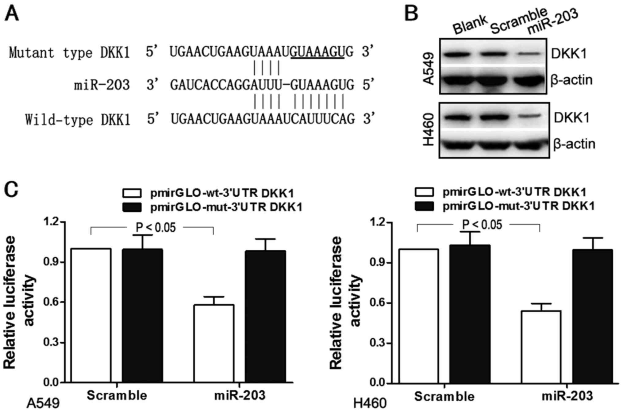
Dual luciferase reporter assay
Dual luciferase reporter gene assay was used to
verify whether miR-203 targets DKK1. First, point mutations at the
seed region of the DKK1 3′ untranslated region (3′UTR) were
established. These had complementary base pairing between miR-203
and the DKK1 mRNA 3′UTR sequence. Then, overlap PCR was used to
collect mutant copies of the DKK1 3′UTR (Fig. 2A). Wild-type DKK1 3′UTR was
amplified from human genomic DNA. Secondly, recombinant reporter
gene vectors pmirGLO-wt-3′UTR DKK1 and pmirGLO-mut-3′UTR DKK1 were
constructed (the combined vectors were purchased from Shanghai
Biological Technology Co., Ltd.), and then co-transfected with
scramble or miR-203 mimics (Shanghai GenePharma Co., Ltd.,
Shanghai, China) into A549 and H460 cells, respectively, using
Lipofectamine™ 2000 (Invitrogen) according to the manufacturer's
recommendations. The confluence of the cells was 70–80%. The
luciferase activity of the cells was determined using a luciferase
assay kit (Promega, Madison, WI, USA) 24 h after transfection.
Lentivirus transfection
Pre-miR-203 and negative control lentivirus vectors
were constructed and packaged by Shanghai GenePharma Co., Ltd. and
transfected into A549 and H460 cells into 6-well plates,
respectively. After transfection, the cells demonstrated puromycin
resistance, and different concentrations of puromycin
(Sigma-Aldrich, St. Louis, MO, USA) were used to select transfected
and stably expressed cells 48 h after transfection. We finally used
puromycin at the concentration of 2 µg/ml for 14 days to select the
stably transfected cells. qRT-PCR was used to verify the expression
of miR-203 in the three groups (miR-203, NC and blank).
CCK-8 assay
A Cell Counting Kit-8 (CCK-8; Dojindo, Kumamoto,
Japan) assay was performed to examine cell growth after
transfection in the three groups in accordance with the
manufacturer's recommendations. After 24 h of treatment with
different concentrations of DDP ranging from 4 to 20 µM, absorbance
of the cells was measured using a fluorescence microplate reader,
representing the proliferative activity of the cells. The
inhibition rate was calculated using the following formula:
Inhibition rate = (control group - experimental group)/control
group × 100%. IC50 is the half inhibitory rate, and
stands for the concentration of DDP when the inhibition rate is
50%.
Cell apoptosis assays
The apoptosis of A549 and H460 cells was measured by
flow cytometry (BD Biosciences, San Diego, CA, USA) using an
Annexin V-FITC/propidium iodide (PI) staining assay. Each group of
cells (miR-203, NC and blank) was divided into two subgroups, one
treated with 4 µM DDP and the other not treated with DDP. Apoptotic
cells were calculated by gating PI- and Annexin V-positive cells
using flow cytometry with fluorescence-activated cell-sorting
(FACS). The activity of caspase-3/−7 in each subgroup was
determined using a caspase-3/−7 kit (Promega).
Animal experiment
Mice were bred in a temperature-controlled sterile
environment with a 12-h light and dark cycle. Twenty BALB/c nude
mice (female, 4–6 weeks old) were purchased from Beijing Vital
River Laboratory Animal Technology Center (Beijing, China). Two
groups of cells (5×10⁶ in 0.2 ml of DMEM) after
lentivirus transfection (miR-203, NC) were subcutaneously injected
under the left forelegs of the mice which were randomly distributed
(n=10/group). In each group, half of the mice randomly received DDP
(2 mg/kg in 0.2 ml of normal saline for 5 days) by intraperitoneal
injection one week after implantation, and the others received 0.2
ml of normal saline alone. Thus, 4 groups were generated and the
growth conditions of the xenografts were measured and recorded
using an in vivo small animal imaging instrument every week
for a total of 4 weeks, represented by the luciferase signals.
Then, the mice were sacrificed by cervical dislocation. Each tumor
was extracted and weighed.
siRNA transfection
siRNA-DKK1 was purchased from Shanghai GenePharma
Co., Ltd. DKK1 lacking the 3′UTR (pcDNA3.1-DKK1 vector) was
constructed by Shanghai Biological Technology Co., Ltd. siRNA-DKK1,
miR-203 mimics, scramble and pcDNA3.1-DKK1 were transfected into
A549/H460 cells alone or the combination using Lipofectamine™ 2000
in accordance with the manufacturers recommendations. The samples
were divided into corresponding groups and CCK-8 assays
(IC50) were performed to assess the effects of miR-203
and DKK1 on DDP sensitivity in the A549/H460 cell lines.
Statistical analysis
Data were analyzed using SPSS 21 software. Data
showing a normal distribution are expressed as mean ± standard
deviation (SD). One-way ANOVA was used for comparison across three
or more groups; a two-tailed unpaired Student's t-test was used to
compare measurement data from pairs of independent samples. Pearson
correlation analysis was used to evaluate the correlation of two
groups. P<0.05 was considered to indicate a statistically
significant result.
Results
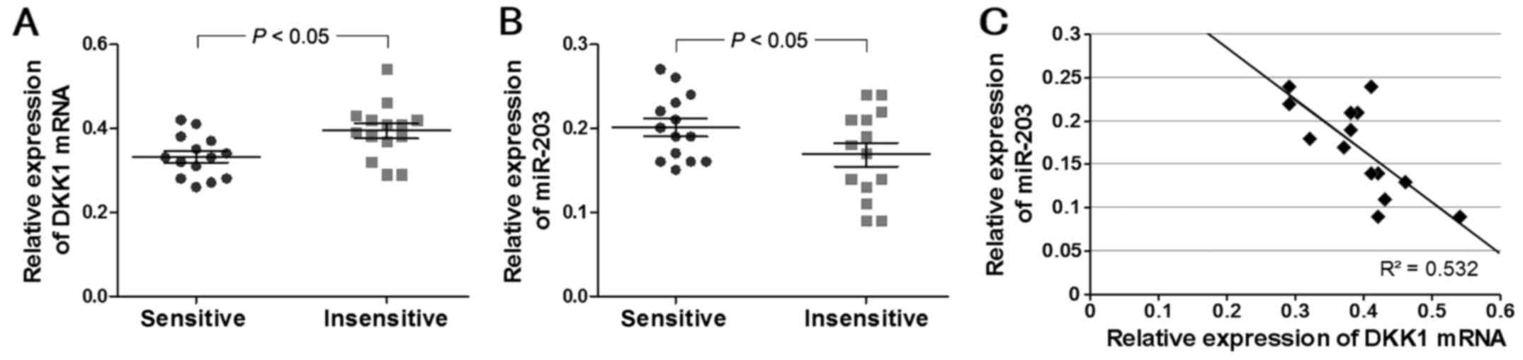
Expression of DKK1 mRNA and miR-203 in
cisplatin-sensitive and -insensitive lung adenocarcinoma
tissues
qRT-PCR was used to examine the expression levels of
DKK1 mRNA and miR-203 in lung cancer tissues from 30 patients with
advanced-stage lung adenocarcinoma; the clinical characteristics of
the patients are shown in Table I.
The results showed that in DDP-sensitive NSCLC samples, miR-203 was
expressed at a higher level when compared with the level noted in
the DDP-insensitive samples (P<0.05; Fig. 1B), while DKK1 mRNA was expressed at
a lower level in the DDP-sensitive NSCLC samples (P<0.05;
Fig. 1A). In addition, statistical
analysis revealed that expression of DKK1 mRNA was negatively
correlated with miR-203 (P<0.05; Fig. 1C; R2=0.532). The
different levels of expression of miR-203 and DKK1 in the
DDP-sensitive and -insensitive samples suggest that miR-203 is
associated with the mechanism of cisplatin resistance.
 | Table I.Clinicopathological characteristics
of the lung adenocarcinoma patients (n=30). |
Table I.
Clinicopathological characteristics
of the lung adenocarcinoma patients (n=30).
|
| Gender | Age (years) |
Differentiation | TNM stage |
|---|
|
|
|
|
|
|
|---|
| Variables | Male | Female | ≥60 | <60 | Well | Moderate/poor | III | IV |
|---|
| No. of samples | 17 | 13 | 19 | 11 | 14 | 16 | 12 | 18 |
| Drug-sensitive | 8 | 7 | 9 | 6 | 8 | 7 | 7 | 8 |
|
Drug-insensitive | 9 | 6 | 10 | 5 | 6 | 9 | 5 | 10 |
Confirmation of DKK1 as a target gene
of miR-203 in A549 and H460 cells
In the present study, DKK1 was predicted to be a
target gene of miR-203 by searching target gene prediction
databases (microRNA.org, miRDB and TargetScan). A
dual luciferase reporter gene assay was used to determine whether
miR-203 targets DKK1. Western blot analysis showed that there was
lower DKK1 expression in the miR-203 group than that in the blank
and scramble groups of the A549 and H460 cells (P<0.05; Fig. 2B), which suggests that upregulation
of miR-203 may lower the expression level of DKK1 protein. Under a
dual reporter gene assay, the relative luciferase activity showed a
nearly 1-fold decrease in wild-type DKK1 with upregulated miR-203.
However, the relative luciferase activity showed no significant
difference in the group with mutant DKK1 with upregulated miR-203
(P>0.05; Fig. 2C). The results
were observed both in the A549 and H460 cells, which indicate that
miR-203 may downregulate the expression level of DKK1 by binding to
the 3′UTR of DKK1 mRNA.
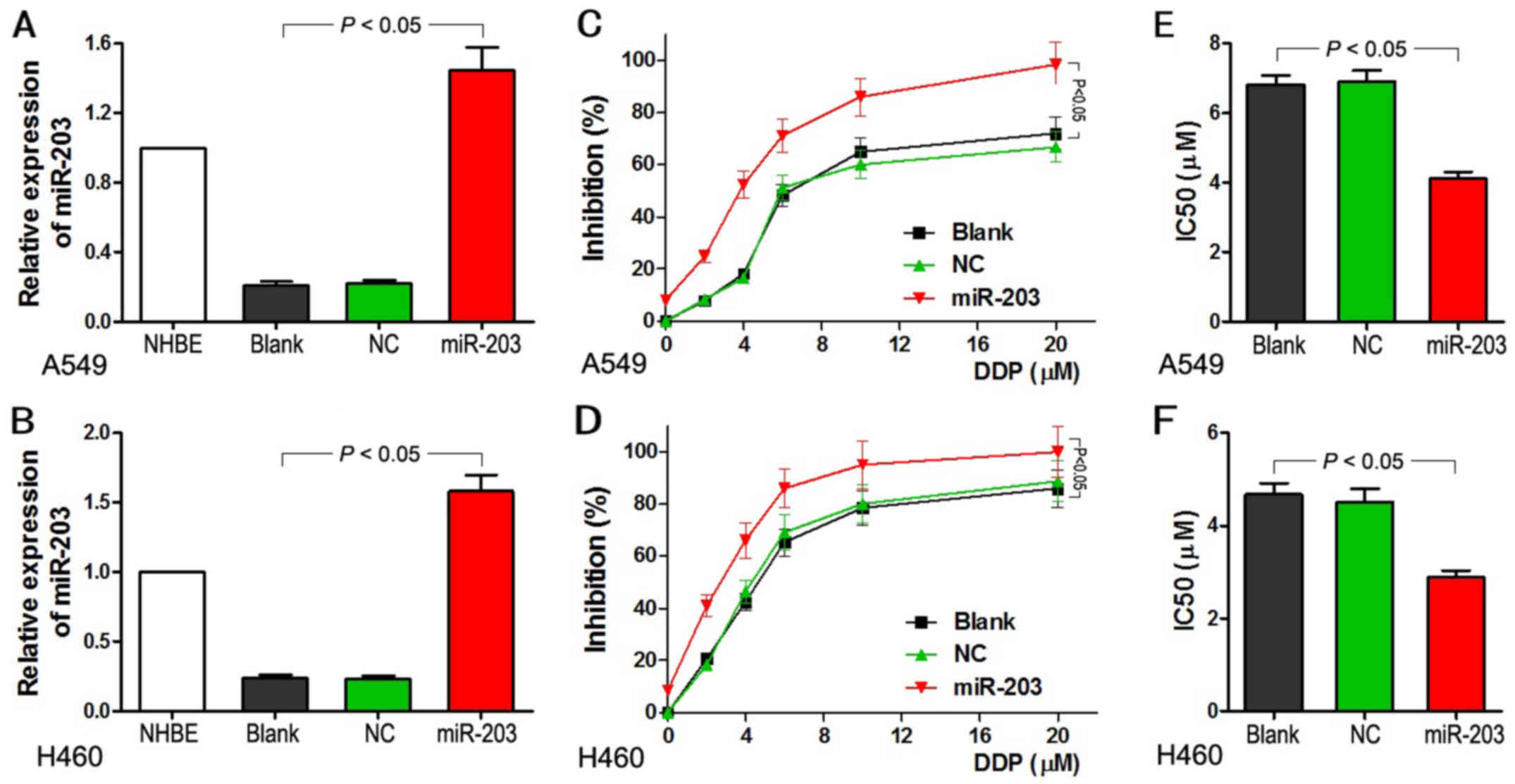
Upregulation of miR-203 increases
cisplatin sensitivity of A549 and H460 cells
qRT-PCR was used to examine the relative expression
of miR-203 in the different groups after A549 and H460 cells were
transfected with the lentiviral vectors. Cells in the group
transfected with miR-203 revealed a ~7- to 8-fold higher expression
of miR-203 when compared with the level in the blank and NC groups
(Fig. 3A and B). Following
treatment with different concentrations of DDP, ranging from 4 to
20 µM, the cells with miR-203 overexpression demonstrated a
significant inhibition of cell growth not observed in the blank and
NC groups (P<0.05). Inhibition of cell growth became more
extensive upon increases in the concentration of DDP (Fig. 3C and D). Furthermore, the
IC50 value for the miR-203 group was significantly lower
than that in the other two groups (P<0.05; Fig. 3E and F). IC50 reflects
the sensitivity of DDP in lung cancer cells. These results indicate
that upregulation of miR-203 increased the sensitivity of lung
adenocarcinoma A549 and H460 cells to DDP.
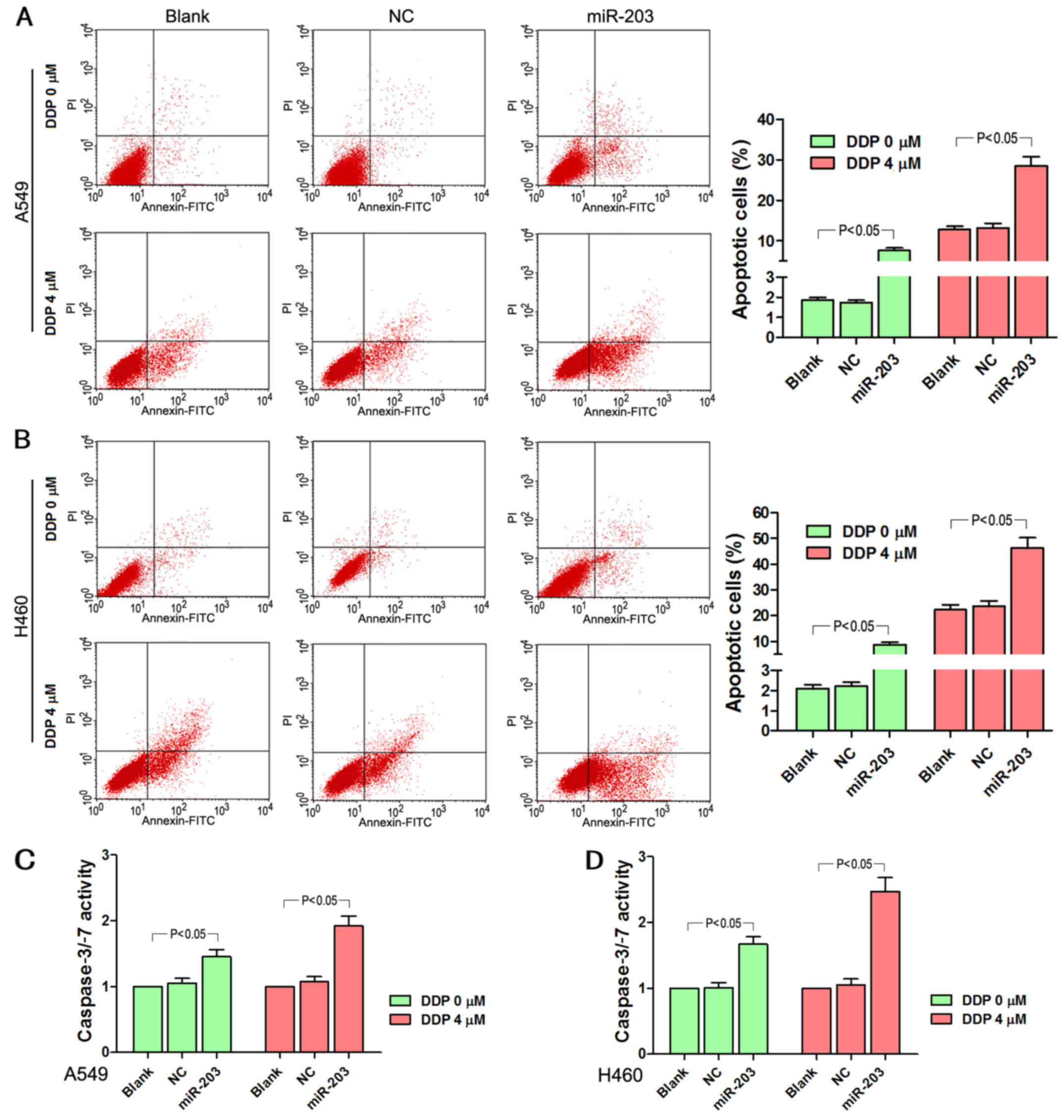
Upregulation of miR-203 increases the
apoptosis of A549 and H460 cells
Cell apoptosis was detected using flow cytometry,
and caspase-3/−7 activity was measured using a caspase-3/−7 kit.
The results indicated that overexpression of miR-203 significantly
increased cell apoptosis of the A549 and H460 cells with and
without DDP treatment (P<0.05; Fig.
4A and B). Similar results were observed when the activity of
caspase-3/−7 was examined (P<0.05; Fig. 4C and D). The increase in
caspase-3/−7 activity was more significant following treatment with
DDP. Upregulation of miR-203 was found to increase the activity of
caspase-3/−7 and promote apoptosis in the A549 and H460 cells.
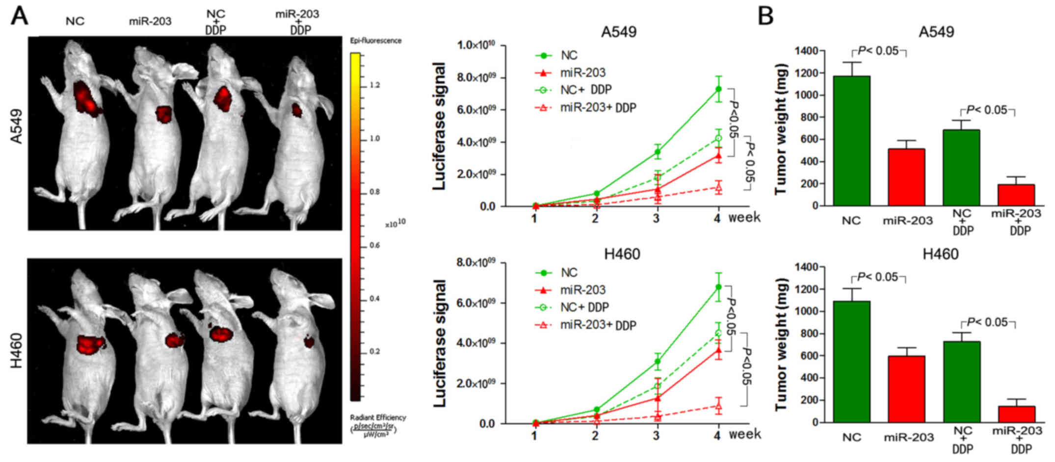
Upregulation of miR-203 inhibits tumor
growth of the nude mouse tumor xenografts
A nude mouse tumor xenograft model was established
to study the effects of miR-203 on tumor growth. The luciferase
signal detected by in vivo small animal imaging was in the
present study considered representative of tumor growth. The tumor
growth curve (Fig. 5A) clearly
showed that overexpression of miR-203 inhibited the tumor growth,
and this function was also observed under the treatment with DDP.
Over time, the tumor inhibition effect of miR-203 and miR-203
combined with DDP was more significant. Moreover, tumors with
upregulated miR-203 were more sensitive to DDP and showed a more
pronounced decline in tumor weight (P<0.05; Fig. 5B). In this way, upregulation of
miR-203 inhibits tumor growth and increases the sensitivity to DDP
of the nude mouse tumor xenograft.
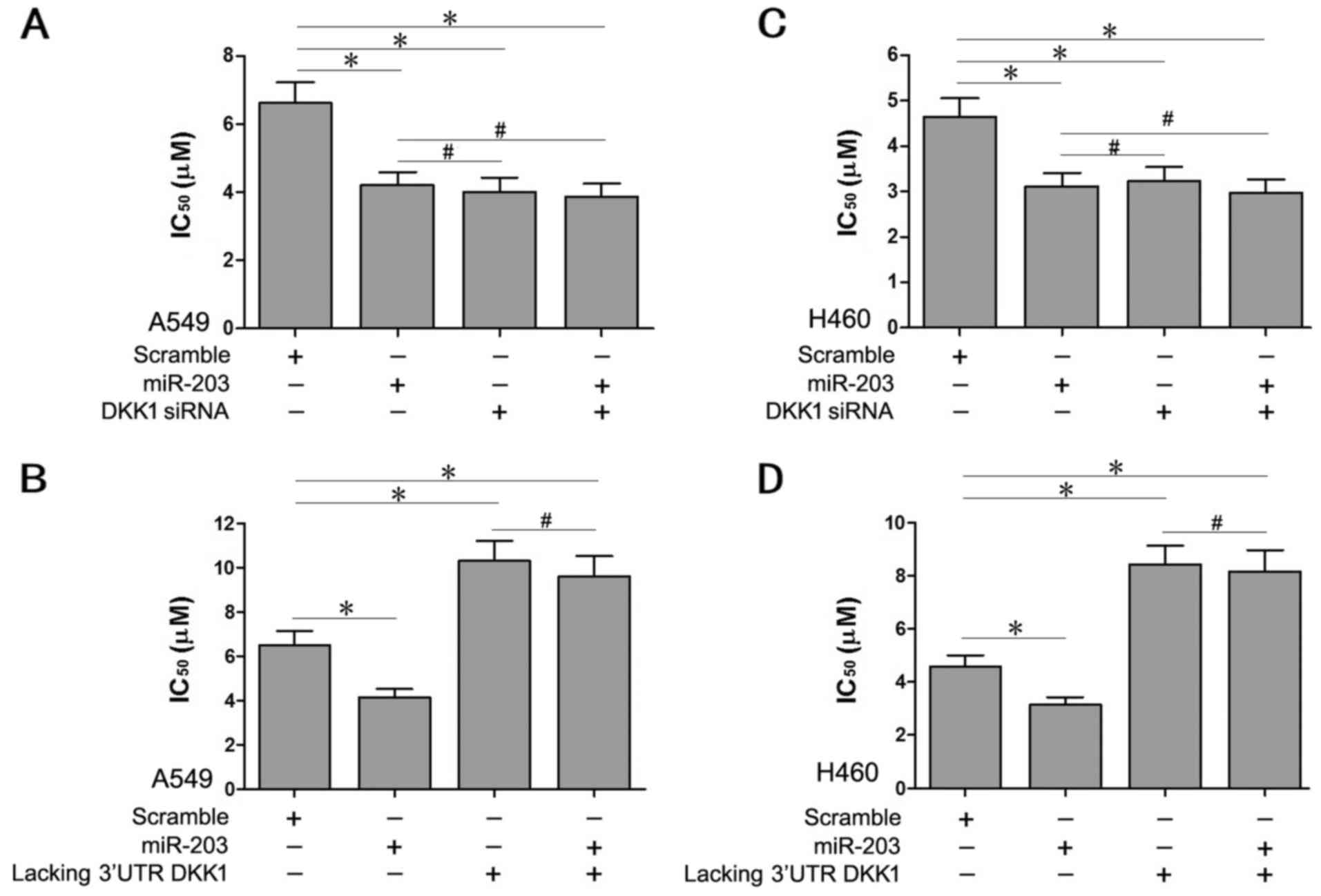
Upregulation of miR-203 sensitizes
A549 and H460 cells to DDP through DKK1
A great deal is known concerning the effects of
miR-203 on the inhibition of cell growth and DDP sensitization.
More experiments were designed to determine how miR-203 exerts its
function. Both overexpression of miR-203 and knockdown of DKK1
resulted in the sensitization to DDP with a lower IC50.
In response to DKK1 knockdown, overexpression of miR-203 had no
added effects on the sensitivity of the cells (P<0.05; Fig. 6A and C). Overexpression of miR-203
increased the sensitivity of the A549 and H460 cells to DDP
compared to the control group, while DKK1 lacking 3′UTR decreased
the sensitivity (P<0.05). In addition, miR-203 was not found to
sensitize cells to DKK1 lacking the 3′UTR (P>0.05; Fig. 6B and D). In summary, miR-203 was
found to exert the function of sensitization to DDP exclusively by
targeting the 3′UTR of DKK1.
Discussion
In the present study, miR-203 and DKK1 were
differentially expressed in DDP-sensitive and -insensitive lung
adenocarcinoma tissues, and a negative correlation was observed
between them, suggesting that miR-203 and DKK1 may predict DDP
sensitivity. Cell and animal experiments were designed to further
investigation the effects of miR-203 and DKK1 on cell
proliferation, apoptosis and xenograft growth and the relationship
between miR-203 and DKK1. Overexpression of miR-203 was found to
inhibit the growth of A549/H460 cells, and also to increase the
activity of caspase-3/−7 and promote cell apoptosis, which were
considered to contribute to sensitize the response of lung cancer
cells to DDP. Dual luciferase reporter assay verified DKK1 as a
target gene of miR-203, which was also identified by restore assay.
Restore assay showed that miR-203 was not able to sensitize cells
with DKK1 lacking the 3′UTR. miR-203 could bind to the 3′UTR of
DKK1 and then regulate the characteristics of lung cancer
cells.
The present study indicates that DKK1 may
participate in the mechanism underlying cisplatin resistance, which
is consistent with the findings of previous studies (23–27).
Since platinum-based chemotherapy is still the main strategy for
treating advanced lung cancer, research into drug resistance is
ongoing. However, the mechanism underlying the action of DDP is
quite complex and may involve many factors and signaling pathways.
More and more evidence has shown that abnormal expression of
miRNAs, such as miR-184 (30),
miR-1244 (31), miR-196a (32) and miR-141 (33), may be related to DDP resistance in
tumor cells. Various studies indicate that BCL2-associated
athanogene-1 (BAG-1) (30,34,35)
may promote sensitivity to chemotherapy in NSCLC patients. DNA
damage and DNA repair-associated genes and factors have been
identified as being involved in DDP resistance (36–38).
Spindle and kinetochore-associated complex subunit 1 (SKA1) is
considered to regulate the ERK1/2 and the Akt-mediated signaling
pathways in NSCLC cells (39).
Reports indicate that numerous signaling pathways, such as the Wnt
(23), p38 MAPK (40), PI3K/Akt (41) and TGFβ/Smad2/STAT3/STAT5 pathways
(42), are involved in the complex
mechanism of DDP resistance. It is also worth mentioning that cell
autophagy by hypoxia induced or exacerbated by FOXM1 (43) via the JNK/mitochondrial pathway
(44), may decrease the
susceptibility of lung cancer cells to cisplatin-induced apoptosis
(45). Large numbers of surveys
indicate the complexity of the mechanism underlying the regulation
of cisplatin resistance.
In conclusion, miR-203 and DKK1 were found to be
expressed at differential levels in DDP-sensitive and -insensitive
lung adenocarcinoma tissues; there was also a negative correlation
between them. Overexpression of miR-203 increased the cisplatin
sensitivity of A549/H460 cell lines by targeting the 3′UTR of DKK1.
Whereas miRNAs have complicated interactions with various mRNA
targets, and the mechanisms underlying the regulation of cisplatin
resistance are intricate and complex, further fundamental research
is needed to be conducted before clinical applications become
practical.
Acknowledgements
The authors are grateful to all the staff at the
Study Centre who contributed to the present study. The present
study was supported by grants from the Ministry of Major Science
and Techology of Henan (201401005).
References
|
1
|
Siegel RL, Miller KD and Jemal A: Cancer
statistics, 2016. CA Cancer J Clin. 66:7–30. 2016. View Article : Google Scholar : PubMed/NCBI
|
|
2
|
Zhang R, Zhang Y, Wen F, Wu K and Zhao S:
Analysis of pathological types and clinical epidemiology of 6,058
patients with lung cancer. Zhongguo Fei Ai Za Zhi. 19:129–135.
2016.(In Chinese). PubMed/NCBI
|
|
3
|
Miller KD, Siegel RL, Lin CC, Mariotto AB,
Kramer JL, Rowland JH, Stein KD, Alteri R and Jemal A: Cancer
treatment and survivorship statistics, 2016. CA Cancer J Clin.
66:271–289. 2016. View Article : Google Scholar : PubMed/NCBI
|
|
4
|
Ettinger DS, Wood DE, Akerley W, Bazhenova
LA, Borghaei H, Camidge DR, Cheney RT, Chirieac LR, D'Amico TA,
Dilling TJ, et al: NCCN Guidelines Insights: Non-Small Cell Lung
Cancer, Version 4. 2016. J Natl Compr Canc Netw. 14:255–264.
2016.PubMed/NCBI
|
|
5
|
Ettinger DS, Wood DE, Akerley W, Bazhenova
LA, Borghaei H, Camidge DR, Cheney RT, Chirieac LR, D'Amico TA,
Demmy TL, et al: National comprehensive cancer network: Non-Small
Cell Lung Cancer, version 6.2015. J Natl Compr Canc Netw.
13:515–524. 2015.PubMed/NCBI
|
|
6
|
Polo V and Besse B: Maintenance strategies
in stage IV non-small-cell lung cancer (NSCLC): In which patients,
with which drugs? Ann Oncol. 25:1283–1293. 2014. View Article : Google Scholar : PubMed/NCBI
|
|
7
|
Davidoff AJ, Tang M, Seal B and Edelman
MJ: Chemotherapy and survival benefit in elderly patients with
advanced non-small-cell lung cancer. J Clin Oncol. 28:2191–2197.
2010. View Article : Google Scholar : PubMed/NCBI
|
|
8
|
Bartel DP: MicroRNAs: Genomics,
biogenesis, mechanism, and function. Cell. 116:281–297. 2004.
View Article : Google Scholar : PubMed/NCBI
|
|
9
|
Hollis M, Nair K, Vyas A, Chaturvedi LS,
Gambhir S and Vyas D: MicroRNAs potential utility in colon cancer:
Early detection, prognosis, and chemosensitivity. World J
Gastroenterol. 21:8284–8292. 2015. View Article : Google Scholar : PubMed/NCBI
|
|
10
|
Lu J, Getz G, Miska EA, Alvarez-Saavedra
E, Lamb J, Peck D, Sweet-Cordero A, Ebert BL, Mak RH, Ferrando AA,
et al: MicroRNA expression profiles classify human cancers. Nature.
435:834–838. 2005. View Article : Google Scholar : PubMed/NCBI
|
|
11
|
Chen Y, Gao Y, Zhang K, Li C, Pan Y, Chen
J, Wang R and Chen L: MicroRNAs as regulators of cisplatin
resistance in lung cancer. Cell Physiol Biochem. 37:1869–1880.
2015. View Article : Google Scholar : PubMed/NCBI
|
|
12
|
Zhou P, Jiang N, Zhang GX and Sun Q:
MiR-203 inhibits tumor invasion and metastasis in gastric cancer by
ATM. Acta Biochim Biophys Sin. 48:696–703. 2016. View Article : Google Scholar : PubMed/NCBI
|
|
13
|
Zheng J, Wang F, Lu S and Wang X: LASP-1,
regulated by miR-203, promotes tumor proliferation and
aggressiveness in human non-small cell lung cancer. Exp Mol Pathol.
100:116–124. 2016. View Article : Google Scholar : PubMed/NCBI
|
|
14
|
Tang R, Zhong T, Dang Y, Zhang X, Li P and
Chen G: Association between downexpression of MiR-203 and poor
prognosis in non-small cell lung cancer patients. Clin Transl
Oncol. 18:360–368. 2016. View Article : Google Scholar : PubMed/NCBI
|
|
15
|
Lin J, Lin Y, Fan L, Kuang W, Zheng L, Wu
J, Shang P, Wang Q and Tan J: miR-203 inhibits cell proliferation
and promotes cisplatin induced cell death in tongue squamous
cancer. Biochem Biophys Res Commun. 473:382–387. 2016. View Article : Google Scholar : PubMed/NCBI
|
|
16
|
Wang C, Wang X, Liang H, Wang T, Yan X,
Cao M, Wang N, Zhang S, Zen K, Zhang C, et al: miR-203 inhibits
cell proliferation and migration of lung cancer cells by targeting
PKCα. PLoS One. 8:e739852013. View Article : Google Scholar : PubMed/NCBI
|
|
17
|
Hu H, Xu Z, Li C, Xu C, Lei Z, Zhang HT
and Zhao J: MiR-145 and miR-203 represses TGF-β-induced
epithelial-mesenchymal transition and invasion by inhibiting SMAD3
in non-small cell lung cancer cells. Lung Cancer. 97:87–94. 2016.
View Article : Google Scholar : PubMed/NCBI
|
|
18
|
Wang N, Liang H, Zhou Y, Wang C, Zhang S,
Pan Y, Wang Y, Yan X, Zhang J, Zhang CY, et al: miR-203 suppresses
the proliferation and migration and promotes the apoptosis of lung
cancer cells by targeting SRC. PLoS One. 9:e1055702014. View Article : Google Scholar : PubMed/NCBI
|
|
19
|
You HY, Xie XM, Zhang WJ, Zhu HL and Jiang
FZ: Berberine modulates cisplatin sensitivity of human gastric
cancer cells by upregulation of miR-203. In Vitro Cell Dev Biol
Anim. 52:857–863. 2016. View Article : Google Scholar : PubMed/NCBI
|
|
20
|
Ru P, Steele R, Hsueh EC and Ray RB:
Anti-miR-203 upregulates SOCS3 expression in breast cancer cells
and enhances cisplatin chemosensitivity. Genes Cancer. 2:720–727.
2011. View Article : Google Scholar : PubMed/NCBI
|
|
21
|
Liu Y, Gao S, Chen X, Liu M, Mao C and
Fang X: Overexpression of miR-203 sensitizes paclitaxel
(Taxol)-resistant colorectal cancer cells through targeting the
salt-inducible kinase 2 (SIK2). Tumour Biol. 37:12231–12239. 2016.
View Article : Google Scholar : PubMed/NCBI
|
|
22
|
Niehrs C: Function and biological roles of
the Dickkopf family of Wnt modulators. Oncogene. 25:7469–7481.
2006. View Article : Google Scholar : PubMed/NCBI
|
|
23
|
Xia Y, He Z, Liu B, Wang P and Chen Y:
Downregulation of Meg3 enhances cisplatin resistance of lung cancer
cells through activation of the WNT/β-catenin signaling pathway.
Mol Med Rep. 12:4530–4537. 2015.PubMed/NCBI
|
|
24
|
Jia X, Li N, Peng C, Deng Y, Wang J, Deng
M, Lu M, Yin J, Zheng G, Liu H, et al: miR-493 mediated DKK1
down-regulation confers proliferation, invasion and
chemo-resistance in gastric cancer cells. Oncotarget. 7:7044–7054.
2016.PubMed/NCBI
|
|
25
|
Huang Y, Yang X, Zhao F, Shen Q, Wang Z,
Lv X, Hu B, Yu B, Fan J and Qin W: Overexpression of Dickkopf-1
predicts poor prognosis for patients with hepatocellular carcinoma
after orthotopic liver transplantation by promoting cancer
metastasis and recurrence. Med Oncol. 31:9662014. View Article : Google Scholar : PubMed/NCBI
|
|
26
|
Li S, Qin X, Guo X, Cui A, He Y, Wei S,
Wang X and Shan B: Dickkopf-1 is oncogenic and involved in invasive
growth in non small cell lung cancer. PLoS One. 8:e849442013.
View Article : Google Scholar : PubMed/NCBI
|
|
27
|
Salim H, Zong D, Hååg P, Novak M, Mörk B,
Lewensohn R, Lundholm L and Viktorsson K: DKK1 is a potential novel
mediator of cisplatin-refractoriness in non-small cell lung cancer
cell lines. BMC Cancer. 15:6282015. View Article : Google Scholar : PubMed/NCBI
|
|
28
|
Menezes ME, Devine DJ, Shevde LA and
Samant RS: Dickkopf1: A tumor suppressor or metastasis promoter?
Int J Cancer. 130:1477–1483. 2012. View Article : Google Scholar : PubMed/NCBI
|
|
29
|
Hirata H, Hinoda Y, Nakajima K, Kawamoto
K, Kikuno N, Ueno K, Yamamura S, Zaman MS, Khatri G, Chen Y, et al:
Wnt antagonist DKK1 acts as a tumor suppressor gene that
induces apoptosis and inhibits proliferation in human renal cell
carcinoma. Int J Cancer. 128:1793–1803. 2011. View Article : Google Scholar : PubMed/NCBI
|
|
30
|
Tung MC, Lin PL, Cheng YW, Wu DW, Yeh SD,
Chen CY and Lee H: Reduction of microRNA-184 by E6 oncoprotein
confers cisplatin resistance in lung cancer via increasing Bcl-2.
Oncotarget. 7:32362–32374. 2016.PubMed/NCBI
|
|
31
|
Li W, Wang W, Ding M, Zheng X, Ma S and
Wang X: MiR-1244 sensitizes the resistance of non-small cell lung
cancer A549 cell to cisplatin. Cancer Cell Int. 16:302016.
View Article : Google Scholar : PubMed/NCBI
|
|
32
|
Li JH, Luo N, Zhong MZ, Xiao ZQ, Wang JX,
Yao XY, Peng Y and Cao J: Inhibition of microRNA-196a might reverse
cisplatin resistance of A549/DDP non-small-cell lung cancer cell
line. Tumour Biol. 37:2387–2394. 2016. View Article : Google Scholar : PubMed/NCBI
|
|
33
|
Fu WF, Chen WB, Dai L, Yang GP, Jiang ZY,
Pan L, Zhao J and Chen G: Inhibition of miR-141 reverses cisplatin
resistance in non-small cell lung cancer cells via upregulation of
programmed cell death protein 4. Eur Rev Med Pharmacol Sci.
20:2565–2572. 2016.PubMed/NCBI
|
|
34
|
Wang Y, Ha M, Liu J, Li P, Zhang W and
Zhang X: Role of BCL2-associated athanogene in resistance to
platinum-based chemotherapy in non-small-cell lung cancer. Oncol
Lett. 11:984–990. 2016.PubMed/NCBI
|
|
35
|
Qiu T, Zhou L, Wang T, Xu J, Wang J, Chen
W, Zhou X, Huang Z, Zhu W, Shu Y, et al: miR-503 regulates the
resistance of non-small cell lung cancer cells to cisplatin by
targeting Bcl-2. Int J Mol Med. 32:593–598. 2013.PubMed/NCBI
|
|
36
|
Xu S, Huang H, Chen YN, Deng YT, Zhang B,
Xiong XD, Yuan Y, Zhu Y, Huang H, Xie L, et al: DNA damage
responsive miR-33b-3p promoted lung cancer cells survival and
cisplatin resistance by targeting p21WAF1/CIP1. Cell
Cycle. 15:2920–2930. 2016. View Article : Google Scholar : PubMed/NCBI
|
|
37
|
Wang S, Liu F, Zhu J, Chen P, Liu H, Liu Q
and Han J: DNA repair genes ERCC1 and BRCA1 expression in non-small
cell lung cancer chemotherapy drug resistance. Med Sci Monit.
22:1999–2005. 2016. View Article : Google Scholar : PubMed/NCBI
|
|
38
|
Im JY, Lee KW, Won KJ, Kim BK, Ban HS,
Yoon SH, Lee YJ, Kim YJ, Song KB and Won M: DNA damage-induced
apoptosis suppressor (DDIAS), a novel target of NFATc1, is
associated with cisplatin resistance in lung cancer. Biochim
Biophys Acta. 1863:40–49. 2016. View Article : Google Scholar : PubMed/NCBI
|
|
39
|
Shen L, Yang M, Lin Q, Zhang Z, Miao C and
Zhu B: SKA1 regulates the metastasis and cisplatin resistance of
non-small cell lung cancer. Oncol Rep. 35:2561–2568.
2016.PubMed/NCBI
|
|
40
|
Qi K, Li Y, Li X, Lei X, Wang B, Zhang L
and Chu X: Id4 promotes cisplatin resistance in lung cancer through
the p38 MAPK pathway. Anticancer Drugs. 27:970–978. 2016.
View Article : Google Scholar : PubMed/NCBI
|
|
41
|
Hu CF, Huang YY, Wang YJ and Gao FG:
Upregulation of ABCG2 via the PI3K-Akt pathway contributes to
acidic microenvironment-induced cisplatin resistance in A549 and
LTEP-a-2 lung cancer cells. Oncol Rep. 36:455–461. 2016. View Article : Google Scholar : PubMed/NCBI
|
|
42
|
Sun W, Ma Y, Chen P and Wang D:
MicroRNA-10a silencing reverses cisplatin resistance in the
A549/cisplatin human lung cancer cell line via the transforming
growth factor-β/Smad2/STAT3/STAT5 pathway. Mol Med Rep.
11:3854–3859. 2015.PubMed/NCBI
|
|
43
|
Wang Y, Wen L, Zhao SH, Ai ZH, Guo JZ and
Liu WC: FoxM1 expression is significantly associated with
cisplatin-based chemotherapy resistance and poor prognosis in
advanced non-small cell lung cancer patients. Lung Cancer.
79:173–179. 2013. View Article : Google Scholar : PubMed/NCBI
|
|
44
|
Liu Y, Chen X, Gu Y, Zhu L, Qian Y, Pei D,
Zhang W and Shu Y: FOXM1 overexpression is associated with
cisplatin resistance in non-small cell lung cancer and mediates
sensitivity to cisplatin in A549 cells via the JNK/mitochondrial
pathway. Neoplasma. 62:61–71. 2015. View Article : Google Scholar : PubMed/NCBI
|
|
45
|
Wu HM, Jiang ZF, Ding PS, Shao LJ and Liu
RY: Hypoxia-induced autophagy mediates cisplatin resistance in lung
cancer cells. Sci Rep. 5:122912015. View Article : Google Scholar : PubMed/NCBI
|