Introduction
Breast cancer is the the most common malignancy in
women worldwide with the highest mortality rate of 12.9 per 100,000
population in both the developed and developing areas (1,2). Based
on the expression status of ER, PR, HER2 receptor, p53 and Ki-67,
the St. Gallen Consensus listed four broad categories of breast
cancer: basal-like breast cancer, the Her2-enriched, luminal A and
luminal B subtypes (3). These
molecular subtypes markedly influence the clinical treatment
strategies, response to therapies, distribution of risk factors and
the patients prognosis (4,5).
Triple negative breast cancer (TNBC) refers to the
breast cancer that has no expression of ER, PR nor HER2 receptor,
which makes up ~15–20% of all the breast cancers with a
disproportionate share of mortality (6). As previous studies depicted, a
significant part (>90%) of basal-like breast cancers were
triple-negative subtypes and 55–81% of TNBCs were BLBCs (7). BLBC is very frequently associated with
TNBC. Prognosis of BLBC and TNBC is the poorest among all the
subtypes (8). Owing to aggressive
clinical courses, higher possibility of distant recurrence and
significant shorter survivals (9),
TNBC and BLBC are challenging public health concerns. With the
advance of biology technologies especially the next generation
sequencing, it is obvious that cellular microenvironment, gene
mutations and patients innate hallmarks have great influence on
disease pathophysiology, treatment sensitivity and recurrence
(10). However, the molecular
mechanism of breast cancer remains to be elaborated.
Currently, diagnostic and therapeutic strategies for
breast cancer are emerging prominently including surgery,
neo-adjuvant chemotherapy, endocrine therapy, radiotherapy and
molecular targeted therapy (11).
Without the molecular targets (ER, PR and Her2) of therapeutic
agents for breast cancer, TNBC is relatively sensitive to cytotoxic
chemotherapy. Besides, chemotherapy is always combined with
neo-adjuvant, adjuvant and metastatic setting (7). However, after the chemotherapy,
residual tumor cells mostly survive and provoke recurrent tumor
growth (12), which contributes to
high recurrence risk and mortality. Yet, there are obstacles and
limitations. A targeted therapy relies on a given tumor marker from
a host of randomized clinical trails. Besides, it is suitable to
the specific population not specific individual patient. Rodler
et al (13) revealed that
veliparib (ABT-888) combined with cisplatin and vinorelbine
possesses antineoplastic activity in TNBC, especially the BRCA
mutation-associated TNBC. Growing clinical and biological features
are proposed to combine the better predictive carcinoma
characteristics and behavior to obtain a more individualized
treatment strategy. Concomitantly, new tumor marker development is
essential for clinical practice, disease subtyping, predictive
diagnosis and prognosis. It is certainly worth screening and
validating reliable predictive biomarkers for breast cancer,
particularly the TNBC and basal-like subtypes.
Accumulating evidence demonstrates that telomerase
plays a pivotal role in maintaining telomere homeostasis and cell
immortalization and that telomerase-targeted cancer therapy seems
to be effective with less adverse side-effects (14). Pin2/TRF1-interacting telomerase
inhibitor 1 (Pinx1) is a haploinsufficient tumor suppressor
localized at human chromosome 8p23, the most common deletion region
correlated to tumor differentiation and tumorigenicity (15). Tian et al (16) recently showed cervical squamous cell
carcinoma patients with PinX1 expression are more sensitive to
paclitaxel chemotherapy and PinX1 could be a novel biomarker for
CSCC patients who might benefit from paclitaxel. Liu et al
(17) reported that PinX1
suppressed the proliferation of bladder urothelial carcinoma cells
via inhibiting activity of telomerase. Also, Li et al
(18) addressed the function of
PinX1 in renal cell carcinomas (RCC) that PinX1 depressed the
migration and invasion of RCC. However, there exist few studies in
terms of the association between PinX1 expression and its clinical
characteristics. Moreover, the concrete molecular mechanism of
PinX1 contributing to breast cancer remains poorly understood.
In the present study, we identified that PinX1 was
downregulated in breast cancer tissues compared with the adjacent
counterparts, especially in the basal-like subtype. Overexpression
of PinX1 inhibited the proliferation rates and migration ability,
while increased the apoptosis rates of human basal-like breast
cancer. Thus, PinX1 is likely a feasible biomarker and a molecular
target of basal-like breast cancer.
Materials and methods
Patients and tissue specimens
In the analysis of PinX1 expression by qRT-PCR and
western blotting, 26 fresh breast cancer tissues and the paired
non-tumor tissue samples were selected from breast cancer patients
without preoperative treatment who had undergone surgical
intervention during February 2013 to July 2015. The tissue
specimens for TMA construction and IHC analysis were previously
described (19). All the tissues
above were obtained with written informed consent of patients. The
Institute Research Medical Ethics Committee of the Nanfang Hospital
approved the utilization of these breast cancer samples for
research purpose only.
Cell cultures and plasmids
Breast cancer cell line MDA-MB-231 was obtained from
laboratory preservation. It was cultured in Dulbeccos modified
Eagles medium (DMEM; Gibco, Carlsbad, CA, USA) containing 10% fetal
bovine serum (FBS; Gibco) at 37°C and 5% CO2. E.
coli DH5α (Invitrogen, Carlsbad, CA, USA) was maintained in
flasks with LB medium. The plasmid pSpCas9(BB)-2A-GFP (PX458) for
gene knockout was aquired from Feng Zhang Lab (#48138; Addgene,
Cambridge, MA, USA). pCDNA3.1(+) (Invitrogen) and pEGFP-N1 vectors
(Clontech Laboratories, Inc., Mountain View, CA, USA) were used for
gene amplification in mammalian cells.
Overexpression and knockout of PinX1
in MDA-MB-231 cells
For PinX1-overexpressed stable cells, the PinX1 ORF
was inserted into pEGFP-N1 vector. Then MDA-MB-231 cells were
transfected separately with the recombined and empty vectors by
Lipofectamine 2000 (Invitrogen). After 24 h, cells were reseeded at
1:4 dilution with a new selective cell culture containing 1300
µg/ml G418 (Sigma-Aldrich, St. Louis, MO, USA) and the selective
culture was renewed every 2 days. After 2–3 weeks, the cell clones
were obtained by an inverted microscope (Olympus IX71; Olympus,
Tokyo, Japan) and grown in maintaining culture with 300 µg/ml of
G418 for later experiments. For the knockout of PinX1, three
sgRNAs were designed according to the Feng Zhang Lab at http://crispr.mit.edu/ and the sequences were as
follows: sgRNA-1, forward, caccgccatcttctctagcatccgc and reverse,
aaacgcggatgctagag aagatggc; sgRNA-2, forward,
caccgtgatggtagctccgagtccc and reverse, aaacgggactcggagctaccatcac;
sgRNA-3, forward, cac cgccgaactgaacacttgccat and reverse,
aaacatggcaagtgttcagttc ggc. In addition, sgRNA-3 was selected for
its better knockout effects. sgRNA-3 was then cloned into PX458 and
MDA-MB-231 cells were transfected with the recombined vector by
Lipofectamine 2000. Cells were reseeded into 96-well plates at a
density of a single cell/well after 24-h transfection by flow
cytometry (BD Biosciences, San Jose, CA, USA). Cell clones were
harvested in 2–3 weeks and detected by western blot analysis later
to obtain the knockout cells.
RNA extraction and qRT-PCR assays
The PinX1 expression level of 26 pairs of breast
malignancy tissues and the normal counterparts were investigated
via qRT-PCR. Total RNA of these samples were isolated by RNAiso
Plus reagent (Takara Bio, Dalian, China) and dissolved in
DEPC-treated water. The RNA concentration and purity were tested by
NanoDrop 2000 spectrophotometer (Thermo Fisher Scientific, Waltham,
MA, USA). The denaturing agarose gel electrophoresis was then used
to qualify the integrity of total RNA. The qualified RNA were
subsquently reverse-transcribed by PrimeScript® RT
reagent kit (Takara Bio) depending on the manufacturers
instructions. SYBR® Premix Ex Taq™ II (Tli RNase H Plus)
kit (Takara Bio) was used for the qRT-PCR on the ABI 7500 real-time
PCR system (Applied Biosystems, Foster City, CA, USA) with holding
stage of 95°C for 10 min, followed by 40 cycles of 95°C for 30 sec
and 60°C for 34 sec, followed by the melt curve stage. The forward
PinX1 primer was 5-ccagagg agaacgaaaccacg-3 and the reverse
was 5-acctgcgtctcagaaa tgtca-3. As an internal reference gene, the
forward primer of GAPDH was 5-ctgggctacactgagcacc-3 and the
reverse was 5-aagtggtcgttgagggcaatg-3. The relative expression
levels of PinX1 was determined by the comparative Ct (ΔΔct) method
(20) and the ΔCt was calculated as
ΔCt = Ctgene - Ctreference. Each experiment
was tested at least in three independent biological repeats.
Immunoblot analysis
The extracted total proteins of breast cancer
tissues and the paired normal samples were lysed in the enhanced
Radio Immunoprecipitation Assay (RIPA) buffer mixed with 1 mM PMSF
(Beyotime Institute of Biotechnology, Haimen, China). Protein
concentrations were measured by BCA protein assay kit (Beyotime
Institute of Biotechnology). Then 40 µg of each protein specimen
was separated by 10% SDS-PAGE. The separated proteins on the gel
were transferred onto polyvinylidene fluoride membrane (Millipore,
Billerica, MA, USA) by a wet transfer apparatus (Bio-Rad
Laboratories, Inc., Hercules, CA, USA). The blots were then blocked
by 5% non-fat dry milk dissolved in Tris-buffered saline with
Tween-20 (TBST) at room temperature for 1 h. Thereafter, the
membranes were, respectively, incubated with primary antibodies
diluted by TBST for PinX1 (1:1,000, goat anti-human polyclonal;
Santa Cruz Biotechnology, Santa Cruz, CA, USA) and GAPDH (1:10,000,
anti-human, conjugated with HRP; Proteintech Group, Inc., Rosemont,
IL, USA) at 4°C overnignt. After washing with TBST for three times,
the membranes for PinX1 detection were incubated with rabbit
anti-goat IgG conjugated to HRP (1:10,000; Multi Sciences Biotech,
Co., Ltd., Hangzhou, China) for 1 h at room temperature. Following
3 washes with TBST, these membranes were visualized by Immobilon
Western Chemiluminescent HRP Substrate (Millipore) and exposed to
X-ray film. Proteins were quantified by Adobe Photoshop and GAPDH
was a loading control. The transfected MDA-MB-231 cells were also
lysed in RIPA. The primary antibody for cleaved caspase-3 (1:1000,
rabbit anti-human polyclonal; Cell Signaling Technology, Danvers,
MA, USA) and the goat anti-rabbit IgG conjugated to HRP were also
used in the present study.
Construction of TMA and IHC
In the present study, the construction of TMA in
general was referred to standard protocols as previously described
(19). The qualified TMA slides
were dewaxed by xylene and then rehydrated in distinct gradient of
alcohol; 3% hydrogen peroxide was used to compromise the endogenous
peroxidase activity of the tissue microarrays for 20 min at room
temperature. Thereafter, the slides were immersed in a boiled
citrate buffer (50 mM, pH 6.0) for 10 min. After three washes of
0.01 M phosphate-buffered saline (PBS) (pH 7.4). The slides were
then blocked by 5% normal goat serum for 30 min, followed with
incubation of primary antibody for PinX1 (1:200, goat polyclonal;
Santa Cruz Biotechnology) at 4°C overnight and secondary antibody
labeled with polymer peroxidase (ZSGB-Bio, Beijing, China).
Furthermore, the slides were treated with DAB Horseradish
Peroxidase Color Development kit (Beyotime Institute of
Biotechnology) followed by hematoxylin counterstaining. In this
study, the negative controls were obtained by using PBS instead of
anti-PinX1 antibody.
Immunohistochemistry evaluation
Each IHC slide for PinX1 positivity was assessed by
staining intensity and the proportion of positive carcinoma cells
across the total carcinoma cells semi-quantitatively. Three
pathologists who were blinded to the patient clinical data
evaluated the slides independently according to the percentage of
positive tumor cells. In case of disagreement, the assessments were
rescored until consensus was reached. The scores were distributed
by 5% increments (0, 5, 10…100%).
Selection of optimal cut-off
value
ROC analysis was applied to obtain the PinX1
positivity threshold according to the 0, 1-criterion. The
clinicopathological parameters were classified into the following
bivariate variables: age (≤50 and >50), clinical stage (I–II and
III), pT stage (T1-T2 and T3-T4), pN stage (N0 and N1-N2),
histology grade (I and II–III), PR status (positive and negative),
ER status (positive and negative), Her2 status (positive and
negative) and p53 status (positive and negative). Based on the
PinX1 scores and the dichotomized clinicopathological features, the
sensitivity and specificity for each test outcome was plotted to
generate a ROC curve. The inherent validity of each diagnostic test
was measured by the area under the curve (AUC) which combined
sensitivity and specificity. As an optimal cut-off value, the PinX1
score was close to the point (0.0 and 0.1) and maximized the
sensitivity and specificity. Consequently, tumors were identified
as positive with a score above the cut-off threshold while it was
negative with an score below the threshold.
CCK-8 cell proliferation assay
The cell viability of MDA-MB-231 cells with
different treatments was measured by the Cell Counting kit-8
(CCK-8; Dojindo Laboratories, Kumamoto, Japan). A total of
2×10³ cells/well were plated into 96-well culture plate
and incubated at 37°C for 24, 48 and 72 h, respectively. At each
specific time-point, the old medium was replaced by 100 µl fresh
medium containing 10 µl CCK-8 solution and cells were incubated for
another 2 h. Values of OD450nm were measured by
Multiskan FC (Thermo Fisher Scientific).
Cell migration assay
For the wound healing assay, cells with different
treatments were plated in 6-well plates at a density of
5×10⁵/well with a monolayer confluence overnight. Then,
10 µl pipette tips were used to make wounds on the single layers.
The floated cells were washed away with 1X PBS three times, and
cells were incubated at 37°C after adding fresh DMEM without serum.
Then images of the wounds at the indicated periods (0, 12, 24 and
36 h) were taken. The 24-well Transwell chambers (BD Biosciences)
was used to assess the migration ability of MDA-MB-231 cells. A
total of 100 µl 5×10⁴ cells/well was seeded in the upper
chamber with serum-free DMEM. In addition, 500 µl DMEM containing
20% FBS was added to the lower chamber. After 24-h incubation,
cells were fixed by 100% methanol for 20 min and dyed by 0.1%
crystal violate (Amresco, Solon, OH, USA) for 30 min. The migration
rate was analyzed after the upper cells were removed.
Apoptotic rate analysis by flow
cytometry
MDA-MB-231 cells (2.5×10⁵/well) were
plated in 12-well plates overnight before transfected with
pCDNA3.1(+) and PinX1-pCDNA3.1(+) by Lipofectamine 2000. After 48-h
incubation, cells were harvested for apoptosis analysis by flow
cytometry (BD FACSCalibur). For the PinX1 knockout and the
wild-type cells, appropriate cells were plated in 12-well plates as
well; 40 mM of etoposide was added to cells 24 h later. After 24 h
of incubation, cells were harvested and washed by 1X PBS twice.
Then cells were resuspended by 500 µl binding buffer, stained by
Annexin V-FITC and propidium iodide (PI) according to the Annexin
V-FITC apoptosis detection kit (Nanjing KeyGen Biotech, Co., Ltd.,
Nanjing, China) and detected by flow cytometry.
Statistical analyses
Statistics shown were analyzed by IBM SPSS
Statistics 20.0 (SPSS, Inc., Chicago, IL, USA) and GraphPad Prism
5.0. The differences of PinX1 expression between the paired tissues
were tested by the Student's paired t-test. The correlation of
PinX1 positivity with the clinicopathological parameters was
evaluated by the Chi-squared test and the Fishers exact test. With
two-tailed tests, P<0.01 or P<0.05 was considered to be
statistically significant. The data expressed are mean ± SEM.
Results
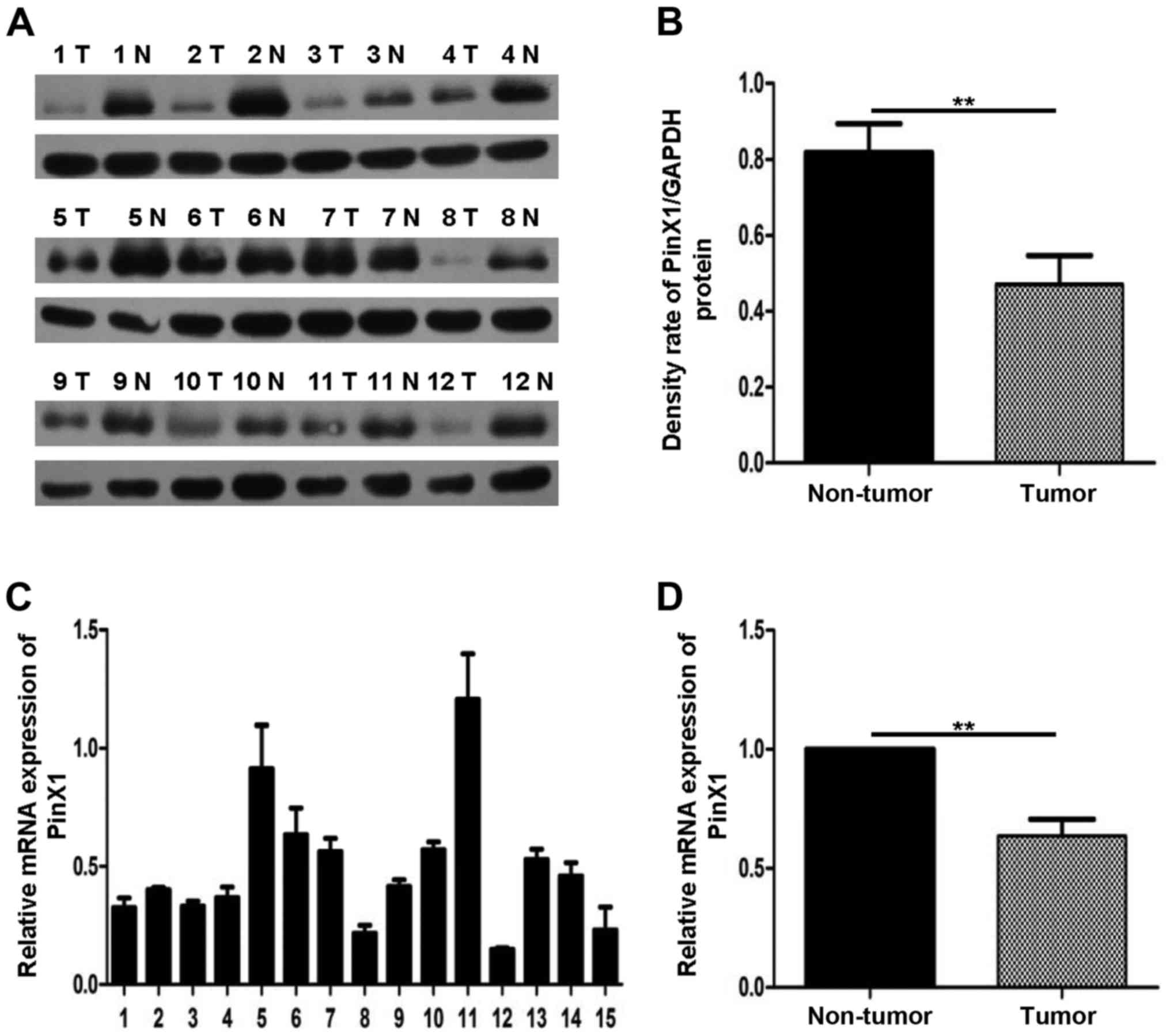
The expression of PinX1 mRNA and
protein declines substantially in human breast cancer tissues
To explore the role of PinX1 in
tumorigenesis, we detected the PinX1 mRNA levels of breast
cancer and the paired adjacent normal breast tissues. The
quantitative real-time PCR (qRT-PCR) assay showed that PinX1
mRNA expression was decreased in 21 out of 26 sample pairs and the
fold-changes of the 21 sample pairs were <1. The qRT-PCR result
of 15 sample pairs is shown in Fig.
1A. In comparison with the paired adjacent non-tumor breast
tissues, the expression of PinX1 in breast cancer tissues
was reduced significantly by 0.362 (Fig. 1B; P<0.05).
Similarly, western blot analysis further confirmed
the lower PinX1 expression between 26 paired normal breast tissues
and the adjacent counterpart tumor (Fig. 1C, 12 pairs of the total 26 pairs).
The densitometry quantification results indicated that PinX1
expression was downregulated in breast cancer tissues and the
difference between the two paired groups were of favorably
statistical significance (Fig. 1D;
P<0.05).
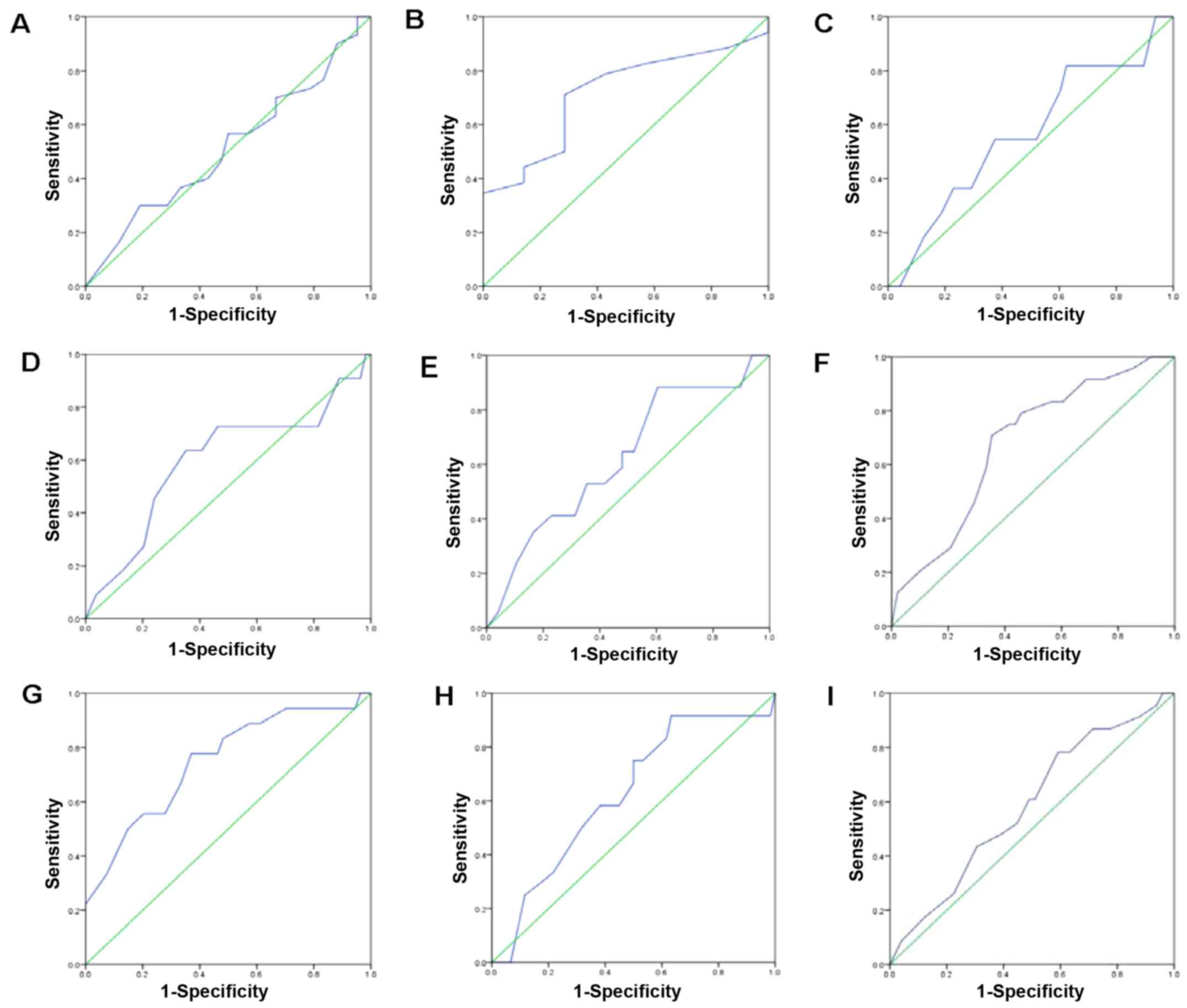
IHC analysis of PinX1 expression level
in breast cancer tissues
We also used IHC to investigate the PinX1 expression
in breast tissues. We found that PinX1 expression varied from
breast cancer to normal breast tissues (Fig. 2). The ROC was used to obtain the
optimal cut-off value, a threshold for positive and negative PinX1
expression. As is shown in Fig. 3,
the ROC curve for PR status is the closest to (0.0 and 0.1) with a
maximized sensitivity and specificity. In addition, the optimal
cut-off threshold to separate the positive from negative expression
of PinX1 was determined as 62.5%. Based on the cut-off value, the
association between PinX1 protein expression and the different
subtypes of breast cancer was elucidated. The positive expression
percentage of PinX1 was 76.9% (10/14) in Luminal A and Luminal B
subtypes, 50% (5/10) in Her2-overexpressed and 27.3% (9/33) in
basal-like (Table I) subtypes. In
the malignant breast carcinomas, PinX1 expression tended to be
negative. The results indicated that PinX1 expression was possibly
correlated with breast cancer subtypes (P<0.05).
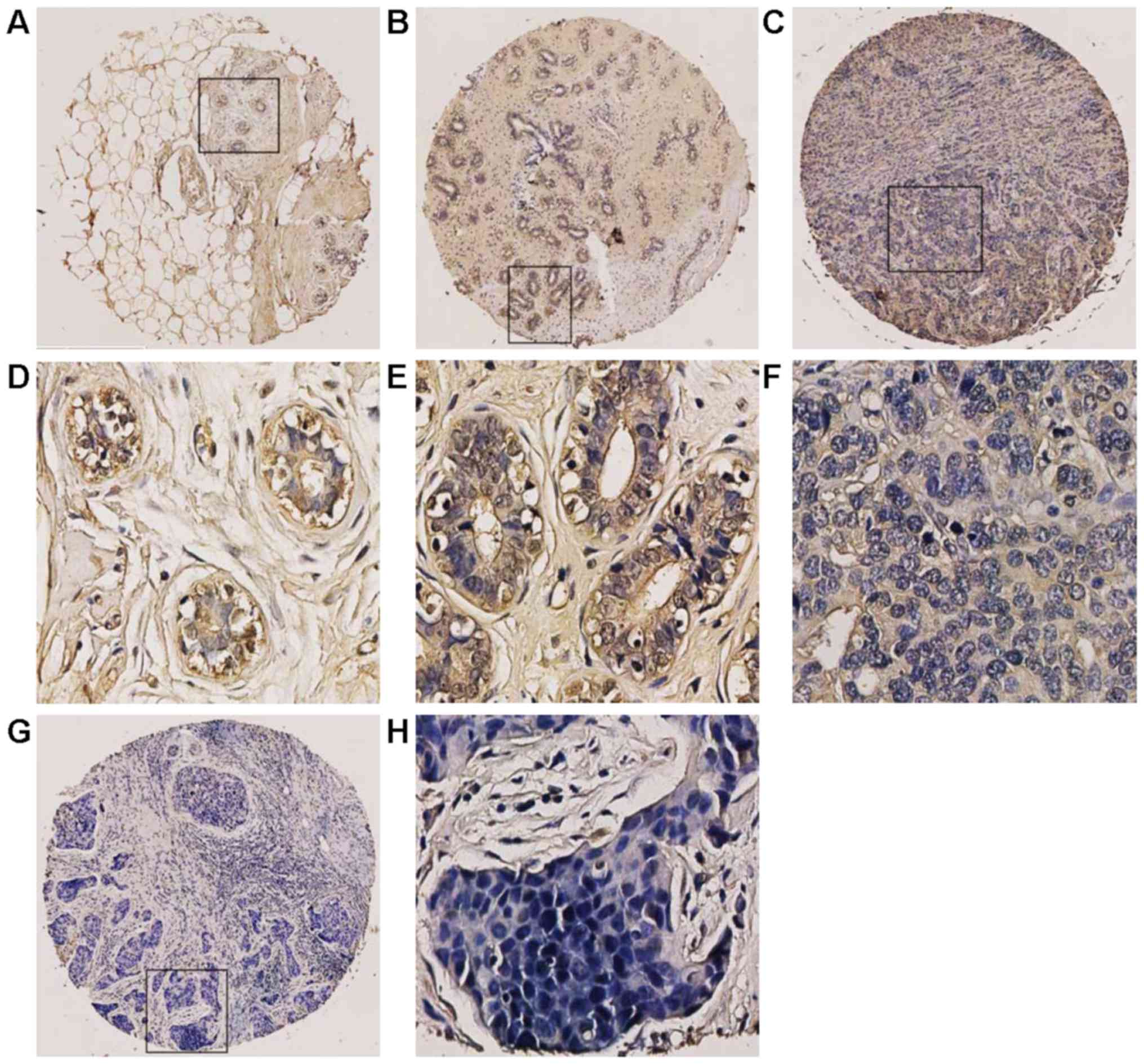 | Figure 2.Immunohistochemical staining of PinX1
was analysed in breast cancer tissues and the normal breast
tissues. (A) A cancer adjacent normal breast tissue (case 1) with
high expression of PinX1, where almost all tumor cells were
positively stained (magnification, ×100). (B) A fibroadenoma tissue
(case 3) with >65% of tumor cells positive staining of PinX1
(magnification, ×100). (C) An invasive ductal carcinoma (case 43)
exhibited low expression of PinX1, where <45% of tumor cells
were positive staining of PinX1 (magnification, ×100). (G) Low
expression of PinX1 was obtained in an invasive ductal carcinoma
(case 29), where <10% of tumor cells were positive staining of
PinX1 (magnification, ×100). Respectively, (D-H) revealed the
higher magnification (magnification, ×400) of the specific areas
boxed in (A, B, C and G). |
 | Table I.Association of PinX1 expression with
subtypes of breast cancer. |
Table I.
Association of PinX1 expression with
subtypes of breast cancer.
| Subtypes | Cases | Negative (%) | Positive (%) |
P-valuea |
|---|
| Luminal A/B | 13 | 3
(23.1) | 10 (76.9) | 0.008 |
|
HER2+ | 10 | 5
(50.0) | 5
(50.0) |
|
| Basal like | 33 | 24 (72.7) | 9
(27.3) |
|
Expression of PinX1 associated with
clinicopathological parameters of breast cancer
The association between PinX1 expression in 59
invasion ductal breast carcinomas and their clinical pathological
characteristics are listed in Table
II. The summary demonstrated that decreased PinX1 expression
was correlated with the histology grade of breast cancer
(P<0.05). Besides, PinX1 expression was positively associated
with PR status (P<0.05) and ER status (P<0.05). However,
there were no significant relationship between PinX1 expression and
other clinicopathological parameters including age at surgery,
clinical stage, TNM stage, Her2 status and p53 status
(P>0.05).
| Table II.Association of PinX1 expression with
clinicopathological parameters in 59 invasive ductal carcinoma
patients. |
Table II.
Association of PinX1 expression with
clinicopathological parameters in 59 invasive ductal carcinoma
patients.
|
| PinX1 staining |
|
|
|---|
|
|
|
|
|
|---|
|
Characteristics | Low (%) | High (%) | Total |
P-valueb |
|---|
| Age at surgery
(years)a |
|
≤50 | 19 (61.3) | 12 (38.7) | 31 | 0.746 |
|
>50 | 16 (57.1) | 12 (42.9) | 28 |
|
| Histology
grade |
| I | 0 (0) | 4 (100) | 4 | 0.028c |
|
II–III | 34 (61.8) | 21 (38.2) | 55 |
|
| Clinical stage |
|
I–II | 30 (62.5) | 18 (37.5) | 48 | 0.485 |
|
III | 5
(45.5) | 6
(54.5) | 11 |
|
| pT status |
|
T1-T2 | 28 (59.6) | 19 (40.4) | 47 | 1.000 |
|
T3-T4 | 47 (58.3) | 5
(41.7) | 12 |
|
| pN status |
| N0 | 27 (64.3) | 15 (35.7) | 42 | 0.222 |
|
N1-N2 | 8
(47.1) | 9
(52.9) | 17 |
|
| ER status |
|
Negative | 29 (72.5) | 11 (27.5) | 40 | 0.003 |
|
Positive | 6
(31.6) | 13 (68.4) | 19 |
|
| PR status |
|
Negative | 32 (66.7) | 16 (33.3) | 48 | 0.040 |
|
Positive | 3
(27.3) | 8
(72.7) | 11 |
|
| HER2 status |
|
Negative | 30 (63.8) | 17 (37.2) | 47 | 0.287 |
|
Positive | 5
(41.7) | 7
(58.3) | 12 |
|
| p53 status |
|
Negative | 10 (55.6) | 8
(45.4) | 18 | 0.696 |
|
Positive | 25 (61.0) | 16 (39.0) | 41 |
|
Then analyses of the positive staining of PinX1 were
stratified by different breast cancer subtypes in the breast cancer
tissues. Among all the 33 basal-like/triple-negative breast cancer
cases, we found that the mean positive staining was 81.1% and the
mean negative staining was 28.1%. Significantly, PinX1 was
downregulated in basal-like/triple-negative breast cancer
(P<0.01), which indicated that PinX1 may be correlated with the
development and progression of basal-like breast cancer.
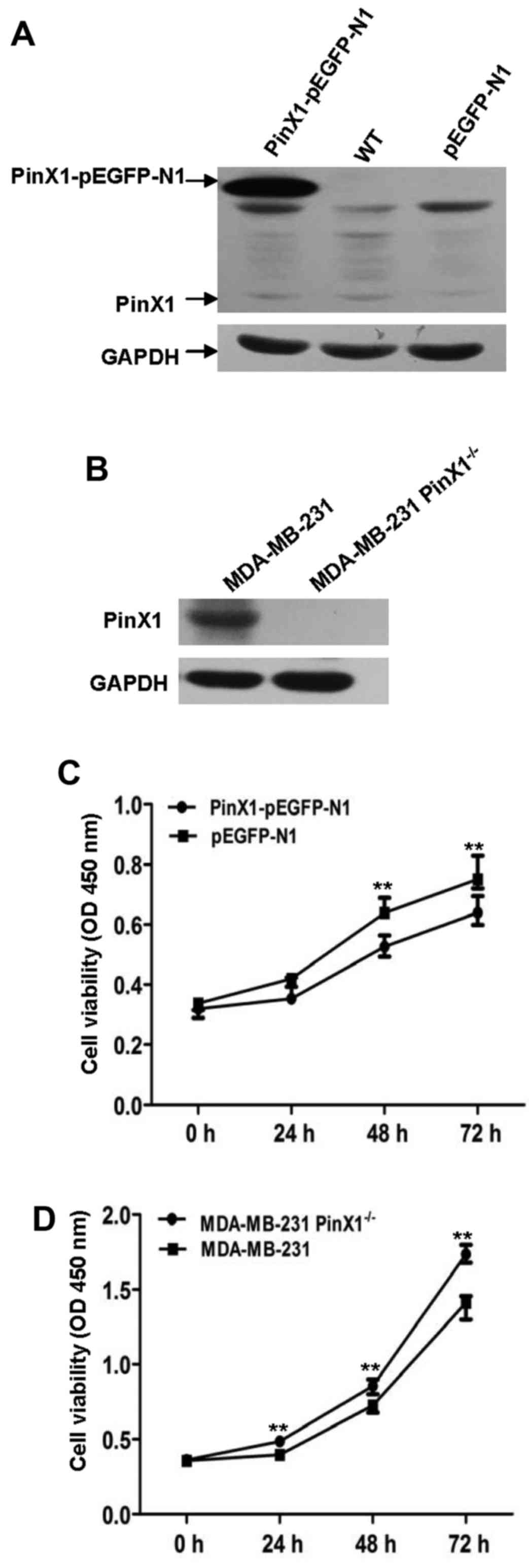
PinX1 attenuates proliferation in a
human basal-like breast cancer cell line
To clarify the probable role of PinX1 in basal-like
breast cancer, we constructed the MDA-MB-231 cell lines that stably
overexpressed PinX1 (PinX1-pEGFP-N1) and the negative control
(pEGFP-N1). We adopted CRISPR-Cas9 system (21) to knock out the PinX1 gene of
MDA-MB-231 cells. PinX1 expression levels were detected by
immunoblot analysis (Fig. 4A and
B). Then Cell Counting kit-8 (CCK- 8) assay was employed to
investigate the impact of PinX1 on cell proliferation rates. The
results showed that overexpression of PinX1 significantly inhibited
the proliferation of MDA-MB-231 cells while PinX1 knockout promoted
cell proliferation (Fig. 4C and
D).
PinX1 suppresses the migration ability
of MDA-MB-231 cells
Subsequently, the wound healing assay was used to
assess the migration ability of MDA-MB-231 cells that received
distinct treatments. As was shown in Fig. 5A and B, the migration rate was
inhibited when the PinX1 was overexpressed. However, it was
significantly reversed when the PinX1 was knocked out. In the
Transwell assay, different experimental groups of cells were fixed
and stained by crystal violet after 24-h incubation. Then, the
number of cells penetrating the membrane was analysed by a digital
light microscope. Results suggested that the cell number/field was
drastically increased in the PinX1 knockout group (Fig. 5D). On the contrary, the cell
migration ability was markedly impaired when PinX1 was upregulated
(Fig. 5C).
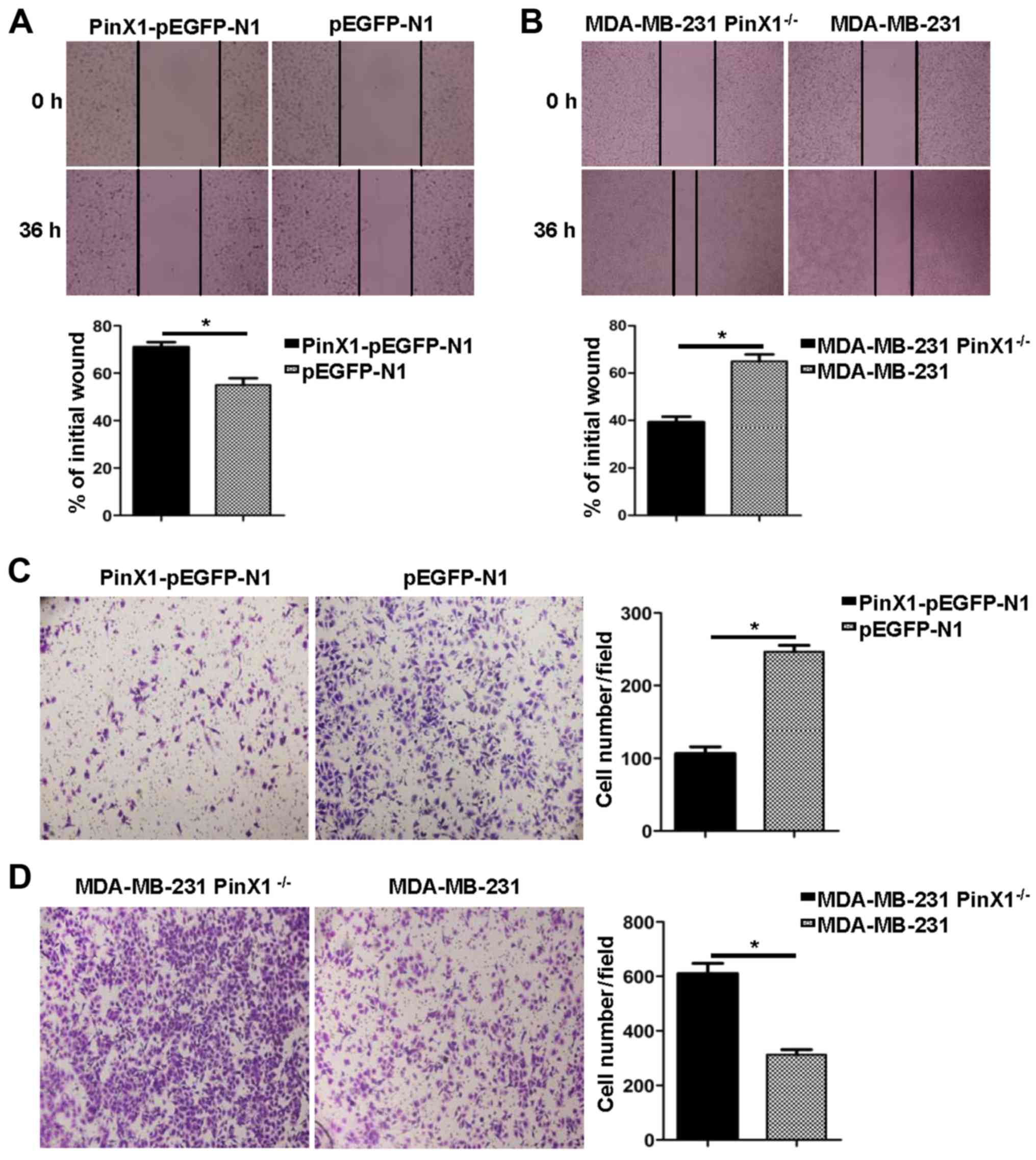 | Figure 5.PinX1 suppresses the migration
ability of MDA-MB-231 cells in vitro. (A) The migration
ability of the PinX1-overexpressed and the control groups were
tested by wound healing assay in a 36-h recovery time (top,
magnification, ×20). Bottom, statistical analysis of the migration
rates, presented relative to the initial wound area. (B) The
migration ability of the PinX1 knockout and the wild-type groups
was tested by wound healing assay in a 36-h recovery time (top,
magnifications, ×10). Bottom, statistical analysis of the migration
rates, presented relative to the initial wound area. (C and D)
Transwell assay was also used to measure the migration ability of
the different experimental groups (left, magnifications, ×10).
Right, statistical analyses of the migrated cell number per field.
At least three independent experiments were performed (*P<0.05,
two-tailed unpaired t-test) and the data expressed are mean ±
SEM. |
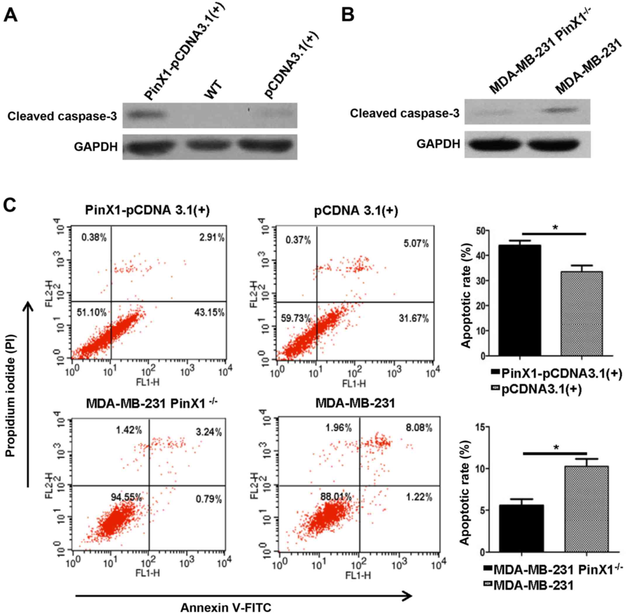
Overexpression of PinX1 provoked
apoptosis of MDA-MB-231 in vitro
Previous reports indicated that PinX1 could regulate
the telomerase activity and induce apoptosis of nasopharyngeal
carcinoma cells (22). Whether
PinX1 expression levels were correlated with apoptosis of breast
cancer is still unclear. Here we detected the expression levels of
cleaved caspase-3, a marker for cell apoptosis by western blot
analysis. After transfected with PinX1-pCDNA3.1(+) and pCDNA3.1(+)
for 48 h, MDA-MB-231 cells were harvested for later analysis. The
PinX1 knockout cells were exposed to etoposide (40 mM) for 24 h. We
observed a higher expression level of cleaved caspase-3 in the
PinX1 overexpressed group [PinX1-pCDNA3.1(+)] compared with the
negative control [pCDNA3.1(+)] (Fig.
6A). There was a higher expression of cleaved caspase-3 in
wild-type MDA-MB-231 than the PinX1 knockout cells (Fig. 6B). Consistently, the flow cytometric
analysis revealed that overexpression of PinX1 significantly
augmented the number of apoptotic cells in MDA-MB-231 and PinX1
knockout markedly decreased the proportion of apoptotic cells
(Fig. 6C). These findings indicated
that PinX1 may play an essential role in breast cancer cell
apoptosis.
Discussion
Breast cancer is the most etiologically
heterogeneous malignancy in women encompassing distinct clinical,
morphological and molecular entities (23). Unlike the other subtypes of breast
cancer, TNBC is highly aggressive. Patients diagnosed of TNBC are
younger. Although TNBC is chemosensitive, a higher risk of relapse
and an increased tendency to metastasize to visceral organs make
TNBC a challenge for current oncologists (24–26).
However, recent clinical therapy for TNBC remains limited. Though
chemotherapy has shown significant results in clinical practice,
previous research revealed that TNBC was not subjected to hormone
therapy or Her2-directed agents (i.e. trastuzumab) with the lack of
hormone receptors or Her2 receptors (27,28).
Besides, Wu et al (29)
reported that neo-adjuvant docetaxel plus epirubicin chemotherapy
increased pathological complete remission (pCR) rate of TNBC
patients with significantly worse survival. During the last
decades, with the widespread use of high-throughput technologies,
precision medicine contributes to specific molecular drivers unique
to specific individual for targeted therapies. The occurrence of
various therapeutic targets (i.e. BRCA1, PARP-1 and EGFR) benefit
TNBC patients markedly (30).
Nonetheless, as a cancer marker, overexpressed Ki-67 fails to
prognosticate DFS or OS in basal-like breast cancer (31). Tumor-targeted biomarkers of good
prognostic significance, analytic performance, and clinical utility
are urgent to be identified and validated to prompt patient
survival and life quality.
The phenomenon that telomerase activity in cancer is
highly elevated is known for several decades (32). Increasing research supports the
potential of telomerase as a clinical therapeutic target. In
general, telomere and telomerase influence the tumor formation in
different manner (33). Telomere
shortening and eventual uncapping are signals for cell to become
senescent and stop division via p53 and Rb pathways while rare
cells may escape from death and compromise the p53 and Rb pathways
with a telomere maintenance mechanism, typically in the way of
prompted telomerase expression. What is worse, these survived cells
secrete tumor-stimulating factors abetting cancers derived from
neighboring cells (34). As a
shelterin-related factor, PinX1 binds directly with both TRF1 and
telomerase. Originally, PinX1 was found to be an intrinsic
telomerase inhibitor and a conceivable tumor suppressor. However,
PinX1 expression patterns are contradictory between different types
of carcinomas. It was reported that PinX1 was negatively expressed
in colorectal, prostate and gastric carcinomas while it was
overexpressed in oesophageal squamous cell carcinomas (19,35–37),
demonstrating that abnormalities and effects of PinX1 in
tumorigenesis are complex and probably tumor-type-specific. In the
present study, we explored potential significance of PinX1 in
breast malignancy. We investigated the PinX1 expression levels in
26 pairs of breast cancer samples and the adjacent normal
counterparts by western blotting and qRT-PCR. We found that PinX1
was downregulated in breast cancer tissues compared with the paired
normal breast tissue samples (P<0.05), which was in consistent
with a previous study.
IHC remains an indispensable research tool in
routine tumor clinicopathology, which is widely used to study
diagnostic and prognostic characteristics of neoplasms (38). To further confirm the function of
PinX1 in breast cancer, IHC for PinX1 was performed in 75 breast
cancer specimens. We applied a specific scoring system to evaluate
PinX1 immunoreactivity, depending on the percentage of PinX1
positive cells. To make a reliable and reproducible assessment
without predetermination and arbitrariness, three pathologists
scored separately until they reached a consensus outcome.
Subsequently, ROC curve analysis helped to obtain the optimal
cut-off value for PinX1 positivity according to distinct
clinicopathological parameters. Our results suggested that reduced
PinX1 expression was correlated with advanced histology grade.
Besides, negative PinX1 expression was consistent with the negative
status of ER and PR. Moreover, stratification by four subtypes of
breast cancer was performed. The data indicated that PinX1
expression levels were correlated with the subtypes of breast
cancer. In addition, PinX1 was significantly downregulated in
basal-like breast cancer. Consequently, we hypothesized that low
expression of PinX1 probably play a critical role in the
development and progression of basal-like/triple-negative breast
cancer.
To validate our hypotheses in basal-like breast
cancer, we constructed knocked out and stably overexpressed PinX1
in the MDA-MB-231 basal-like breast cancer cell line. Then
influence of PinX1 on cell proliferation rates were detected by
CCK-8 assay. We found that overexpression of PinX1 decreased the
proliferation rate of MDA-MB-231 cells, while knockout of PinX1
expression level markedly reversed the effects. Besides, we
performed wound healing assay and trans- well assay to further
explore whether PinX1 was associated with the migration of
basal-like breast cancer. Consistently, high expression level of
PinX1 suppressed the number of migratory cells. It has been
demonstrated that overexpression of PinX1 induced esophageal
epithelial cell apoptosis by downregulating telomerase activity
(39). To further confirm the
relationship between PinX1 and breast cancer apoptosis, we
performed immunoblot analysis to assess the cleaved caspase-3 (a
marker for apoptosis) expression levels. Notably, the expression
level of cleaved caspase-3 was increased with the higher expression
of PinX1. The cleaved caspase-3 was downregulated when PinX1 was
knocked out. The flow cytometric analysis of apoptosis supported
the conclusion that PinX1 promoted also basal-like cell
apoptosis.
We confirmed the significant role of PinX1 as a
breast cancer suppressor. PinX1 was deregulated in breast cancer
tissues while it was upregulated in the normal samples. Among
distinguishing subtypes of breast cancer tissues, PinX1 expression
was reduced in the basal-like subtype compared with other subtypes.
Moreover, overexpression of PinX1 inhibited the proliferation rates
and migration ability and increased the apoptosis rates of a human
basal-like cancer. Our data clarified the importance of PinX1 in
basal-like breast cancer and could have a fundamental impact on the
development of basal-like breast cancer. PinX1 may be a potential
therapic target of basal-like breast cancer. However, the
underlying mechanisms of PinX1 contributing to basal-like breast
cancer remain to be further studied.
Acknowledgments
The present study was supported by the National
Natural Science Foundation of China (81372154, 81672588) and the
Science and Technology Planning Project of Guangdong Province,
China (2013B021800146).
Glossary
Abbreviations
Abbreviations:
|
ER
|
estrogen receptor
|
|
PR
|
progesterone receptor
|
|
HER2 receptor
|
human epidermal growth factor
receptor-2 receptor
|
|
BLBC
|
basal-like breast cancer
|
|
TNBC
|
triple-negative breast cancer
|
|
BRCA
|
breast cancer susceptibility gene
|
|
Pinx1
|
Pin2/TRF1-interacting telomerase
inhibitor 1
|
|
qRT-PCR
|
quantitative real-time polymerase
chain reaction
|
|
TMA
|
tissue microarray
|
|
AUC
|
the area under the curve
|
|
CCK-8
|
Cell Counting kit-8
|
|
pCR
|
pathological complete remission
|
|
EGFR
|
epidermal growth factor receptor
|
|
PARP-1
|
poly(ADP-ribose) polymerase 1
|
|
OS
|
overall survival
|
|
DFS
|
disease-free survival
|
|
TRF1
|
telomeric repeat binding factor 1
|
References
|
1
|
Taherian-Fard A, Srihari S and Ragan MA:
Breast cancer classification: linking molecular mechanisms to
disease prognosis. Brief Bioinform. 16:461–474. 2015. View Article : Google Scholar : PubMed/NCBI
|
|
2
|
Ferlay J, Soerjomataram I, Dikshit R, Eser
S, Mathers C, Rebelo M, Parkin DM, Forman D and Bray F: Cancer
incidence and mortality worldwide: Sources, methods and major
patterns in GLOBOCAN 2012. Int J Cancer. 136:E359–E386. 2015.
View Article : Google Scholar : PubMed/NCBI
|
|
3
|
Goldhirsch A, Winer EP, Coates AS, Gelber
RD, Piccart-Gebhart M, Thürlimann B, Senn HJ, Albain KS, André F,
Bergh J, et al: Panel members: Personalizing the treatment of women
with early breast cancer: Highlights of the St Gallen International
Expert Consensus on the Primary Therapy of Early Breast Cancer
2013. Ann Oncol. 24:2206–2223. 2013. View Article : Google Scholar : PubMed/NCBI
|
|
4
|
Vallejos CS, Gómez HL, Cruz WR, Pinto JA,
Dyer RR, Velarde R, Suazo JF, Neciosup SP, León M, de la Cruz MA,
et al: Breast cancer classification according to
immunohistochemistry markers: Subtypes and association with
clinicopathologic variables in a Peruvian hospital database. Clin
Breast Cancer. 10:294–300. 2010. View Article : Google Scholar : PubMed/NCBI
|
|
5
|
Ramirez-Fort MK, Case EC, Rosen AC, Cerci
FB, Wu S and Lacouture ME: Rash to the mTOR inhibitor everolimus:
Systematic review and meta-analysis. Am J Clin Oncol. 37:266–271.
2014. View Article : Google Scholar : PubMed/NCBI
|
|
6
|
Gelmon K, Dent R, Mackey JR, Laing K,
McLeod D and Verma S: Targeting triple-negative breast cancer:
Optimising therapeutic outcomes. Ann Oncol. 23:2223–2234. 2012.
View Article : Google Scholar : PubMed/NCBI
|
|
7
|
Bianchini G, Balko JM, Mayer IA, Sanders
ME and Gianni L: Triple-negative breast cancer: Challenges and
opportunities of a heterogeneous disease. Nat Rev Clin Oncol.
13:674–690. 2016. View Article : Google Scholar : PubMed/NCBI
|
|
8
|
Perou CM: Molecular stratification of
triple-negative breast cancers. Oncologist. 15 Suppl 5:39–48. 2010.
View Article : Google Scholar : PubMed/NCBI
|
|
9
|
De Laurentiis M, Cianniello D, Caputo R,
Stanzione B, Arpino G, Cinieri S, Lorusso V and De Placido S:
Treatment of triple negative breast cancer (TNBC): Current options
and future perspectives. Cancer Treat Rev. 36 Suppl 3:S80–S86.
2010. View Article : Google Scholar : PubMed/NCBI
|
|
10
|
Eroles P, Bosch A, Pérez-Fidalgo JA and
Lluch A: Molecular biology in breast cancer: Intrinsic subtypes and
signaling pathways. Cancer Treat Rev. 38:698–707. 2012. View Article : Google Scholar : PubMed/NCBI
|
|
11
|
Esposito A: Highlights from the 14th St
Gallen International Breast Cancer Conference 2015 in Vienna:
Dealing with classification, prognostication, and prediction
refinement to personalize the treatment of patients with early
breast cancer. E Cancer Med Sci. 9:5182015.
|
|
12
|
Nelson ER, Li S, Kennedy M, Payne S,
Kilibarda K, Groth J, Bowie M, Parilla-Castellar E, De Ridder G,
Marcom PK, et al: Chemotherapy enriches for an invasive
triple-negative breast tumor cell subpopulation expressing a
precursor form of N-cadherin on the cell surface. Oncotarget.
7:84030–84042. 2016.PubMed/NCBI
|
|
13
|
Rodler ET, Kurland BF, Griffin M, Gralow
JR, Porter P, Yeh RF, Gadi VK, Guenthoer J, Beumer JH, Korde L, et
al: Phase I study of veliparib (ABT-888) combined with cisplatin
and vinorelbine in advanced triple-negative breast cancer and/or
BRCA mutation-associated breast cancer. Clin Cancer Res.
22:2855–2864. 2016. View Article : Google Scholar : PubMed/NCBI
|
|
14
|
Li Y and Tergaonkar V: Noncanonical
functions of telomerase: Implications in telomerase-targeted cancer
therapies. Cancer Res. 74:1639–1644. 2014. View Article : Google Scholar : PubMed/NCBI
|
|
15
|
Zhou XZ, Huang P, Shi R, Lee TH, Lu G,
Zhang Z, Bronson R and Lu KP: The telomerase inhibitor PinX1 is a
major haploinsufficient tumor suppressor essential for chromosome
stability in mice. J Clin Invest. 121:1266–1282. 2011. View Article : Google Scholar : PubMed/NCBI
|
|
16
|
Tian XP, Qian D, He LR, Huang H, Mai SJ,
Li CP, Huang XX, Cai MY, Liao YJ, Kung HF, et al: The
telomere/telomerase binding factor PinX1 regulates paclitaxel
sensitivity depending on spindle assembly checkpoint in human
cervical squamous cell carcinomas. Cancer Lett. 353:104–114. 2014.
View Article : Google Scholar : PubMed/NCBI
|
|
17
|
Liu JY, Qian D, He LR, Li YH, Liao YJ, Mai
SJ, Tian XP, Liu YH, Zhang JX, Kung HF, et al: PinX1 suppresses
bladder urothelial carcinoma cell proliferation via the inhibition
of telomerase activity and p16/cyclin D1 pathway. Mol Cancer.
12:1482013. View Article : Google Scholar : PubMed/NCBI
|
|
18
|
Li HL, Han L, Chen HR, Meng F, Liu QH, Pan
ZQ, Bai J and Zheng JN: PinX1 serves as a potential prognostic
indicator for clear cell renal cell carcinoma and inhibits its
invasion and metastasis by suppressing MMP-2 via NF-κB-dependent
transcription. Oncotarget. 6:21406–21420. 2015. View Article : Google Scholar : PubMed/NCBI
|
|
19
|
Shi R, Zhao Z, Zhou H, Wei M, Ma WL, Zhou
JY and Tan WL: Reduced expression of PinX1 correlates to
progressive features in patients with prostate cancer. Cancer Cell
Int. 14:462014. View Article : Google Scholar : PubMed/NCBI
|
|
20
|
Livak KJ and Schmittgen TD: Analysis of
relative gene expression data using real-time quantitative PCR and
the 2ΔΔCT method. Methods. 25:402–408. 2001. View Article : Google Scholar : PubMed/NCBI
|
|
21
|
Ran FA, Hsu PD, Wright J, Agarwala V,
Scott DA and Zhang F: Genome engineering using the CRISPR-Cas9
system. Nat Protoc. 8:2281–2308. 2013. View Article : Google Scholar : PubMed/NCBI
|
|
22
|
Lai XF, Shen CX, Wen Z, Qian YH, Yu CS,
Wang JQ, Zhong PN and Wang HL: PinX1 regulation of telomerase
activity and apoptosis in nasopharyngeal carcinoma cells. J Exp
Clin Cancer Res. 31:122012. View Article : Google Scholar : PubMed/NCBI
|
|
23
|
Toss A and Cristofanilli M: Molecular
characterization and targeted therapeutic approaches in breast
cancer. Breast Cancer Res. 17:602015. View Article : Google Scholar : PubMed/NCBI
|
|
24
|
Morris PG, Murphy CG, Mallam D, Accordino
M, Patil S, Howard J, Omuro A, Beal K, Seidman AD, Hudis CA, et al:
Limited overall survival in patients with brain metastases from
triple negative breast cancer. Breast J. 18:345–350. 2012.
View Article : Google Scholar : PubMed/NCBI
|
|
25
|
Ogata H, Kikuchi Y, Natori K, Shiraga N,
Kobayashi M, Magoshi S, Saito F, Osaku T, Kanazawa S, Kubota Y, et
al: Liver metastasis of a triple-negative breast cancer and
complete remission for 5 years after treatment with combined
bevacizumab/paclitaxel/carboplatin: Case Report and Review of the
Literature. Medicine (Baltimore). 94:e17562015. View Article : Google Scholar : PubMed/NCBI
|
|
26
|
Bartholomeusz C, Xie X, Pitner MK, Kondo
K, Dadbin A, Lee J, Saso H, Smith PD, Dalby KN and Ueno NT: MEK
inhibitor selumetinib (AZD6244; ARRY-142886) prevents lung
metastasis in a triple-negative breast xancer xenograft model. Mol
Cancer Ther. 14:2773–2781. 2015. View Article : Google Scholar : PubMed/NCBI
|
|
27
|
Burnett JP, Korkaya H, Ouzounova MD, Jiang
H, Conley SJ, Newman BW, Sun L, Connarn JN, Chen CS, Zhang N, et
al: Trastuzumab resistance induces EMT to transform HER2+ PTEN to a
triple negative breast cancer that requires unique treatment
options. Sci Rep. 5:158212015. View Article : Google Scholar : PubMed/NCBI
|
|
28
|
Pal SK, Childs BH and Pegram M: Triple
negative breast cancer: Unmet medical needs. Breast Cancer Res
Treat. 125:627–636. 2011. View Article : Google Scholar : PubMed/NCBI
|
|
29
|
Wu J, Li S, Jia W and Su F: Response and
prognosis of taxanes and anthracyclines neoadjuvant chemotherapy in
patients with triple-negative breast cancer. J Cancer Res Clin
Oncol. 137:1505–1510. 2011. View Article : Google Scholar : PubMed/NCBI
|
|
30
|
Carey LA, Rugo HS, Marcom PK, Mayer EL,
Esteva FJ, Ma CX, Liu MC, Storniolo AM, Rimawi MF, Forero-Torres A,
et al: TBCRC 001: Randomized phase II study of cetuximab in
combination with carboplatin in stage IV triple-negative breast
cancer. J Clin Oncol. 30:2615–2623. 2012. View Article : Google Scholar : PubMed/NCBI
|
|
31
|
Lang JE, Wecsler JS, Press MF and Tripathy
D: Molecular markers for breast cancer diagnosis, prognosis and
targeted therapy. J Surg Oncol. 111:81–90. 2015. View Article : Google Scholar : PubMed/NCBI
|
|
32
|
Kim NW, Piatyszek MA, Prowse KR, Harley
CB, West MD, Ho PL, Coviello GM, Wright WE, Weinrich SL and Shay
JW: Specific association of human telomerase activity with immortal
cells and cancer. Science. 266:2011–2015. 1994. View Article : Google Scholar : PubMed/NCBI
|
|
33
|
Johnson FB: PinX1 the tail on the
chromosome. J Clin Invest. 121:1242–1244. 2011. View Article : Google Scholar : PubMed/NCBI
|
|
34
|
Artandi SE and DePinho RA: Telomeres and
telomerase in cancer. Carcinogenesis. 31:9–18. 2010. View Article : Google Scholar : PubMed/NCBI
|
|
35
|
Deng W, Jiao N, Li N, Wan X, Luo S and
Zhang Y: Decreased expression of PinX1 protein predicts poor
prognosis of colorectal cancer patients receiving 5-FU adjuvant
chemotherapy. Biomed Pharmacother. 73:1–5. 2015. View Article : Google Scholar : PubMed/NCBI
|
|
36
|
Qian D, Zhang B, He LR, Cai MY, Mai SJ,
Liao YJ, Liu YH, Lin MC, Bian XW, Zeng YX, et al: The
telomere/telomerase binding factor PinX1 is a new target to improve
the radiotherapy effect of oesophageal squamous cell carcinomas. J
Pathol. 229:765–774. 2013. View Article : Google Scholar : PubMed/NCBI
|
|
37
|
Wang HB, Wang XW, Zhou G, Wang WQ, Sun YG,
Yang SM and Fang DC: PinX1 inhibits telomerase activity in gastric
cancer cells through Mad1/c-Myc pathway. J Gastrointest Surg.
14:1227–1234. 2010. View Article : Google Scholar : PubMed/NCBI
|
|
38
|
Swanson PE: Immunohistochemistry as a
surrogate for molecular testing: A review. Appl Immunohistochem Mol
Morphol. 23:81–96. 2015. View Article : Google Scholar : PubMed/NCBI
|
|
39
|
Zuo J, Wang DH, Zhang YJ, Liu L, Liu FL
and Liu W: Expression and mechanism of PinX1 and telomerase
activity in the carcinogenesis of esophageal epithelial cells.
Oncol Rep. 30:1823–1831. 2013.PubMed/NCBI
|