Introduction
Liver cancer is the second leading cause of
cancer-related deaths in males worldwide. More than half of these
liver cancer-related deaths occurred in China during 2012 (1). A recent study estimated that
approximately 422,100 Chinese patients died from liver cancer in
2015, which will make it the third leading cause of cancer-related
death in China (2). A
population-based study of 138,852 cancer cases reported that liver
cancer is associated with poor survival with an age-standardized
5-year relative survival of 10.1% in China (3). Liver cancer death rates in Guangxi
Province were the highest in China for both males and females
(4). Hepatocellular carcinoma (HCC)
is the most common type of liver cancer (85–90%) (5). The most prominent parameters
associated with HCC in China include hepatitis B virus (HBV) and C
viral infection, alcoholic liver disease, and
aflatoxin-B1-contaminated food (6).
Previous studies of the Guangxi population reported that high HBV
infection and aflatoxin B1 (AFB1) exposure resulted in a higher HCC
morbidity and mortality in this province than in other provinces in
China (7,8).
Tumor protein p53 (TP53) is a tumor-suppressor
protein involved in transcriptional activation, DNA binding, and
oligomerization domains. TP53 wild-type protein can induce cell
cycle arrest, apoptosis, senescence, DNA repair, and changes in
metabolism (9,10). Wild-type TP53 is an important
tumor-suppressor gene in many types of cancers, especially in HCC,
and its mutation is regarded as oncogenic (10,11),
and affects HCC tumorigenesis and cancer progression (12–16).
AFB1 has been strongly associated with TP53 mutations at
codon 249 in exon 7, and HCC patients in Guangxi have a high rate
(34%) of TP53 mutations at codon 249 in exon 7 (8,17).
Thus, the population in this region presents a unique opportunity
to investigate the relationship of HBV infection, AFB1 exposure,
and TP53 gene mutations with HCC. Recently, meta-analyses
have reported that immunohistochemical characterization of
TP53 expression is associated with a poor prognosis of HCC
(18).
Aldehyde dehydrogenase 1 family member L1 (ALDH1L1),
also known as 10-formyltetrahydrofolate dehydrogenase (FDH), is a
folate metabolism enzyme with tumor suppressor-like properties and
is involved in the regulation of cell proliferation. A previous
study conducted by Oleinik and Krupenko demonstrated that the
antiproliferative effects of FDH in human lung cancer cell line
A549 induced G1 arrest and apoptosis, accompanied by an increase in
TP53 and p21 (19). In addition,
subsequent research by this group also demonstrated that
FDH-induced tumor-suppressor effects were strictly TP53-dependent
in A549 cells and the TP53 pathway was a downstream
mechanism in response to induction of FDH expression (20). Another study of HCC patients in
Guangxi reported that low ALDH1L1 protein expression was a new and
potential prognostic marker for the survival of HCC patients
(21). Using bioinformatic
analyses, we found that expression of ALDH1L1 in the liver
was the highest in various human normal tissues, and was
significantly downregulated in HCC tumor tissues compared to
tissues adjacent to the tumor. Our previous genome-wide association
study also reported that single nucleotide polymorphisms (SNPs)
were associated with positive immunohistochemical characterization
of TP53 expression in Guangxi patients with HBV-related HCC
(22). In the present study, we
determined the association between ALDH1L1 genetic
variations and mRNA expression and the postoperative prognosis in
Chinese HBV-related HCC patients, and its interaction with
TP53.
Materials and methods
Study population
This study was approved by the Ethics Committee of
the First Affiliated Hospital of Guangxi Medical University
(Guangxi, China) with approval number KY-E-032. Fresh specimens of
415 cases of HCC were collected from 2001 to 2013 at the First
Affiliated Hospital of Guangxi Medical University and were
confirmed by pathology. All the patients were positive for serum
HBV surface antigen inspection. The TP53 expression status
in the cancer tissues was detected by immunohistochemistry. The
cancer tissues were collected during surgery and immediately stored
at −80°C for further use. The tumor status was classified using the
Barcelona Clinic Liver Cancer (BCLC) staging system, and the liver
reserve function was determined using the Child-Pugh
classification. Portal vein tumor thrombus (PVTT) was classified
according to a previous study (23).
SNP selection and genotyping
ALDH1L1 tagged SNPs and non-synonymous SNPs
were selected by using an SNP info Web Server (http://snpinfo.niehs.nih.gov/. accessed 20 October
2016). Evaluation of SNP non-synonymous mutations caused by changes
in protein amino acids were determined using SIFT (http://sift.jcvi.org/. accessed October 20, 2016)
(24) and PolPhen 2 (http://genetics.bwh.harvard.edu/pph2/index.shtml.
accessed October 20, 2016) (25).
Bioinformatic analyses of ALDH1L1 SNPs with the tagged SNP
located in the exon region showed that rs2276724 was a
non-synonymous SNP. The influence of SNP non-synonymous mutation
analyses by SIFT also showed that rs2276724 S481G was deleterious
to protein coding. Consistent results from PolyPhen2 also showed
that rs2276724 S481G was possibly deleterious to protein coding.
Thus, rs2276724 was further studied. The transcriptional regulation
of rs2276724 S481G was detected by F-SNP database (http://compbio.cs.queensu.ca/F-SNP/. accessed
February 7, 2017) and the prediction tool Golden Path suggested
that non-synonymous mutation of rs2276724 S481G caused
transcriptional regulation change (26).
Genomic DNA was extracted from surgical tumor
samples using the TIANamp Genomic DNA kit (Tiangen Biotech,
Beijing, China). All samples were genotyped by DNA sequencing using
an ABI Prism 3100 (Applied Biosystems, Shanghai Sangon Biological
Engineering Technology and Services, Shanghai, China) with the
following primers: forward, 5′-GCCCTGTCTTCCCTTCCTGTG-3′ and
reverse, 5′-CCTGAGCCCACTCTGCTGAAAT-3′ for rs2276724. The sequencing
results were analyzed using Chromas software (http://technelysium.com.au/wp/chromas/
accessed October 20, 2016) with a signal/noise >98%.
GEO data and bioinformatic
analysis
Based on the predictive result of F-SNP,
non-synonymous mutation of rs2276724 S481G may affect gene
transcriptional regulation. We hypothesized that ALDH1L1
mRNA expression may contribute to prognostic prediction of
HBV-related HCC. To test this hypothesis, we further analyzed the
association of ALDH1L1 and TP53 at the
transcriptional level to evaluate the effects of ALDH1L1
mRNA expression and the interactions with TP53 on HCC
prognosis after hepatectomy. The profile chip dataset of Chinese
HBV-related HCC from Gene Expression Omnibus (GEO, http://www.ncbi.nlm.nih.gov/geo/. accessed
October 20, 2016) was analyzed and Spearman's correlation
coefficient was used to assess its correlations. The GEO data
selection criteria were set as follows: i), expression profiling
chip; ii), Chinese HBV-related HCC; iii), corresponding survival
profiles was available; and iv), patients undergoing hepatectomy.
By searching the GEO database, we found that only the data of
GSE14520 met the criteria above. Then, the samples were divided
into two groups according to the ALDH1L1 expression in
tumors. The high ALDH1L1 group was composed of samples with
ALDH1L1 expression levels above the median value, and the
low ALDH1L1 group was composed of the remaining samples.
TP53 expression was grouped in the same manner. Both
disease-free survival (DFS) and overall survival (OS) were analyzed
in the different ALDH1L1 expression groups and used for the
joint-effect survival analyses of the TP53 groups. We also
stratified the analyses of associations between different
ALDH1L1 expression levels and clinical features, both for OS
and DFS. A gene interaction analysis web site (GeneMANIA:
http://www.genemania.org/ accessed October 20,
2016) was used for correlation analyses between genes. Online
analysis tool was used to analyze the ALDH1L1 expression in
multiple human normal tissues (http://www.gtexportal.org/home/ accessed October 20,
2016) and in differences of expression of HCC tumor and adjacent
tumor tissues (MERAV, Metabolic gEne Rapid Visualizer: http://merav.wi.mit.edu/. accessed October 20,
2016).
Statistical analysis
Hardy-Weinberg equilibrium (HWE) of the selected
SNPs was estimated using a goodness-of-fit χ2-test. The
binary logistic regression model was used to analyze the genetic
model of ALDH1L1 genotypes for the status of different
TP53 expression levels and for the association between
clinical risk factors with ALDH1L1 genotypes. Survival
analyses were performed using the Kaplan-Meier method with the
log-rank test for different clinical factors and different
genotypes. Cox proportion haphazard regression analyses were used
to calculate the crude or adjusted hazard ratio (HR) and the 95%
confidence interval (CI) in univariate analyses and multivariate
analyses, adjusted for those variables with P<0.1 in later
multivariate analyses. A value of P<0.05 was considered
statistically significant. All statistical analyses were conducted
using SPSS Statistical Software for Windows, version 20.0 (SPSS,
Chicago, IL, USA).
Results
Clinical features and outcomes
Patients were followed up after surgery until the
final follow-up or until death. The final follow-up was conducted
in September 2014. A total of 415 patients successfully completed
the follow-up, with 6.7% of the patients lost in the follow-up. The
duration of the follow-up ranged from 12–125 months, with an
overall median survival time (MST) of 48 months. At the time of
analyses, 192 (46.3%) of the patients had died. A total of 162
patients were negative for TP53 expression and 253 patients
were positive for TP53 expression. Clinical features of all
patients and the association with the OS are shown in Table I. Using Kaplan-Meier analyses, the
biological characteristics of tumor size, tumor number, BCLC stage,
and portal vein tumor thrombus (PVTT) were significantly associated
with the OS (log-rank test, P<0.001) and increased risk of
death. A Child-Pugh classification score for 356 (85.8%) patients
was associated with the OS (log-rank P=0.005). Radical resection
was conducted in 231 patients (55.7%), and was associated with the
OS (log-rank P=0.052), and patients without cirrhosis had a better
prognosis (log-rank P=0.027). Adjuvant antiviral treatment in 143
(34.5%) patients was significantly associated with the OS (log-rank
P=0.019), compared with those without treatment. The other clinical
parameters were not associated with the OS.
 | Table I.Clinical features of the patients
with HBV-related HCC. |
Table I.
Clinical features of the patients
with HBV-related HCC.
| Variables | Patients
(N=415) | No. of events
(%) | MST (months) | HR (95% CI) | Log-rank
p-value |
|---|
| Age (years) |
|
|
|
| 0.149 |
|
≤60 | 367 | 169 (46.0) | 51 | 1 |
|
|
>60 | 48 | 23
(47.9) | 41 | 1.375
(0.888–2.128) |
|
| Sex |
|
|
|
| 0.479 |
|
Male | 375 | 177 (47.2) | 48 | 1 |
|
|
Female | 40 | 15
(37.5) | 42 | 0.828
(0.488–1.404) |
|
| Ethnicity |
|
|
|
| 0.989 |
|
Han | 260 | 122 (46.9) | 47 | 1 |
|
|
Minority | 155 | 70
(45..2) | 50 | 0.998
(0.743–1.341) |
|
| BMI |
|
|
|
| 0.745 |
|
≤25 | 328 | 150 (45.7) | 45 | 1 |
|
|
>25 | 87 | 42
(48.3) | 51 | 0.945
(0.6670–1.333) |
|
| Smoking status |
|
|
|
| 0.107 |
|
None | 263 | 118 (44.9) | 51 | 1 |
|
|
Ever | 152 | 74
(48.7) | 39 | 1.269
(0.947–1.702) |
|
| Drinking
status |
|
|
|
| 0.084 |
|
None | 246 | 107 (43.5) | 51 | 1 |
|
|
Ever | 169 | 85
(50.3) | 40 | 1.284
(0.964–1.710) |
|
| Child-Pugh
score |
|
|
|
| 0.005 |
| A | 356 | 153 (43.0) | 51 | 1 |
|
| B | 59 | 39
(66.1) | 31 | 1.689
(1.159–2.460) |
|
| Cirrhosis |
|
|
|
| 0.027 |
| No | 46 | 16
(34.8) | NA | 1 |
|
|
Yes | 369 | 176 (47.7) | 44 | 1.769
(1.058–2.958) |
|
| Radical
resectiona |
|
|
|
| 0.052 |
|
Yes | 231 | 97
(42.0) | 71 | 1 |
|
|
None | 172 | 89
(51.7) | 40 | 1.330
(0.997–1.774) |
|
| Portal
hypertensionb |
|
|
|
| 0.243 |
| No | 208 | 100 (48.1) | 52 | 1 |
|
|
Yes | 172 | 75
(43.6) | 42 | 1.197
(0.883–1.623) |
|
| Pathological
diagnosisc |
|
|
|
| 0.622 |
| Well
differentiated | 24 |
9 (37.5) | 79 | 1 |
|
|
Moderately differentiated | 341 | 159 (46.6) | 44 | 1.378
(0.703–2.699) |
|
| Poorly
differentiated | 11 |
4 (36.4) | NA | 1.200
(0.369–3.898) |
|
| Adjuvant antiviral
treatment |
|
|
|
| 0.019 |
|
Yes | 143 | 43
(30.0) | NA | 1 |
|
| No | 272 | 149 (54.8) | 41 | 1.501
(1.065–2.116) |
|
| AFP
leveld |
|
|
|
| 0.233 |
|
<400 | 210 | 89
(42.4) | 51 | 1 |
|
|
≥400 | 175 | 85
(48.6) | 42 | 1.197
(0.889–1.612) |
|
| Tumor behavior |
|
|
|
|
|
| Tumor size
(cm) |
|
|
|
| <0.001 |
| ≤5 | 158 | 59
(37.3) | 75 | 1 |
|
|
>5 | 229 | 133 (58.1) | 36 | 1.802
(1.326–2.450) |
|
| Tumor number |
|
|
|
| <0.001 |
|
Single | 302 | 127 (42.0) | 58 | 1 |
|
|
Multiple | 113 | 65
(57.5) | 28 | 1.792
(1.326–2.420) |
|
| Regional
invasion |
|
|
|
| 0.156 |
|
Absence | 353 | 162 (45.9) | 51 | 1 |
|
|
Presence | 62 | 30
(48.4) | 35 | 1.323
(0.895–1.958) |
|
| BCLC stage |
|
|
|
| <0.001 |
| A | 236 | 81
(34.3) | 95 | 1 |
|
| B | 68 | 37
(54.4) | 36 | 2.055
(1.391–3.035) |
|
| C | 111 | 74
(66.7) | 24 | 2.741
(1.994–3.767) |
|
| PVTT |
|
|
|
| <0.001 |
| No | 342 | 139 (40.6) | 73 | 1 |
|
|
Yes | 73 | 53
(72.6) | 18 | 2.801
(2.032–3.861) |
|
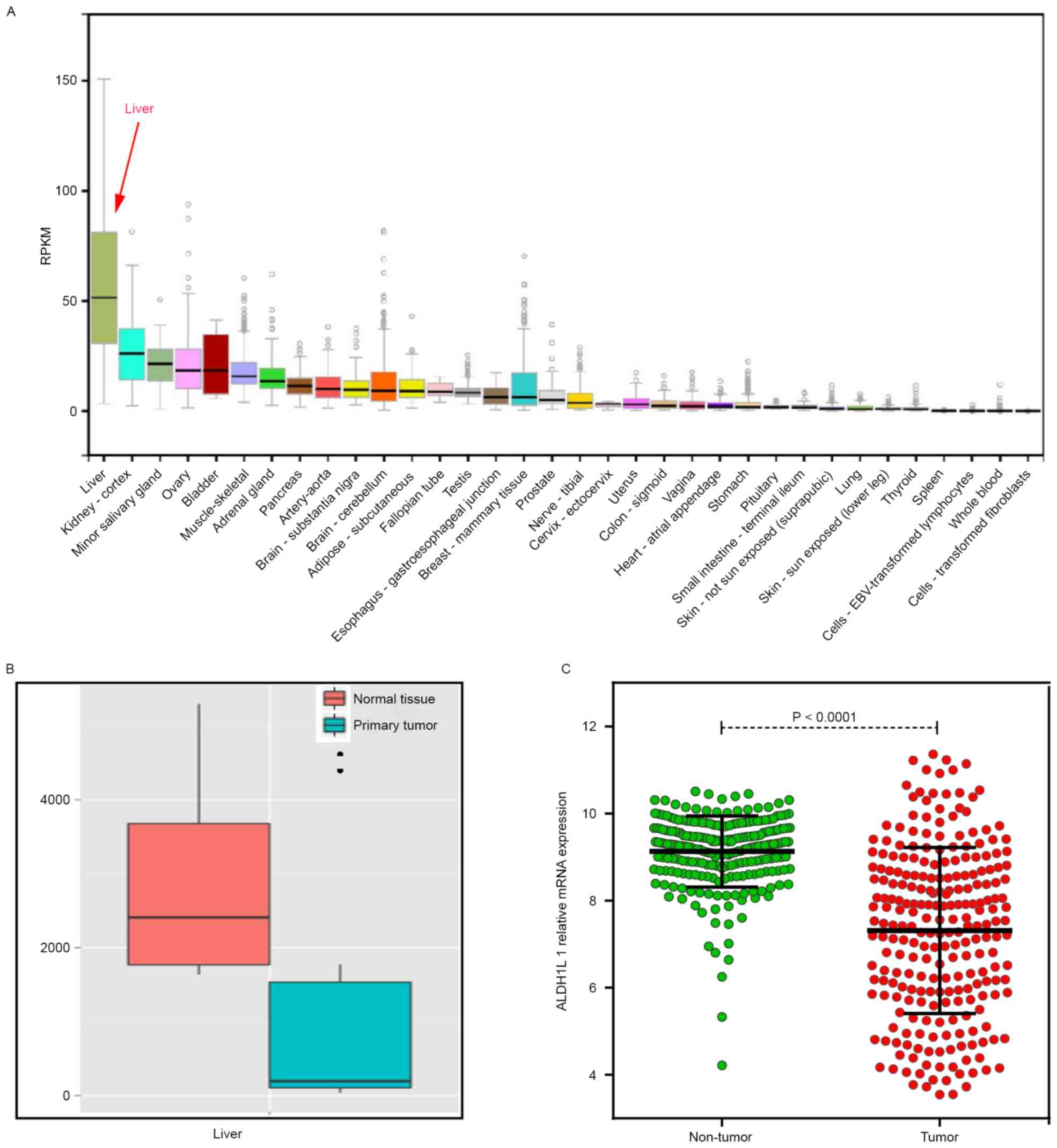
Gene expression analysis
Bioinformatic analysis of ALDH1L1 gene
expression in multiple human normal tissues showed that
ALDH1L1 was the highest expression in normal liver tissue
(Fig. 1A). ALDH1L1
expression was significantly downregulated in HCC tumor tissue, as
determined for MERAV (Fig. 1B) and
GSE14520 (Fig. 1C).
Genetic model analysis of
rs2276724
The success of genotyping for rs2276724 was 100%.
The genotype frequencies met Hardy-Weinberg equilibrium as shown by
the goodness-of-fit χ2-test (rs2276724,
χ2=0.236; P=0.627). The genotype distribution of
rs2276724 in patients with different TP53 expression is
shown in Table II. The binary
logistic regression model was used for adjustment for alcohol
consumption, the Child-Pugh score, tumor size, tumor number, BCLC
stage, radical resection, cirrhosis, adjuvant antiviral treatment,
and PVTT. Using a co-dominant genetic model, the CT genotype of
rs2276724 was significantly reduced with TP53 expression in
HBV-related HCC patients (adjusted P=0.045; adjusted OR=0.644; 95%
CI=0.418–0.990), compared with the TT genotype. The genotype
distributions of rs2276724 in different types of TP53
expression were similar to the four genetic models.
| Table II.Genotype distribution of rs2276724 in
HBV-related HCC patients with different TP53 expression statuses
(genetic model). |
Table II.
Genotype distribution of rs2276724 in
HBV-related HCC patients with different TP53 expression statuses
(genetic model).
| SNP | TP53- negative
(n=162) | TP53-positive
(n=253) | Crude OR (95%
CI) | Crude p-value | Adjusted OR (95%
CI) | Adjusted
P-valuea |
|---|
| rs2276724 |
|
Allele |
| T | 246 | 394 | 1 |
| 1 |
|
| C | 78 | 112 | 0.894
(0.640–1.249) | 0.512 | 0.878
(0.623–1.238) | 0.458 |
| Co-dominant |
| TT | 89 | 156 |
1 |
| 1 |
|
| CT | 68 | 82 | 0.688
(0.455–1.040) | 0.076 | 0.644
(0.418–0.990) | 0.045 |
| CC |
5 | 15 | 1.712
(0.602–4.867) | 0.314 | 1.838
(0.629–5.365) | 0.266 |
| Dominant |
| TT | 89 | 156 | 1 |
| 1 |
|
|
CT+CC | 73 | 97 | 0.758
(0.508–1.131) | 0.175 | 0.725
(0.479–1.097) | 0.128 |
| Recessive |
|
CT+TT | 157 | 238 | 1 |
| 1 |
|
| CC |
5 | 15 | 1.979
(0.705–5.554) | 0.195 | 2.153
(0.747–6.207) | 0.156 |
Genetic polymorphisms in the HCC risk
factors and in stratified analysis
Association between risk factors and rs2276724
genotypes are summarized in Table
III. None of the selected risk factors was associated with
rs2276724 genotypes in the present study. Stratified analyses of
the rs2276724 genotype and different strata of the OS of selected
risk factors are shown in Fig. 2.
In the favorable strata, the CT/CC genotype of rs2276724
significantly decreased the risk of death among patients with tumor
sizes ≤5 cm, Child-Pugh score A, and without a PVTT. Regarding the
invasion in the adverse strata, we also observed a similar effect
that the CT/CC genotype of rs2276724 significantly decreased the
risk of death among patients with BCLC stage B/C, non-radical
resection, and the presence of regional invasion. The genotype
distributions of rs2276724 in other strata showed no
difference.
| Table III.Association between risk factors and
rs2276724 in HBV-related HCC patients. |
Table III.
Association between risk factors and
rs2276724 in HBV-related HCC patients.
| Variables | TT | CT+CC | OR (95% CI) | P-value |
|---|
| Tumor size
(cm) |
|
|
|
|
| ≤5 | 98 | 70 | 1 |
|
|
>5 | 147 | 100 | 0.952
(0.640–1.418) | 0.810 |
| Tumor number |
|
|
|
|
|
Single | 176 | 126 | 1 |
|
|
Multiple | 69 | 44 | 0.891
(0.573–1.386) | 0.608 |
| Child-Pugh
score |
|
|
|
|
| A | 211 | 145 | 1 |
|
| B | 34 | 25 | 1.070
(0.612–1.869) | 0.812 |
| BCLC stage |
|
|
|
|
| A | 136 | 100 | 1 |
|
| B | 43 | 25 | 0.791
(0.453–1.379) | 0.408 |
| C | 66 | 45 | 0.927
(0.586–1.467) | 0.747 |
| Radical
resectiona |
|
|
|
|
|
Yes | 133 | 98 | 1 |
|
|
None | 103 | 69 | 0.909
(0.609–1.358) | 0.642 |
| AFP
levelb |
|
|
|
|
|
<400 | 122 | 88 | 1 |
|
|
≥400 | 109 | 66 | 0.839
(0.557–1.266) | 0.403 |
| Regional
invasion |
|
|
|
|
|
Absence | 209 | 144 | 1 |
|
|
Presence | 36 | 26 | 1.048
(0.606–1.812) | 0.866 |
| PVTT |
|
|
|
|
| No | 199 | 143 | 1 |
|
|
Yes | 46 | 27 | 0.817
(0.485–1.376) | 0.447 |
| Pathological
diagnosisc |
|
|
|
|
| Well
differentiated | 13 | 11 | 1 |
|
|
Moderately differentiated | 198 | 143 | 0.854
(0.372–1.960) | 0.709 |
| Poorly
differentiated | 7 | 4 | 0.675
(0.156–2.930) | 0.600 |
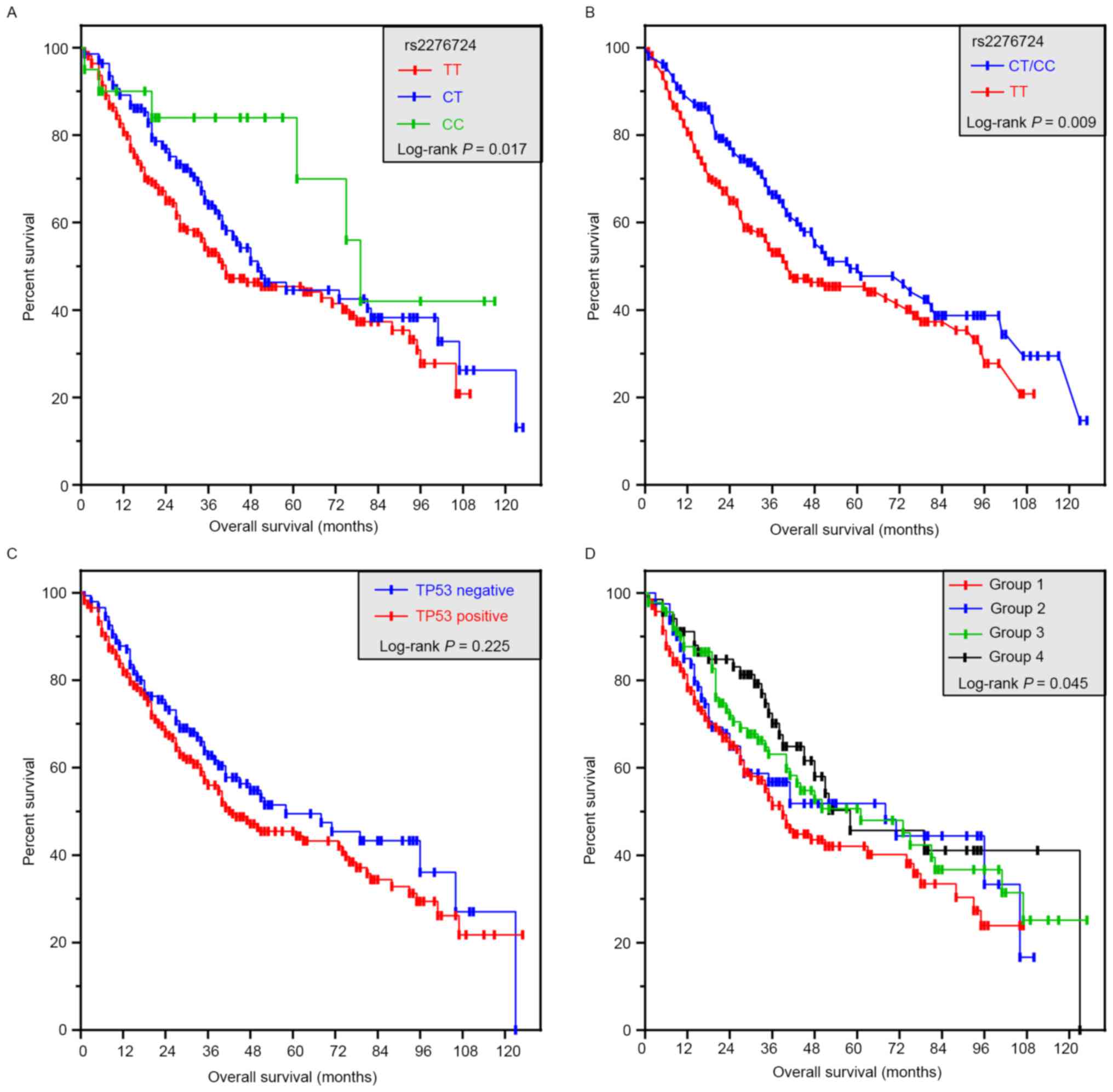
Relationship of rs2276724 and TP53
status with the OS
Using a dominant genetic model, patients with the TT
genotype had a shorter MST compared to those with CT or CC
genotypes of rs9275572 (TT vs. CT vs. CC; 39 vs. 50 vs. 79 months;
log-rank P=0.017; Fig. 3A).
However, the difference was similar after adjustment for alcohol
consumption, Child-Pugh score, tumor size, tumor number, BCLC
stage, radical resection, cirrhosis, adjuvant antiviral treatment,
and a PVTT. In the dominant genetic model, patients with the TT
genotype had a significantly smaller MST than the C allele carriers
(TT vs.CT/CC; 39 vs. 58 months; log-rank P=0.009; Fig. 3B), and the CT/CC genotype of
rs2276724 had a significantly decreased risk of death (adjusted
P=0.040; adjusted HR=0.725; 95% CI=0.533–0.986; Table IV). Haplotype analysis showed that
the C allele was associated with a significantly decreased risk of
death (adjusted P=0.032; adjusted HR=0.747; 95% CI=0.572–0.976;
Table IV), compared with the T
allele. The prognosis for a different status of TP53
expression was similar in the patients (adjusted P=0.280; adjusted
HR=1.183; 95% CI=0.872–1.605; Table
IV and Fig. 3C).
| Table IV.Survival analysis of HBV-related HCC
patients according to rs2276724 and TP53 status. |
Table IV.
Survival analysis of HBV-related HCC
patients according to rs2276724 and TP53 status.
| Variable | Patients
(n=415) | No. of events
(%) | MST (months) | Crude HR (95%
CI) | Crude p-value | Adjusted HR (95%
CI) | Adjusted
p-valuea |
|---|
| rs2276724 |
|
|
|
|
|
|
|
|
Allele |
|
|
|
|
|
|
|
| T | 640 | 309 (48.3) | 41 | 1 |
| 1 |
|
| C | 190 | 75 (39.5) | 73 | 0.692
(0.535–0.893) | 0.005 | 0.747
(0.572–0.976) | 0.032 |
| Genotype |
|
|
|
|
|
|
|
| TT | 245 | 123 (50.2) | 39 | 1 |
| 1 |
|
| CT | 150 | 63
(42.0) | 50 | 0.716
(0.527–0.972) | 0.032 | 0.749
(0.545–1.029) | 0.074 |
| CC | 20 |
6 (30.0) | 79 | 0.421
(0.185–0.958) | 0.039 | 0.554
(0.240–1.278) | 0.166 |
|
CT+CC | 170 | 69
(40.6) | 58 | 0.675
(0.502–0.909) | 0.010 | 0.725
(0.533–0.986) | 0.040 |
| TP53 status |
|
|
|
|
|
|
|
|
Negative | 162 | 68
(42.0) | 58 | 1 |
| 1 |
|
|
Positive | 253 | 124 (49.0) | 41 | 1.199
(0.892–1.612) | 0.229 | 1.183
(0.872–1.605) | 0.280 |
Joint-effect analysis
We further analyzed the TP53 status and rs2276724
genotypes with the mutual association with OS of the HBV-related
HCC patients. TP53-positive patients with CT/CC genotypes had a
significantly longer MST (Table V
and Fig. 3D) as compared to the
TP53-positive patients with TT genotype. After adjustment for
alcohol consumption, Child-Pugh score, tumor size, tumor number,
BCLC stage, radical resection, cirrhosis, adjuvant antiviral
treatment, PVTT in the Cox proportion haphazard regression model,
the TP53-negative patients with CT/CC genotypes showed a
significantly decreased risk of death (adjusted P=0.037; adjusted
HR=0.621; 95% CI=0.396–0.973; Table
V).
| Table V.Joint effect survival analysis of
rs2276724 and different TP53 expression statuses in
HBV-related HCC patients. |
Table V.
Joint effect survival analysis of
rs2276724 and different TP53 expression statuses in
HBV-related HCC patients.
| Group | Genotype | TP53 status | Patients
(n=421) | No. of events
(%) | MST (months) | Crude HR (95%
CI) | Crude p-value | Adjusted HR (95%
CI) | Adjusted
P-valuea |
|---|
| 1 | TT | Positive | 156 | 81 (51.9) | 36 | 1 |
| 1 |
|
| 2 | TT | Negative | 89 | 42 (47.2) | 41 | 0.868
(0.598–1.260) | 0.457 | 0.903
(0.610–1.336) | 0.610 |
| 3 | CT+CC | Positive | 97 | 43 (44.3) | 61 | 0.695
(0.479–1.008) | 0.055 | 0.763
(0.518–1.126) | 0.173 |
| 4 | CT+CC | Negative | 73 | 26 (35.6) | 58 | 0.570
(0.366–0.888) | 0.013 | 0.621
(0.396–0.973) | 0.037 |
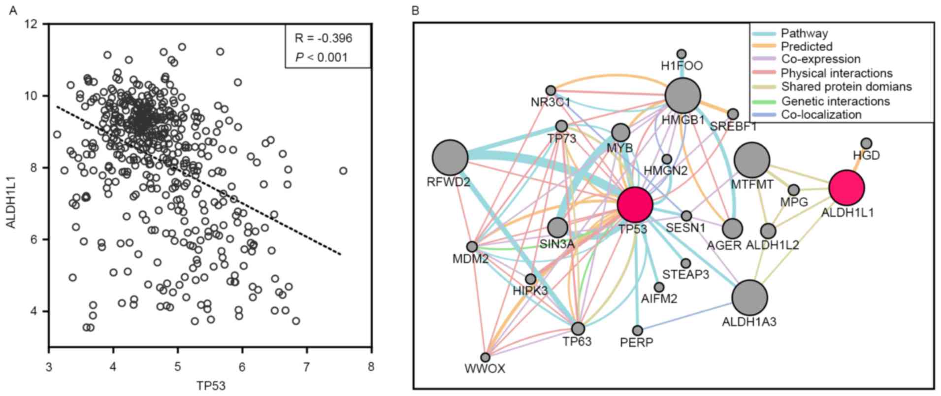
GEO data and gene interaction
analysis
In order to determine the relationship of
ALDH1L1 with TP53 at the transcriptional level, the
GSE14520 database (including 218 Chinese HBV-related HCC patients
with clinical information and prognosis) was used to correlate the
ALDH1L1 and TP53 mRNA expression in HBV-related HCC
patients. The results showed that ALDH1L1 had a weak
negative correlation with TP53 (r=−0.396; P<0.001;
Fig. 4A). Gene interaction analyses
through GenMANIA also showed that ALDH1L1 shared protein domains
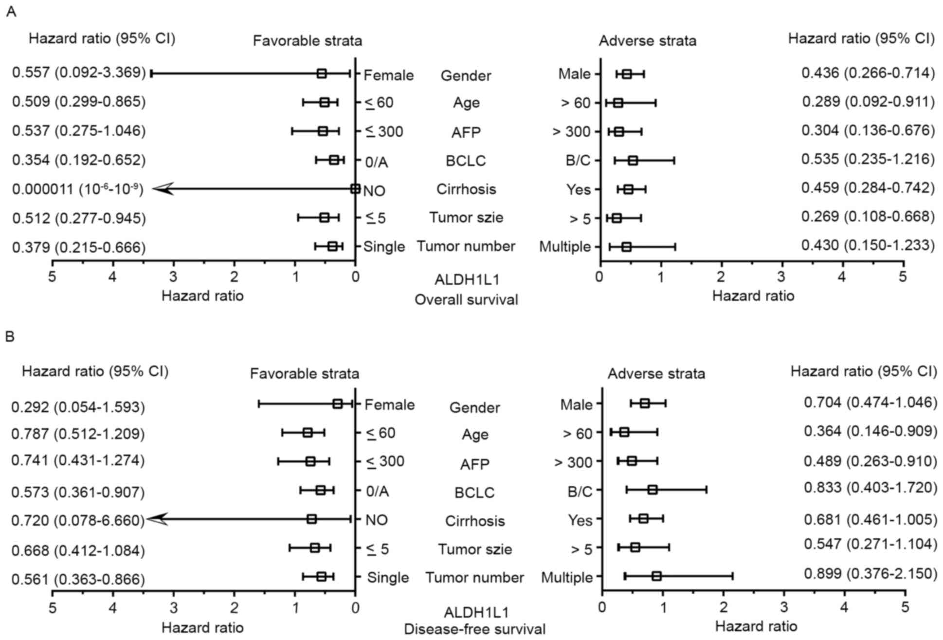
with ALDH1A3, that affected the TP53 pathway (Fig. 4B). Stratified analyses for the DFS
showed that high ALDH1L1 expression significantly decreased
the risk of recurrence among patients >60 years of age, with a
single tumor, BCLC stage 0/A, and AFP >300 ng/ml (Fig. 5A). Regarding the OS, high
ALDH1L1 expression significantly decreased the risk of death
among patients in both male age groups, both tumor size groups with
a single tumor, who were characterized with cirrhosis, BCLC stage
0/A, and AFP >300 ng/ml (Fig.
5B). We further analyzed the effects of ALDH1L1
expression on the DFS and OS of GEO14520 HBV-related HCC patients
by adjusting for age, sex, cirrhosis, BCLC stage and serum AFP
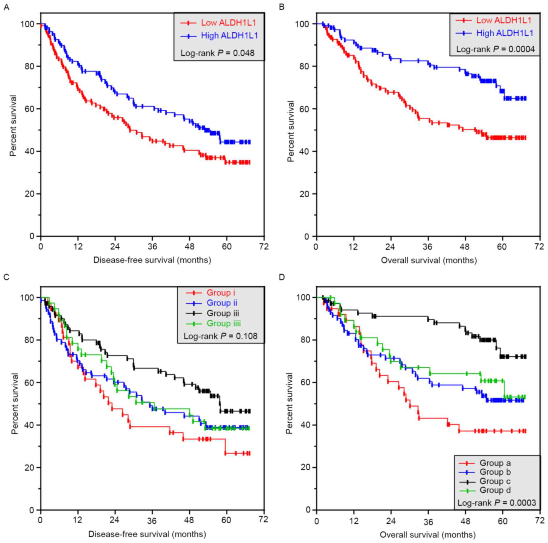
level. The results showed that high ALDH1L1 expression was
significantly associated with a favorable prognosis for both the
DFS and OS (adjusted P=0.04; adjusted HR=0.669; 95% CI=0.456–0.981
for DFS; adjusted P=0.001; adjusted HR=0.446; 95% CI=0.277–0.719
for OS; Table VI and Fig. 6A and B).
| Table VI.Survival analysis between
ALDH1L1 and TP53 mRNA expression in GSE14520
HBV-related HCC patients. |
Table VI.
Survival analysis between
ALDH1L1 and TP53 mRNA expression in GSE14520
HBV-related HCC patients.
|
|
| OS | DFS |
|---|
|
|
|
|
|
|---|
| Gene
expression | Patients
(n=218) | No. of events
(%) | MST (months) | Adjusted HR (95%
CI) | Adjusted
P-valuea | No. of events | MST (months) | Adjusted HR (95%
CI) | Adjusted
P-valuea |
|---|
| ALDH1L1 |
|
Low | 109 | 66 (60.6) | 28 | 1 |
| 54 (49.5) | 51 | 1 |
|
|
High | 109 | 55 (50.5) | 53 | 0.669
(0.456–0.981) | 0.040 | 30 (27.5) | NA | 0.446
(0.277–0.719) | 0.001 |
| TP53 |
|
Low | 109 | 58 (53.2) | 50 | 1 |
| 37 (33.9) | NA | 1 |
|
|
High | 109 | 63 (57.8) | 35 | 1.054
(0.726–1.5229) | 0.783 | 47 (43.1) | NA | 1.137
(0.722–1.791) | 0.580 |
Joint-effect analyses among different ALDH1L1
and TP53 expression groups showed that patients with a high
ALDH1L1 and low TP53 expression were significantly
associated with a favorable prognosis, when compared with patients
with a low ALDH1L1 and TP53 expression for both the
DFS and OS (adjusted P=0.005; adjusted HR=0.460; 95% CI=0.266–0.795
for DFS; adjusted P=0.000011; adjusted HR=0.211; 95% CI=0.105–0.422
for OS; Table VII and Fig. 6C and D). Patients in groups b and d
also had a reduced risk of death, compared with patients with low
ALDH1L1 expression in the low TP53 expression group
(adjusted P=0.023; adjusted HR=0.524; 95% CI=0.300–0.914 for group
b; adjusted P=0.014; adjusted HR=0.434; 95% CI=0.222–0.846 for
group d; Table VII and Fig. 6D).
| Table VII.Joint effect survival analysis
between ALDH1L1 and TP53 mRNA expression level in GSE14520
HBV-related HCC patients. |
Table VII.
Joint effect survival analysis
between ALDH1L1 and TP53 mRNA expression level in GSE14520
HBV-related HCC patients.
| Group | ALDH1L1
expression | TP53
expression | Patients
(n=218) | No. of events
(%) | MST (months) | Adjusted HR (95%
CI) | Adjusted
P-valuea |
|---|
| DFS |
| i | Low | Low | 38 | 25 (65.8) | 23 | 1 |
|
| ii | Low | High | 71 | 41 (57.7) | 35 | 0.675
(0.406–1.122) | 0.129 |
|
iii | High | Low | 71 | 33 (46.5) | 57 | 0.460
(0.266–0.795) | 0.005 |
|
iiii | High | High | 38 | 22 (57.9) | 36 | 0.614
(0.342–1.101) | 0.102 |
| OS |
| a | Low | Low | 38 | 22 (57.9) | 30 | 1 |
|
| b | Low | High | 71 | 32 (45.1) | NA | 0.524
(0.300–0.914) | 0.023 |
| c | High | Low | 71 | 15 (21.1) | NA | 0.211
(0.105–0.422) | 0.000011 |
| d | High | High | 38 | 15 (39.5) | NA | 0.434
(0.222–0.846) | 0.014 |
Discussion
ALDH1L1 has been widely accepted as an astroglial
marker in the brain (27,28) and is also expressed in neural stem
cells (29), but is not a suitable
marker for enteric glial cells (30). Due to the cell-specificity of
ALDH1L1 in nerve cells, ALDH1L1 polymorphisms have also been
associated with neurological diseases, such as neural tube defects
(31) and ischemic stroke (32), but ALDH1L1 polymorphisms have
not been investigated in spina bifida (33). It's upregulation is involved in
central nervous system development and reduced proliferation
(34). ALDH1L1 is mainly expressed
in human liver (35), implying that
ALDH1L1 has an important function in this organ. Consistent with
our bioinformatic analyses, ALDH1L1 is significantly
downregulated in various human malignant tumors and cancer cell
lines, including HCC (36). A
similar result for ALDH1L1 downregulation in different tumor
tissues was confirmed by ONCOMINE analyses (37) and other studies (21,38,39),
but in non-small cell lung cancer (NSCLC), ALDH1L1
expression was upregulated (40).
Our bioinformatic analyses also showed that ALDH1L1 was
downregulated in HBV-related HCC tumor tissues. ALDH1L1 is
upregulated in the presence of high concentrations of folate, and
depletion of folate leads to the absence of ALDH1L1,
resulting in cofilin dephosphorylation and inhibition of motility
by protein phosphatase 1 (PP1) and protein phosphatase 2A (PP2A) in
several cell lines. These results suggested that folate promotes a
malignant phenotype in cancer (41). However, a study of oral cancer
reported that folate supplementation decreased the risk of oral
cancer even with alcohol abuse, thus, ALDH1L1 may play a causal
role in oral cancer occurrence (42). In spite of the conflicting results
of these studies, underexpressed ALDH1L1 was associated with
an aggressive histology and/or biological behavior in renal cell
carcinomas and pilocytic astrocytomas (43,44).
ALDH1L1 knockdown in lung cancer cell lines showed that
inhibition of ALDH1L1 expression reduced adenosine
triphosphate (ATP) production by decreasing nicotinamide adenine
dinucleotide (NADH) levels, resulting in cell death (40). Recent studies also reported that
high expression of ALDH1L1 is correlated with better
survival in HCC (21),
neuroblastoma (45) and breast
cancer (BC) (46). However,
survival analyses of gastric cancer showed the opposite result that
high expression of ALDH1L1 was associated with a worse
prognosis (37,47). Furthermore, no significant
relationship was observed between ALDH1L1 mRNA expression
and OS in NSCLC (48). As
previously mentioned, ALDH1L1 may play a different role as a
tumor-suppressor during oncogenesis. Genetic variation analyses
have reported that rs2276731 and rs2002287 of ALDH1L1 can
affect the risk of BC morbidity (n=1007) (49), but this conclusion was not found for
the risk of prostate cancer (n=2288) including other ALDH1L1 SNPs
(50). A study of HCC and lung
cancer reported that ALDH1L1 mRNA and protein levels
correlated with the methylation status of the CpG island, and
modicum ALDH1L1 CpG island methylation was sufficient to
significantly decrease ALDH1L1 expression, suggesting that
the mechanism of action of ALDH1L1 involves downregulation
in cancers (51). A follow-up study
in Chinese Kazakh patients with esophageal squamous cell carcinoma
also showed that ALDH1L1 is involved in a one carbon
metabolic process that plays a key role in DNA methylation
(39). A study carried out by
Oleinik and Krupenko also reported that inducible ALDH1L1
expression in A549 cells induced G1 cell cycle arrest and
apoptosis. These anti-proliferative and apoptotic effects result in
activation of TP53, followed by the TP53-mediated
transcriptional activation of a downstream target of p21 (19), to function as a potent
cyclin-dependent kinase inhibitor. Further studies of the
relationship between ALDH1L1 and TP53 showed that
expression of ALDH1L1 induced suppressor effects that were
p53-dependent, and a TP53 deficit resulted in suppressor effects
(20). This ALDH1L1-induced
p53-dependent apoptosis also responded to folate stress, resulting
in upregulation of ceramide synthesis (52).
A previous study of Guangxi HCC patients reported
that ALDH1L1 expression was associated with the prognosis of
HCC (21). HBV-related HCC patients
in Guangxi were associated with a high morbidity of HBV-infection
(53). The present study
characterized ALDH1L1 polymorphism in HBV-related HCC
patients and its association with TP53 expression.
Bioinformatic analyses showed that rs2276724 S481G in
ALDH1L1 affected gene expression and was possibly
deleterious to patients. We further analyzed the distribution of
rs2276724 genotypes in different TP53 expression groups and
its possible association with the prognosis of HBV-related HCC
patients. The results suggested that the occurrence of rs2276724
was similar between different TP53 expression groups when
analyzed in different genetic models. Survival analyses showed that
the C allele was associated with a decreased risk of death in
HBV-related HCC patients, when compared to the T allele. Through
stratified analyses, the C allele carriers of rs2276724 had
significantly decreased risk of death among patients with a tumor
size ≤5 cm, a Child-Pugh score, and without a PVTT, BCLC stage B/C,
non-radical resection, and the presence of regional invasion.
Joint-effect analyses showed that the CT/CC of rs2276724 in
TP53-negative patients was associated with a significantly
decreased risk of mortality, compared to the TT of rs2276724 with
TP53-positive patients. We then used the Chinese HBV-HCC mRNA
expression profiling chip from the GSE14520 dataset to evaluate the
prognosis of ALDH1L1 expression in Chinese HBV-related HCC
patients, and found that low ALDH1L1 expression predicted a
poor prognosis for Chinese HBV-related HCC patients with low
expression of HBV-related HCC tumor tissues. High ALDH1L1
expression significantly decreased the risk of HCC recurrence among
patients with an age >60 years, a single tumor, BCLC stage 0/A,
AFP >300 ng/ml, and showed a decreased risk of mortality among
the male HCC patients in both age groups, both tumor size groups, a
single tumor with cirrhosis, with BCLC stage 0/A, and an AFP
>300 ng/ml. Gene interaction analyses showed that GeneMANIA
ALDH1L1 and TP53 expression mRNA levels were
negatively correlated in Chinese HBV-related HCC patients, and
further showed that ALDH1L1 shared a protein domain with
ALDH1A3 that was involved in the TP53 pathway. We combined
the analyses of ALDH1L1 and TP53 expression in
HBV-related HCC patients to show that high ALDH1L1 with low
TP53 expression was associated with a significantly
decreased risk of HBV-related HCC recurrence and mortality when
compared with low ALDH1L1 and low TP53 expression.
Patients with high TP53 expression also had a significantly
decreased risk of HBV-related HCC death, compared with low
ALDH1L1 and low TP53-expressing patients.
In conclusion, the present study showed, for the
first time, that prognosis can be predicted for the rs2276724
genotypes of ALDH1L1 in HBV-related HCC patients and their
associations with TP53 expression. The CT/TT genotype of
rs2276724 may have a protective survival value and may be a
potential prognostic marker in patients with HBV-related HCC
receiving hepatic resection. We also confirmed that a decrease in
ALDH1L1 expression predicts a poor prognosis for patients
with HBV-related HCC. The expression of ALDH1L1 and
genotypes of rs2276724 may therefore play a role in TP53
expression in HBV-related HCC of Chinese hepatic resection
patients. Due to the limitations of the relatively small sample
sizes and the long period of specimen collection, we did not
analyze the association among rs2276724 genotypes and mRNA
expression. Further well-designed, comprehensive, and large sample
size studies are therefore needed to confirm our results.
Acknowledgements
This study was supported in part by the National
Nature Science Foundation of China (nos. 81560535, 81072321,
30760243, 30460143 and 30560133), 2009 Program for New Century
Excellent Talents in University (NCET), Guangxi Nature Sciences
Foundation (no. GuiKeGong 1104003A-7), Guangxi Health Ministry
Medicine Grant (Key-Scientific Research-Grant Z201018) and
Self-raised Scientific Research Projects of the Guangxi Zhuang
Autonomous Region Health and Family Planning Commission (Z2016318).
The authors thank Professor Xiao Qin, Xigang Chen, Bin Chen,
Zhixiong Su, Ming Su, Zhang Wen, Jingning Lu, Ning Peng, Hai Zhu
who provided HCC samples for this study, and who are from the
Department of Hepatobiliary Surgery, the First Affiliated Hospital
of Guangxi Medical University. Thanks also go to researchers
Jiaquan Li and Ying Gui from Guangxi Medical University for their
contribution to specimen management and Professor Jiahong Dong from
Beijing Tsinghua Changgung Hospital for his contribution to thesis
guidance. In addition, we also would like to acknowledge the
helpful comments on this paper received from our reviewers, and the
contributors of GSE14520 for sharing their dataset on open
access.
References
|
1
|
Torre LA, Bray F, Siegel RL, Ferlay J,
Lortet-Tieulent J and Jemal A: Global cancer statistics, 2012. CA
Cancer J Clin. 65:87–108. 2015. View Article : Google Scholar : PubMed/NCBI
|
|
2
|
Chen W, Zheng R, Baade PD, Zhang S, Zeng
H, Bray F, Jemal A, Yu XQ and He J: Cancer statistics in China,
2015. CA Cancer J Clin. 66:115–132. 2016. View Article : Google Scholar : PubMed/NCBI
|
|
3
|
Zeng H, Zheng R, Guo Y, Zhang S, Zou X,
Wang N, Zhang L, Tang J, Chen J, Wei K, et al: Cancer survival in
China, 2003–2005: A population-based study. Int J Cancer.
136:1921–1930. 2015. View Article : Google Scholar : PubMed/NCBI
|
|
4
|
Zhou M, Wang H, Zhu J, Chen W, Wang L, Liu
S, Li Y, Wang L, Liu Y, Yin P, et al: Cause-specific mortality for
240 causes in China during 1990–2013: A systematic subnational
analysis for the Global Burden of Disease Study 2013. Lancet.
387:251–272. 2016. View Article : Google Scholar : PubMed/NCBI
|
|
5
|
El-Serag HB and Rudolph KL: Hepatocellular
carcinoma: Epidemiology and molecular carcinogenesis.
Gastroenterology. 132:2557–2576. 2007. View Article : Google Scholar : PubMed/NCBI
|
|
6
|
Wang FS, Fan JG, Zhang Z, Gao B and Wang
HY: The global burden of liver disease: The major impact of China.
Hepatology. 60:2099–2108. 2014. View Article : Google Scholar : PubMed/NCBI
|
|
7
|
Xu L, Qian G, Tang L, Su J and Wang JS:
Genetic variations of hepatitis B virus and serum aflatoxin-lysine
adduct on high risk of hepatocellular carcinoma in Southern
Guangxi, China. J Hepatol. 53:671–676. 2010. View Article : Google Scholar : PubMed/NCBI
|
|
8
|
Qi LN, Bai T, Chen ZS, Wu FX, Chen YY, De
Xiang B, Peng T, Han ZG and Li LQ: The p53 mutation spectrum in
hepatocellular carcinoma from Guangxi, China: Role of chronic
hepatitis B virus infection and aflatoxin B1 exposure. Liver Int.
35:999–1009. 2015. View Article : Google Scholar : PubMed/NCBI
|
|
9
|
Yin Y, Stephen CW, Luciani MG and Fåhraeus
R: p53 stability and activity is regulated by Mdm2-mediated
induction of alternative p53 translation products. Nat Cell Biol.
4:462–467. 2002. View
Article : Google Scholar : PubMed/NCBI
|
|
10
|
Hainaut P and Wiman KG: 30 years and a
long way into p53 research. Lancet Oncol. 10:913–919. 2009.
View Article : Google Scholar : PubMed/NCBI
|
|
11
|
Soussi T and Wiman KG: TP53: An oncogene
in disguise. Cell Death Differ. 22:1239–1249. 2015. View Article : Google Scholar : PubMed/NCBI
|
|
12
|
Petitjean A, Achatz MI, Borresen-Dale AL,
Hainaut P and Olivier M: TP53 mutations in human cancers:
Functional selection and impact on cancer prognosis and outcomes.
Oncogene. 26:2157–2165. 2007. View Article : Google Scholar : PubMed/NCBI
|
|
13
|
Liu J, Ma Q, Zhang M, Wang X, Zhang D, Li
W, Wang F and Wu E: Alterations of TP53 are associated with a poor
outcome for patients with hepatocellular carcinoma: Evidence from a
systematic review and meta-analysis. Eur J Cancer. 48:2328–2338.
2012. View Article : Google Scholar : PubMed/NCBI
|
|
14
|
Staib F, Hussain SP, Hofseth LJ, Wang XW
and Harris CC: TP53 and liver carcinogenesis. Hum Mutat.
21:201–216. 2003. View Article : Google Scholar : PubMed/NCBI
|
|
15
|
Hussain SP, Schwank J, Staib F, Wang XW
and Harris CC: TP53 mutations and hepatocellular carcinoma:
Insights into the etiology and pathogenesis of liver cancer.
Oncogene. 26:2166–2176. 2007. View Article : Google Scholar : PubMed/NCBI
|
|
16
|
Villanueva A and Hoshida Y: Depicting the
role of TP53 in hepatocellular carcinoma progression. J Hepatol.
55:724–725. 2011. View Article : Google Scholar : PubMed/NCBI
|
|
17
|
Gouas D, Shi H and Hainaut P: The
aflatoxin-induced TP53 mutation at codon 249 (R249S): Biomarker of
exposure, early detection and target for therapy. Cancer Lett.
286:29–37. 2009. View Article : Google Scholar : PubMed/NCBI
|
|
18
|
Ji YN, Wang Q and Xue J: TP53
immunohistochemical expression is associated with the poor outcome
for hepatocellular carcinoma: Evidence from a meta-analysis. Tumour
Biol. 35:1653–1659. 2014. View Article : Google Scholar : PubMed/NCBI
|
|
19
|
Oleinik NV and Krupenko SA: Ectopic
expression of 10-formyltetrahydrofolate dehydrogenase in A549 cells
induces G1 cell cycle arrest and apoptosis. Mol Cancer Res.
1:577–588. 2003.PubMed/NCBI
|
|
20
|
Oleinik NV, Krupenko NI, Priest DG and
Krupenko SA: Cancer cells activate p53 in response to
10-formyltetrahydrofolate dehydrogenase expression. Biochem J.
391:503–511. 2005. View Article : Google Scholar : PubMed/NCBI
|
|
21
|
Chen XQ, He JR and Wang HY: Decreased
expression of ALDH1L1 is associated with a poor prognosis in
hepatocellular carcinoma. Med Oncol. 29:1843–1849. 2012. View Article : Google Scholar : PubMed/NCBI
|
|
22
|
Liao X, Han C, Qin W, Liu X, Yu L, Lu S,
Chen Z, Zhu G, Su H, Mo Z, et al: Genome-wide association study
identified PLCE1- rs2797992 and EGFR- rs6950826 were associated
with TP53 expression in the HBV-related hepatocellular
carcinoma of Chinese patients in Guangxi. Am J Transl Res.
8:1799–1812. 2016.PubMed/NCBI
|
|
23
|
Kondo K, Chijiiwa K, Kai M, Otani K,
Nagaike K, Ohuchida J, Hiyoshi M and Nagano M: Surgical strategy
for hepatocellular carcinoma patients with portal vein tumor
thrombus based on prognostic factors. J Gastrointest Surg.
13:1078–1083. 2009. View Article : Google Scholar : PubMed/NCBI
|
|
24
|
Kumar P, Henikoff S and Ng PC: Predicting
the effects of coding non-synonymous variants on protein function
using the SIFT algorithm. Nat Protoc. 4:1073–1081. 2009. View Article : Google Scholar : PubMed/NCBI
|
|
25
|
Adzhubei IA, Schmidt S, Peshkin L,
Ramensky VE, Gerasimova A, Bork P, Kondrashov AS and Sunyaev SR: A
method and server for predicting damaging missense mutations. Nat
Methods. 7:248–249. 2010. View Article : Google Scholar : PubMed/NCBI
|
|
26
|
Lee PH and Shatkay H: An integrative
scoring system for ranking SNPs by their potential deleterious
effects. Bioinformatics. 25:1048–1055. 2009. View Article : Google Scholar : PubMed/NCBI
|
|
27
|
Yang Y, Vidensky S, Jin L, Jie C,
Lorenzini I, Frankl M and Rothstein JD: Molecular comparison of
GLT1+ and ALDH1L1+ astrocytes in vivo in
astroglial reporter mice. Glia. 59:200–207. 2011. View Article : Google Scholar : PubMed/NCBI
|
|
28
|
Feresten AH, Barakauskas V, Ypsilanti A,
Barr AM and Beasley CL: Increased expression of glial fibrillary
acidic protein in prefrontal cortex in psychotic illness. Schizophr
Res. 150:252–257. 2013. View Article : Google Scholar : PubMed/NCBI
|
|
29
|
Foo LC and Dougherty JD: Aldh1L1 is
expressed by postnatal neural stem cells in vivo. Glia.
61:1533–1541. 2013. View Article : Google Scholar : PubMed/NCBI
|
|
30
|
Boesmans W, Rocha NP, Reis HJ, Holt M and
Vanden Berghe P: The astrocyte marker Aldh1L1 does not reliably
label enteric glial cells. Neurosci Lett. 566:102–105. 2014.
View Article : Google Scholar : PubMed/NCBI
|
|
31
|
Wu L, Lu X, Guo J, Zhang T, Wang F and Bao
Y: Association between ALDH1L1 gene polymorphism and neural
tube defects in the Chinese Han population. Neurol Sci.
37:1049–1054. 2016. View Article : Google Scholar : PubMed/NCBI
|
|
32
|
Williams SR, Yang Q, Chen F, Liu X, Keene
KL, Jacques P, Chen WM, Weinstein G, Hsu FC, Beiser A, et al:
Genomics and Randomized Trials Network; Framingham Heart Study:
Genome-wide meta-analysis of homocysteine and methionine metabolism
identifies five one carbon metabolism loci and a novel association
of ALDH1L1 with ischemic stroke. PLoS Genet.
10:e10042142014. View Article : Google Scholar : PubMed/NCBI
|
|
33
|
Franke B, Vermeulen SH,
Steegers-Theunissen RP, Coenen MJ, Schijvenaars MM, Scheffer H, den
Heijer M and Blom HJ: An association study of 45 folate-related
genes in spina bifida: Involvement of cubilin (CUBN) and tRNA
aspartic acid methyltransferase 1 (TRDMT1). Birth Defects Res A
Clin Mol Teratol. 85:216–226. 2009. View Article : Google Scholar : PubMed/NCBI
|
|
34
|
Anthony TE and Heintz N: The folate
metabolic enzyme ALDH1L1 is restricted to the midline of the early
CNS, suggesting a role in human neural tube defects. J Comp Neurol.
500:368–383. 2007. View Article : Google Scholar : PubMed/NCBI
|
|
35
|
Hong M, Lee Y, Kim JW, Lim JS, Chang SY,
Lee KS, Paik SG and Choe IS: Isolation and characterization of cDNA
clone for human liver 10-formyltetrahydrofolate dehydrogenase.
Biochem Mol Biol Int. 47:407–415. 1999.PubMed/NCBI
|
|
36
|
Krupenko SA and Oleinik NV:
10-formyltetrahydrofolate dehydrogenase, one of the major folate
enzymes, is down-regulated in tumor tissues and possesses
suppressor effects on cancer cells. Cell Growth Differ. 13:227–236.
2002.PubMed/NCBI
|
|
37
|
Shen JX, Liu J, Li GW, Huang YT and Wu HT:
Mining distinct aldehyde dehydrogenase 1 (ALDH1) isoenzymes in
gastric cancer. Oncotarget. 7:25340–25349. 2016. View Article : Google Scholar : PubMed/NCBI
|
|
38
|
Dmitriev AA, Kashuba VI, Haraldson K,
Senchenko VN, Pavlova TV, Kudryavtseva AV, Anedchenko EA, Krasnov
GS, Pronina IV, Loginov VI, et al: Genetic and epigenetic analysis
of non-small cell lung cancer with NotI-microarrays. Epigenetics.
7:502–513. 2012. View Article : Google Scholar : PubMed/NCBI
|
|
39
|
Chen Y, Yin D, Li L, Deng YC and Tian W:
Screening aberrant methylation profile in esophageal squamous cell
carcinoma for Kazakhs in Xinjiang area of China. Mol Biol Rep.
42:457–464. 2015. View Article : Google Scholar : PubMed/NCBI
|
|
40
|
Kang JH, Lee SH, Lee JS, Nam B, Seong TW,
Son J, Jang H, Hong KM, Lee C and Kim SY: Aldehyde dehydrogenase
inhibition combined with phenformin treatment reversed NSCLC
through ATP depletion. Oncotarget. 7:49397–49410. 2016. View Article : Google Scholar : PubMed/NCBI
|
|
41
|
Oleinik NV, Krupenko NI and Krupenko SA:
ALDH1L1 inhibits cell motility via dephosphorylation of cofilin by
PP1 and PP2A. Oncogene. 29:6233–6244. 2010. View Article : Google Scholar : PubMed/NCBI
|
|
42
|
Hwang PH, Lian L and Zavras AI: Alcohol
intake and folate antagonism via CYP2E1 and ALDH1: Effects on oral
carcinogenesis. Med Hypotheses. 78:197–202. 2012. View Article : Google Scholar : PubMed/NCBI
|
|
43
|
Dmitriev AA, Rudenko EE, Kudryavtseva AV,
Krasnov GS, Gordiyuk VV, Melnikova NV, Stakhovsky EO, Kononenko OA,
Pavlova LS, Kondratieva TT, et al: Epigenetic alterations of
chromosome 3 revealed by NotI-microarrays in clear cell renal cell
carcinoma. Biomed Res Int. 2014:7352922014. View Article : Google Scholar : PubMed/NCBI
|
|
44
|
Rodriguez FJ, Giannini C, Asmann YW,
Sharma MK, Perry A, Tibbetts KM, Jenkins RB, Scheithauer BW, Anant
S, Jenkins S, et al: Gene expression profiling of NF-1-associated
and sporadic pilocytic astrocytoma identifies aldehyde
dehydrogenase 1 family member L1 (ALDH1L1) as an underexpressed
candidate biomarker in aggressive subtypes. J Neuropathol Exp
Neurol. 67:1194–1204. 2008. View Article : Google Scholar : PubMed/NCBI
|
|
45
|
Hartomo TB, Van Huyen Pham T, Yamamoto N,
Hirase S, Hasegawa D, Kosaka Y, Matsuo M, Hayakawa A, Takeshima Y,
Iijima K, et al: Involvement of aldehyde dehydrogenase 1A2 in the
regulation of cancer stem cell properties in neuroblastoma. Int J
Oncol. 46:1089–1098. 2015.PubMed/NCBI
|
|
46
|
Wu S, Xue W, Huang X, Yu X, Luo M, Huang
Y, Liu Y, Bi Z, Qiu X and Bai S: Distinct prognostic values of
ALDH1 isoenzymes in breast cancer. Tumour Biol. 36:2421–2426. 2015.
View Article : Google Scholar : PubMed/NCBI
|
|
47
|
Li K, Guo X, Wang Z, Li X, Bu Y, Bai X,
Zheng L and Huang Y: The prognostic roles of ALDH1 isoenzymes in
gastric cancer. Onco Targets Ther. 9:3405–3414. 2016.PubMed/NCBI
|
|
48
|
You Q, Guo H and Xu D: Distinct prognostic
values and potential drug targets of ALDH1 isoenzymes in
non-small-cell lung cancer. Drug Des Devel Ther. 9:5087–5097. 2015.
View Article : Google Scholar : PubMed/NCBI
|
|
49
|
Stevens VL, McCullough ML, Pavluck AL,
Talbot JT, Feigelson HS, Thun MJ and Calle EE: Association of
polymorphisms in one-carbon metabolism genes and postmenopausal
breast cancer incidence. Cancer Epidemiol Biomarkers Prev.
16:1140–1147. 2007. View Article : Google Scholar : PubMed/NCBI
|
|
50
|
Stevens VL, Rodriguez C, Sun J, Talbot JT,
Thun MJ and Calle EE: No association of single nucleotide
polymorphisms in one-carbon metabolism genes with prostate cancer
risk. Cancer Epidemiol Biomarkers Prev. 17:3612–3614. 2008.
View Article : Google Scholar : PubMed/NCBI
|
|
51
|
Oleinik NV, Krupenko NI and Krupenko SA:
Epigenetic silencing of ALDH1L1, a metabolic regulator of
cellular proliferation, in cancers. Genes Cancer. 2:130–139. 2011.
View Article : Google Scholar : PubMed/NCBI
|
|
52
|
Hoeferlin LA, Fekry B, Ogretmen B,
Krupenko SA and Krupenko NI: Folate stress induces apoptosis via
p53-dependent de novo ceramide synthesis and up-regulation of
ceramide synthase 6. J Biol Chem. 288:12880–12890. 2013. View Article : Google Scholar : PubMed/NCBI
|
|
53
|
Yeh FS, Yu MC, Mo CC, Luo S, Tong MJ and
Henderson BE: Hepatitis B virus, aflatoxins, and hepatocellular
carcinoma in southern Guangxi, China. Cancer Res. 49:2506–2509.
1989.PubMed/NCBI
|