Introduction
Glioma is the most common and aggressive type of
primary malignant brain tumour derived from glial cells and
accounts for ~35–61% of all intracranial tumours (1). It can be classified into the following
subtypes according to histology: astrocytomas, anaplastic
astrocytomas, glioblastomas and other subtypes (1). The biological characteristics of
glioma are high recurrence rates, uncontrollable invasiveness,
strong angiogenesis and widespread hypoxia (2–5).
Currently, therapeutic treatments for glioma include surgical
excision, chemotherapy, radiation and biological therapies
(6), among which surgery is the
most preferred treatment. Radiation therapy and chemotherapy are
considered effectual supplementary treatment after surgery to
prevent recurrence and metastasis (7). Despite the considerable progress in
the treatment of predominant glioma, the average survival time of
glioma patients is 9–12 months only (8,9).
Therefore, understanding the molecular mechanisms of glioma
tumourigenesis and progression are essential to the development of
novel strategies for glioma therapy.
MicroRNAs (miRNAs) belong to a large group of
evolutionarily conserved, short, endogenous and non-coding RNA
molecules with ~19–22 nucleotides in length (10). miRNAs negatively modulate gene
expression transcriptionally or post-transcriptionally by binding
to the 3′ untranslated regions (3′UTRs) of their target genes,
thereby inducing their degradation or inhibiting their translation
(11). Increasing evidence has
demonstrated that miRNAs have been implicated in the regulation of
various physiological and pathological processes including cell
growth, the cell cycle, cell division, apoptosis, invasion,
metastasis and angiogenesis (12–15).
Over the past decade, aberrant expression of miRNAs has been
implicated in a wide range of human types of cancer, such as
miR-548b in glioma (16), miR-19b
in gastric cancer (17), miR-181b
in colorectal cancer (18) and
miR-126 in hepatocellular carcinoma (19). An increasing number of studies have
indicated that the dysregulation of miRNAs is closely associated
with tumour occurrence and development (20,21).
Specifically, downregulated miRNAs act as tumour suppressors in
tumourigenesis by downregulating oncogenes, whereas other
overexpressed miRNAs may function as tumour promoters via negative
regulation of tumour suppressors (22,23).
These findings demonstrated that miRNAs are potential therapeutic
targets for cancer diagnosis, treatment and prognosis.
miR-202, located in 10q26, has been studied in
several types of human cancers (22,24,25),
however information concerning miR-202 in glioma is insufficient.
The present study aimed to investigate miR-202 expression in
glioma, to determine its correlation with clinicopathological
parameters and identify the biological roles of miR-202 in glioma.
The molecular mechanism underlying its tumour-suppressive effect
was also elucidated. The results of the present study may
contribute towards identifying a novel therapeutic target for the
treatment of glioma.
Materials and methods
Tissue samples and cell lines
The present study was approved by the Ethics
Committee of The First Affiliated Hospital of Henan University of
Science and Technology (Henan, China). Written consent was also
acquired from all glioma patients. Glioma and paired adjacent
normal tissues were obtained from 43 glioma patients who underwent
surgery at the Department of Neurosurgery of The First Affiliated
Hospital of Henan University of Science and Technology. None of
these patients were treated with neoadjuvant radiotherapy and
adjuvant chemotherapy.
Five glioma cell lines (A172, U87, U251, U373 and
LN229) and primary normal human astrocytes (NHAs) were purchased
from the American Type Culture Collection (ATCC; Manassas, VA,
USA). All cells were grown in Dulbecco's modified Eagle's medium
(DMEM) supplemented with 10% fetal bovine serum (FBS) (both from
Invitrogen, Carlsbad, CA, USA) in a humidified incubator at 37°C
with 5% CO2.
Cell transfection
miR-202 mimics and miRNA negative control (miR-NC)
were obtained from GenePharma (Shanghai, China). Small interfering
RNA targeting metadherin (MTDH) (si-MTDH) and its negative control
(si-NC) were synthesised by RiboBio (Guangzhou, China). MTDH
overexpressed plasmid (pCDNA3.1-MTDH) and blank plasmid (pCDNA3.1)
were purchased from the Chinese Academy of Sciences (Changchun,
China). For functional assays, the cells were seeded into 6-well
plates and transfected with miR-202 mimics, miR-NC, si-MTDH, si-NC,
pCDNA3.1-MTDH or pCDNA3.1 using Lipofectamine 2000 reagent
(Invitrogen) according to the manufacturer's protocol.
Reverse transcription-quantitative
polymerase chain reaction (RT-qPCR)
Total RNA was extracted form tissues or cells using
TRIzol reagent (Invitrogen). A NanoDrop® ND-1000
spectrophotometer was used to determine the purity and
concentration of total RNA. TaqMan MicroRNA assay (Applied
Biosystems, Foster City, CA, USA) was performed to detect miR-202
expression, with U6 used as the endogenous control. For mRNA
expression, reverse transcription was performed using M-MLV Reverse
Transcription system (Promega Corporation, Madison, WI, USA),
followed by qPCR with SYBR Premix Ex Taq (Takara, Dalian, China).
β-actin was used as an internal control for MTDH mRNA expression.
The relative expression was analysed using the 2−ΔΔCt
method (26). Primers are shown in
Table I.
 | Table I.RT-qPCR primers. |
Table I.
RT-qPCR primers.
| Gene |
| Sequences
(5′→3′) |
|---|
| miR-202 | F |
CCTCCCAGGCTCACGAGGCT |
|
| R |
GGTGCAGGTGCACTGGTGC |
| U6 | F |
CTCGCTTCGGCAGCACATATACT |
|
| R |
ACGCTTCACGAATTTGCGTGTC |
| MTDH | F |
TGCAGCCGAGGAATAAAGGA |
|
| R |
CTGTGCATAAGATCCAAGGAATTG |
| GAPDH | F |
ATAGCACAGCCTGGATAGCAACGTAC |
|
| R |
CACCTTCTACAATGAGCTGCGTGTG |
Cell Counting Kit-8 (CCK-8) assay
CCK-8 assay (Dojindo, Kumamoto, Japan) was performed
to examine the cell proliferative capacity. Briefly, transfected
cells were trypsinised, collected and re-seeded into 96-well plates
at a density of 3,000 cells in 200 µl of medium in each well.
Subsequently, the cells were incubated at 37°C with 5%
CO2 for 24, 36, 48 and 72 h. At each time-point, a CCK-8
assay was performed according to the manufacturer's instructions.
CCK-8 reagent (10 µl) was added into each well and incubated at
37°C for another 4 h. The absorbance at 450 nm (OD450) was assessed
using a microplate reader (Bio-Rad, Hercules, CA, USA).
Cell migration and invasion
assays
Cell migration and invasion assays were used to
determine the ability of glioma cell metastasis. Transfected cells
were trypsinised, collected and re-suspended in FBS-free culture
medium at 48 h post-transfection. For the migration assay,
1×10⁵ cells were added into the upper chamber of a
Transwell plate (BD Biosciences, Franklin Lakes, NJ, USA). The
lower chamber was filled with 500 µl culture medium containing 20%
FBS as a chemoattractant. After incubation at 37°C with 5%
CO2 for 48 h, the non-migrated cells were carefully
removed with cotton swabs. The migrated cells were fixed, stained,
washed and dried in air. Finally, migrated cells were photographed
and counted in five random fields using an inverted microscope
(Olympus Corporation, Tokyo, Japan). Cell invasion assays were
conducted in a similar manner to the cell migration assays except
that Transwell chambers were pre-coated with Matrigel (BD
Biosciences, San Jose, CA, USA).
Bioinformatic analysis
Potential targets of miR-202 were analysed using
publicly available algorithms: microRNA (www.microrna.org/microrna/home.do) and TargetScan
(www.targetscan.org).
Western blotting
Total protein was isolated from tissues and cells
using ice-cold RIPA lysis buffer (150 mm NaCl, 1% NP-40, 0.5%
deoxycholate and 1% SDS). The concentration of total protein was
detected using BCA assay kit (Beyotime Institute of Biotechnology,
Haimen, China) according to the manufacturer's instructions. Equal
amounts of proteins were resolved on 10% sodium dodecyl
sulfate-polyacrylamide gel electrophoresis (SDS-PAGE), and
transferred to polyvinylidene fluoride membranes (Millipore,
Billerica, MA, USA). The membranes were blocked with 5% skimmed
milk in Tris-buffered saline containing 0.1% Tween-20 (TBST) at
room temperature for 1 h, incubated with primary antibodies at 4°C
overnight and washed in TBST three times. The primary antibodies
used in the present study included mouse anti-human monoclonal MTDH
(1:1,000 dilution; sc-517220), rabbit anti-human polyclonal AKT
(sc-8312; 1:1,000 dilution), mouse anti-human monoclonal p-AKT
(sc-514032; 1:1,000 dilution), β-catenin (sc-59737; 1:1,000
dilution), p-β-catenin (sc-57534; 1:1,000 dilution) and mouse
anti-human monoclonal GAPDH (1:1,000 dilution; sc-137179) (all from
Santa Cruz Biotechnology, Santa Cruz, CA, USA). Subsequently, the
membranes were probed with goat anti-mouse horseradish peroxidase
(HRP)-conjugated secondary antibody (1:4,000 dilution; Santa Cruz
Biotechnology) for 1 h at room temperature. Finally, the protein
bands were visualized using enhanced chemiluminescence (ECL;
Pierce, Rockford, IL, USA). GAPDH was used as a loading
control.
Luciferase reporter assay
Luciferase reporter plasmids, pGL3-MTDH-3′UTR
wild-type (Wt) and pGL3-MTDH-3′UTR mutant (Mut), were synthesised
and confirmed by GenePharma. HEK293T cells (Cell Bank of the
Chinese Academy of Sciences, Shanghai, China) were seeded into
24-well plates at a density of 40–50% confluency. The cells were
incubated at 37°C with 5% CO2 overnight and transfected
with a reporter plasmid, along with miR-20 mimics or miR-NC using
Lipofectamine 2000. Following 48 h of incubation, the cells were
harvested and the luciferase activity was determined using a
Dual-Luciferase Reporter Assay System (Promega, Manheim, Germany).
Renilla luciferase activity was used for normalization.
Statistical analysis
All data are expressed as the mean ± SD and analysed
using Student's t-tests or one-way ANOVA with SPSS 19.0 software
(SPSS, Inc., Chicago, IL, USA). The correlation between miR-202 and
MTDH mRNA expression was analysed using Spearman's correlation
analysis. P<0.05 was considered to indicate statistically
significant differences.
Results
miR-202 is downregulated in glioma
tissue and cell lines
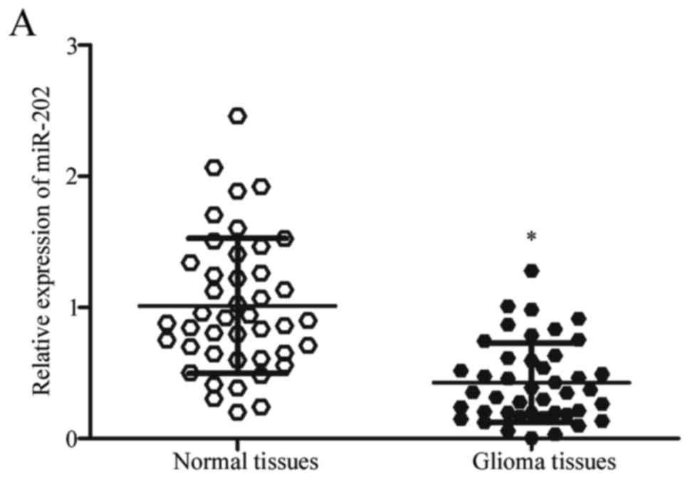
Firstly, we assessed miR-202 expression in glioma
and paired adjacent normal tissues using RT-qPCR. As shown in
Fig. 1A, the expression of miR-202
in glioma tissues was lower than that in paired adjacent normal
tissues (P<0.05). In addition, decreased miR-202 expression was
observed in five glioma cell lines (A172, U87, U251, U373 and
LN229) compared with primary NHA (Fig.
1B; P<0.05). These results revealed that miR-202 was
downregulated in both glioma tissue and cell lines.
Correlation between the expression of
miR-202 and the clinicopathological features in glioma
patients
We then examined the correlation between the
expression of miR-202 and the clinicopathological parameters of
glioma patients. As shown in Table
II, the expression levels of miR-202 were closely correlated
with the KPS score (P=0.037) and WHO grade (P=0.009). However, no
significant correlations were found between the expression level of
miR-202 and other clinicopathological factors, including sex, age
or tumour size (all P>0.05).
| Table II.Correlation between the expression of
miR-202 and the clinicopathological characteristics in patients
with glioma. |
Table II.
Correlation between the expression of
miR-202 and the clinicopathological characteristics in patients
with glioma.
|
|
| miR-202
expression |
|
|---|
|
|
|
|
|
|---|
| Clinicopathological
characteristics | No. of cases | Low | High | P-value |
|---|
| Sex |
|
|
| 0.927 |
|
Male | 14 | 8 | 6 |
|
|
Female | 29 | 17 | 12 |
|
| Age (years) |
|
|
| 0.576 |
|
<55 | 26 | 16 | 10 |
|
|
≥55 | 17 | 9 | 8 |
|
| Tumour size
(cm) |
|
|
| 0.515 |
|
<3 | 19 | 10 | 9 |
|
| ≥3 | 24 | 15 | 9 |
|
| KPS score |
|
|
| 0.037 |
|
<80 | 20 | 15 | 5 |
|
|
≥80 | 23 | 10 | 13 |
|
| WHO grade |
|
|
| 0.009 |
|
I-II | 21 | 8 | 13 |
|
|
III-IV | 22 | 17 | 5 |
|
miR-202 attenuates cell proliferation,
migration and invasion of glioma
To examine the functions of miR-202 in glioma
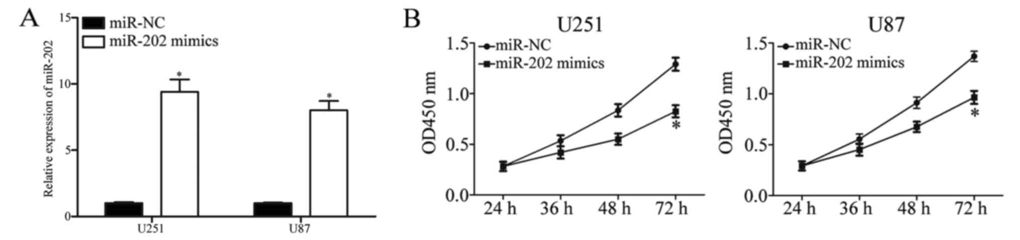
progression, miR-202 mimics were transiently transfected into U251
and U87 cells to increase expression (Fig. 2A; P<0.05). Firstly, we performed
a CCK-8 assay to evaluate the effect of miR-202 on U251 and U87
cells. The results revealed that miR-202 overexpression inhibited
the proliferative capacity of U251 and U87 cells (Fig. 2B; P<0.05). Cell migration and
invasion assays were conducted to assess the role of miR-202 in
metastasis. As shown in Fig. 2C,
the resumption of miR-202 expression decreased the metastatic
abilities in both U251 and U87 cells (P<0.05). Overall, these
results revealed that miR-202 inhibited the cell growth and
metastasis of glioma.
MTDH is a direct target of miR-202 in
glioma
To better understand the molecular mechanism
underlying the tumour-suppressive effect of miR-202 in glioma, the
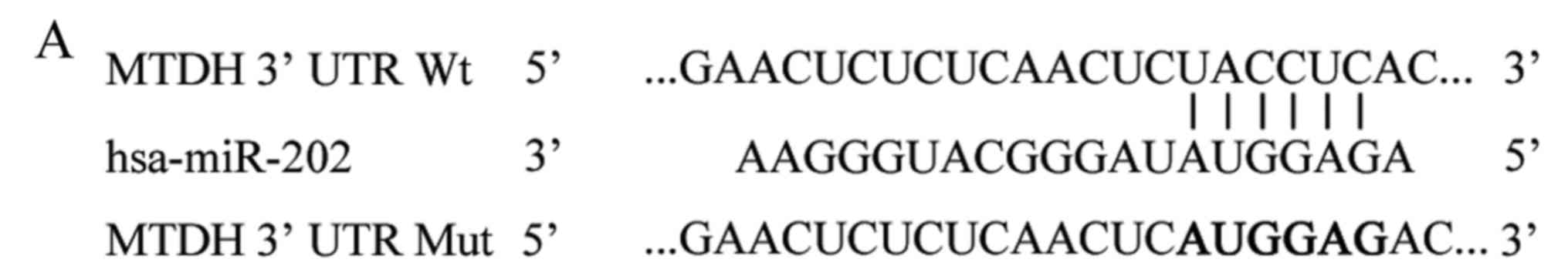
direct target genes of miR-202 were explored. Firstly,
bioinformatic analysis was performed to predict the potential
candidates of miR-202. Among various potential targets, MTDH
attracted our attention since the 3′UTR of MTDH contains putative
target sequences for miR-202 (Fig.
3A). Moreover, MTDH is highly expressed in glioma and involved
in the tumourigenesis and progression of glioma (27–29).
To confirm this hypothesis, a luciferase reporter assay was
performed in HEK293T cells transfected with miR-202 mimics or
miR-NC, along with luciferase reporter plasmids carrying Wt and Mut
sequences of the predicted binding sites. The results revealed that
miR-202 markedly decreased the luciferase activities of
pGL3-MTDH-3′UTR WT (Fig. 3B;
P<0.05), but did not affect the activity of pGL3-MTDH-3′UTR Mut.
Thus, miR-202 could directly target the 3′UTR of MTDH.
To further confirm the influence of miR-202 on the
expression of MTDH, the mRNA and protein expression levels of MTDH
in U251 and U87 cells with miR-202 overexpression were detected
using RT-qPCR and western blotting, respectively. We found that
miR-202 overexpression significantly suppressed endogenous MTDH
mRNA (Fig. 3C; P<0.05) and
protein (Fig. 3D; P<0.05)
expression levels in U251 and U87 cells. Collectively, theses
results revealed that miR-202 negatively regulated the expression
of MTDH by directly binding to the 3′UTR of MTDH.
Inverse correlation between miR-202
and MTDH in glioma tissues
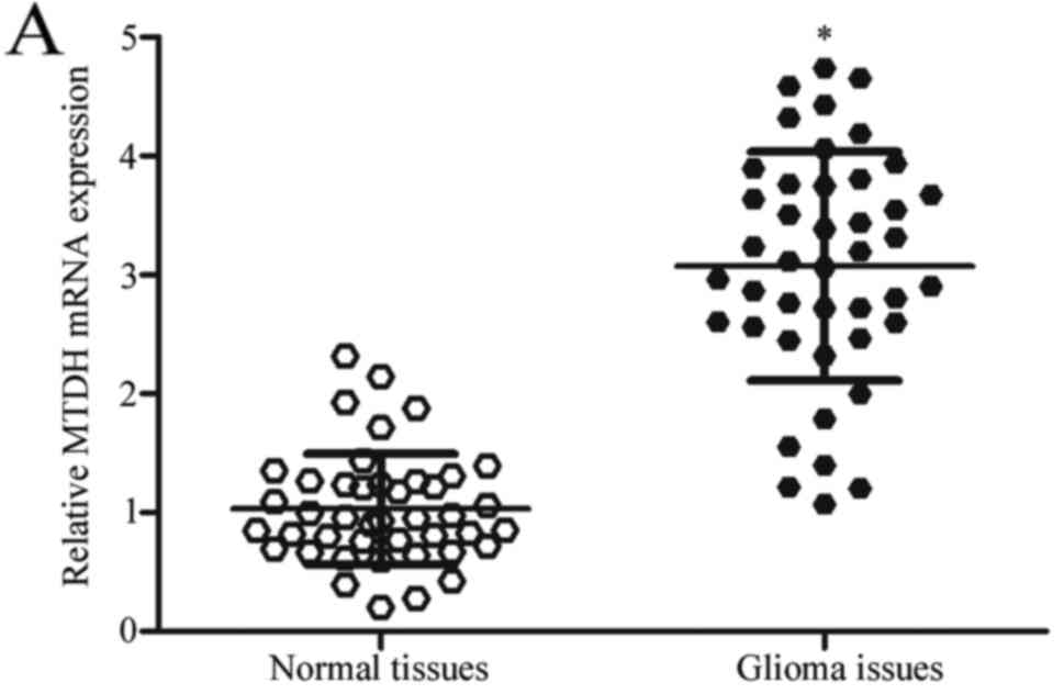
To further investigate the association between
miR-202 and MTDH, the expression of MTDH in glioma tissues was
determined. The results revealed that MTDH mRNA was significantly
increased in glioma tissues compared with that in paired adjacent
normal tissues (Fig. 4A;
P<0.05). Furthermore, MTDH protein expression in glioma tissues
and cell lines was assessed. As shown in Fig. 4B and C, MTDH protein expression was
obviously upregulated in glioma tissues and cell lines compared
with that in adjacent normal tissues and primary NHA, respectively.
Spearman's correlation analysis indicated an inverse correlation
between miR-202 and MTDH mRNA expression in glioma tissues
(Fig. 4D; r=−0.5503; P=0.001).
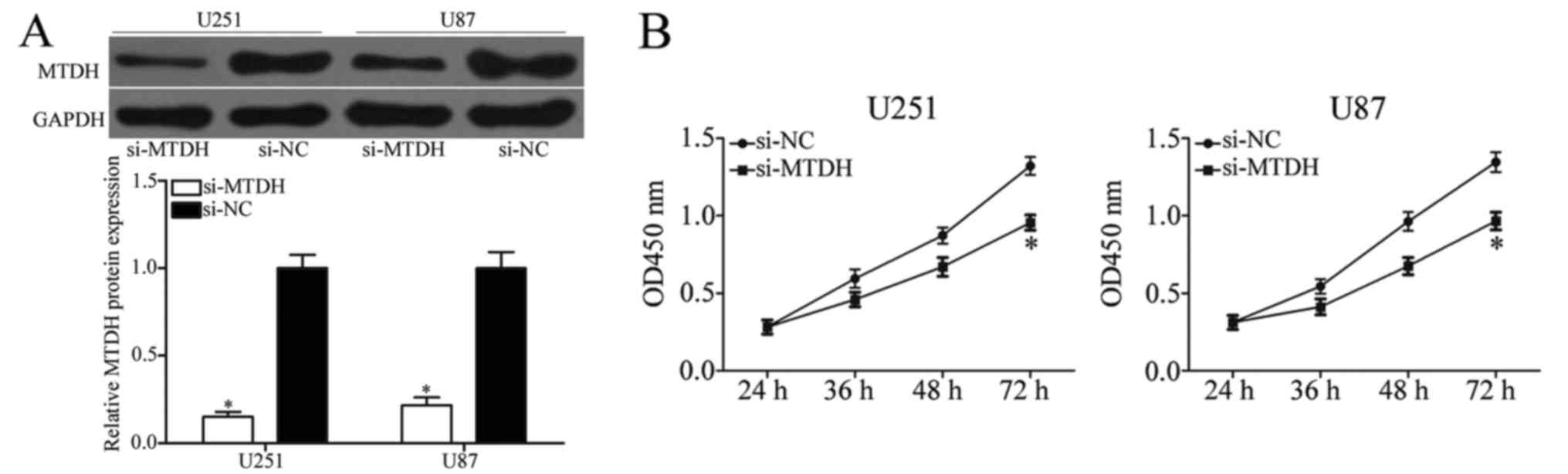
Inhibition of MTDH produces similar
effects to miR-202 overexpression in glioma
MTDH was identified as a direct target of miR-202.
Therefore, we hypothesised that miR-202 suppresses cell growth and
metastasis in glioma via inhibition of MTDH expression. To confirm
this hypothesis, endogenous MTDH expression was knocked down in
U251 and U87 cells using si-MTDH (Fig.
5A; P<0.05). CCK-8, and cell migration and invasion assays
revealed that inhibition of MTDH prevented the proliferation
(Fig. 5B; P<0.05), migration and
invasion (Fig. 5C; P<0.05) of
U251 and U87 cells. These results demonstrated that MTDH knockdown
had similar effects to miR-202 overexpression in glioma and
indicated that MTDH was a direct and functional target of
miR-202.
Restoration of MTDH expression
reverses miR-202 suppression of glioma cell growth and
metastasis
To explore whether miR-202 targeting of MTDH is
responsible for the inhibition of growth and metastasis in glioma,
we utilised rescue experiments. U251 and U87 cells were transfected
with pcDNA3.1-MTDH or pcDNA3.1. As shown in Fig. 6A, MTDH was successfully
overexpressed in pcDNA3.1-transfected U251 and U87 cells
(P<0.05). CCK-8 and cell invasion assays revealed that the
inhibition of the proliferation (Fig.
6B; P<0.05), migration and invasion (Fig. 6C; P<0.05) of miR-202 in U251 and
U87 cells was markedly reversed by MTDH overexpression. These
results demonstrated that the tumour-suppressive effect of miR-202
in glioma cells was partially dependent on MTDH suppression.
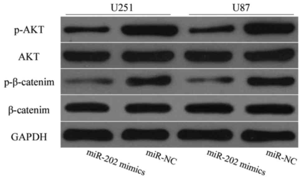
miR-202 inhibits the activation of the
PI3K/Akt and Wnt/β-catenin signalling pathways in glioma
It has been previously reported that MTDH
contributes to the activation of the PI3K/Akt and Wnt/β-catenin
pathways (30,31). Considering the regulatory effect of
miR-202 on MTDH, we hypothesized that miR-202 re-expression may
decrease the PI3K/Akt and Wnt/β-catenin pathways by negatively
regulating MTDH. To confirm this assumption, western blot analysis
was employed to detect AKT, p-AKT, β-catenin and p-β-catenin
expression levels in glioma cells after transfection with miR-202
mimics or miR-NC. As expected, miR-202 overexpression decreased
p-AKT and p-β-catenin expression levels in U251 and U87 cells
(Fig. 7; P<0.05). These results
revealed that miR-202 impaired the PI3K/Akt and Wnt/β-catenin
pathways by regulating MTDH.
Discussion
In recent years, miRNAs have been reported to be
aberrantly expressed in various types of human cancers, such as
glioma (32–34). Dysregulated expression of miRNAs is
often correlated with malignant biological behaviours of glioma,
such as rapid growth, metastasis, apoptosis inhibition, radiation
and chemotherapy resistance (10,35).
Therefore, miRNAs may be used as prognostic markers, and
miRNA-based therapy may be a valuable strategy for cancer
treatment. In the present study, miR-202 was identified as a
tumour-suppressor miRNA for glioma and low expression of miR-202
was detected in glioma tissues and cell lines. Low miR-202
expression was associated with the KPS score and WHO grade of
glioma patients. In addition, the restoration of miR-202 expression
suppressed cell proliferation, migration and invasion in glioma.
MTDH was identified as a direct functional target of miR-202. The
upregulation of miR-202 inhibited the activation of the PI3K/Akt
and Wnt/β-catenin signalling pathways in glioma. Our results
demonstrated that miR-202 was poorly expressed in glioma cells, and
thus may be a potential therapeutic target for glioma patients.
A previous study reported that miR-202 is frequently
dysregulated in multiple types of human tumours. In gastric cancer,
miR-202 is downregulated in tumour tissues and negatively
correlated with tumour size and age (36). In oesophageal squamous cell
carcinoma, miR-202 is poorly expressed in tumour tissues and
inversely correlated with the degree of cell differentiation and
lymph node metastasis. In addition, miR-202 is decreased in the
peripheral blood of oesophageal squamous cell carcinoma patients
and significantly associated with the development, invasion and
metastasis of oesophageal squamous cell carcinoma (37). Moreover, low expression levels of
miR-202 are observed in multiple myeloma (24), colorectal cancer (22), hepatocellular carcinoma (25), lung (38) and cervical cancer (39). The high frequency of miR-202
downregulation in these types of human types of cancer suggests
that miR-202 could be a diagnostic and prognostic marker for
specific cancers.
Abbberantly expressed miR-202 plays an important
role in the initiation and progression of several types of tumours.
Sun et al found that miR-202 overexpression suppresses cell
growth in vitro and in vivo and enhances cell
apoptosis in osteosarcoma (40).
Another study revealed that the restoration of miR-202 expression
suppresses cervical cancer cell growth and metastasis (39). Ma et al reported that ectopic
expression of miR-202 inhibits cell proliferation, migration and
invasion and induces cell apoptosis of oesophageal squamous cell
carcinoma (37,41). Meanwhile, Jiang et al
(38) demonstrated that the
upregulation of miR-202 decreases cell proliferation and improves
the G0/G1 cell cycle arrest and apoptosis in lung cancer. These
findings revealed that miR-202 plays an important role in these
types of cancers, and may be investigated as a potential
therapeutic target for the treatment of specific cancers.
To investigate how miR-202 functions as a tumour
suppressor in glioma, we explored the direct targets of miR-202. To
date, few genes have been validated as direct targets of miR-202,
including Gli1 in gastric cancer (36), BAFF in multiple myeloma (42), ARL5A in colorectal cancer (22), LPR6 in hepatocellular carcinoma
(25), LAMA1 in esophageal squamous
cell carcinoma (41) and CCND1 in
lung cancer (38). In the present
study, we screened potential candidates of miR-202 via
bioinformatic analysis. MTDH was selected for further investigation
since MTDH is highly expressed in glioma and involved in the
tumourigenesis and progression of glioma (27–29).
Luciferase reporter assay further confirmed that the 3′UTR of MTDH
could be directly targeted by miR-202. In addition, endogenous MTDH
expression on mRNA and protein levels was decreased in glioma cells
with miR-202 overexpression. MTDH expression was increased in
glioma tissues and negatively correlated with miR-202 expression.
MTDH knockdown had similar effects to miR-202 overexpression in
glioma. Rescue experiments revealed that upregulation of MTDH
reversed miR-202 suppression of glioma cell growth and metastasis.
These results suggested that miR-202 exerted its tumour-suppressive
effect in glioma partly by negatively regulating MTDH.
Identification of the miR-202 target in glioma is important for
understanding its role in the initiation and progression of
glioma.
MTDH, also known as astrocyte elevated gene-1
(AEG-1) and lysine-rich CEACAM1 co-isolated (LYRIC), was originally
identified as a neuropathology-associated gene in primary human
fetal astrocytes (43). MTDH is
significantly overexpressed in numerous human types of cancer, and
its expression level is correlated with the progression and poor
prognosis of malignant tumours, such as breast (44) and cervical cancer (45), and hepatocellular carcinoma
(46). A number of studies have
ascertained the important roles of MTDH in cell proliferation,
apoptosis regulation, angiogenesis, migration, invasion and
metastasis of various human cancers by activating signal pathways,
including the Ha-Ras and PI3K/Akt, nuclear factor-κB,
ERK/mitogen-activated protein kinase and Wnt/β-catenin and aurora-A
kinase signalling pathways (30,47–49).
In glioma, MTDH is highly expressed in tumour tissues and cell
lines. The expression levels of MTDH are correlated with the
metastasis and histological grade of gliomas (27,28).
Functional assays demonstrated that MTDH acts as an oncogene in
glioma and thus, regulated tumour cell proliferation, apoptosis and
metastasis (27,50,51).
Therefore, targeting MTDH may prolong the survival time and improve
the outcome of patients afflicted with this aggressive and
invariably fatal disease. MTDH may be a useful therapeutic target
for the therapy of glioma patients.
In conclusion, the present study demonstrated that
miR-202 may be associated with carcinogenesis and progression of
glioma by targeting MTDH. Future studies are warranted to explore
whether the potential of miR-202 may be fully realised in glioma
treatments.
References
|
1
|
Zhang C, Bao Z, Zhang W and Jiang T:
Progress on molecular biomarkers and classification of malignant
gliomas. Front Med. 7:150–156. 2013. View Article : Google Scholar : PubMed/NCBI
|
|
2
|
Katakowski M, Buller B, Wang X, Rogers T
and Chopp M: Functional microRNA is transferred between glioma
cells. Cancer Res. 70:8259–8263. 2010. View Article : Google Scholar : PubMed/NCBI
|
|
3
|
Zhou M, Wang H, Zhou K, Luo X, Pan X, Shi
B, Jiang H, Zhang J, Li K, Wang HM, et al: A novel EGFR isoform
confers increased invasiveness to cancer cells. Cancer Res.
73:7056–7067. 2013. View Article : Google Scholar : PubMed/NCBI
|
|
4
|
McNamara MG and Mason WP: Antiangiogenic
therapies in glioblastoma multiforme. Expert Rev Anticancer Ther.
12:643–654. 2012. View Article : Google Scholar : PubMed/NCBI
|
|
5
|
Vaupel P: Hypoxia and aggressive tumor
phenotype: Implications for therapy and prognosis. Oncologist.
13:(Suppl 3). S21–S26. 2008. View Article : Google Scholar
|
|
6
|
Khan UA, Bhavsar A, Asif H, Karabatsou K,
Leggate JR, Sofat A and Kamaly-Asl ID: Treatment by specialist
surgical neurooncologists improves survival times for patients with
malignant glioma. J Neurosurg. 122:297–302. 2015. View Article : Google Scholar : PubMed/NCBI
|
|
7
|
Hong Y, Shang C, Xue YX and Liu YH:
Silencing of Bmi-1 gene enhances chemotherapy sensitivity in human
glioblastoma cells. Med Sci Monit. 21:1002–1007. 2015. View Article : Google Scholar : PubMed/NCBI
|
|
8
|
Clarke J, Butowski N and Chang S: Recent
advances in therapy for glioblastoma. Arch Neurol. 67:279–283.
2010. View Article : Google Scholar : PubMed/NCBI
|
|
9
|
Davis FG and McCarthy BJ: Current
epidemiological trends and surveillance issues in brain tumors.
Expert Rev Anticancer Ther. 1:395–401. 2001. View Article : Google Scholar : PubMed/NCBI
|
|
10
|
Bartel DP: MicroRNAs: Genomics,
biogenesis, mechanism, and function. Cell. 116:281–297. 2004.
View Article : Google Scholar : PubMed/NCBI
|
|
11
|
Lee YS and Dutta A: MicroRNAs in cancer.
Annu Rev Pathol. 4:199–227. 2009. View Article : Google Scholar : PubMed/NCBI
|
|
12
|
Jovanovic M and Hengartner MO: miRNAs and
apoptosis: RNAs to die for. Oncogene. 25:6176–6187. 2006.
View Article : Google Scholar : PubMed/NCBI
|
|
13
|
Miska EA: How microRNAs control cell
division, differentiation and death. Curr Opin Genet Dev.
15:563–568. 2005. View Article : Google Scholar : PubMed/NCBI
|
|
14
|
Magee P, Shi L and Garofalo M: Role of
microRNAs in chemoresistance. Ann Transl Med. 3:3322015.PubMed/NCBI
|
|
15
|
Cellini F, Morganti AG, Genovesi D,
Silvestris N and Valentini V: Role of microRNA in response to
ionizing radiations: Evidences and potential impact on clinical
practice for radiotherapy. Molecules. 19:5379–5401. 2014.
View Article : Google Scholar : PubMed/NCBI
|
|
16
|
Pan Y, Liang W, Zhao X, Liu L, Qing Y and
Li Y: miR-548b inhibits the proliferation and invasion of malignant
gliomas by targeting metastasis tumor-associated protein-2.
Neuroreport. 27:1266–1273. 2016. View Article : Google Scholar : PubMed/NCBI
|
|
17
|
Wang H, Xiong M, Hu Y, Sun Y and Ma Q:
MicroRNA-19b inhibits proliferation of gastric cancer cells by
targeting B-cell CLL/lymphoma 3. Oncol Rep. 36:2079–2086.
2016.PubMed/NCBI
|
|
18
|
Liu Y, Uzair-Ur-Rehman, Guo Y, Liang H,
Cheng R, Yang F, Hong Y, Zhao C, Liu M, Yu M, et al: miR-181b
functions as an oncomiR in colorectal cancer by targeting PDCD4.
Protein Cell. 7:722–734. 2016. View Article : Google Scholar : PubMed/NCBI
|
|
19
|
Hu MH, Ma CY, Wang XM, Ye CD, Zhang GX,
Chen L and Wang JG: MicroRNA-126 inhibits tumor proliferation and
angiogenesis of hepatocellular carcinoma by down-regulating EGFL7
expression. Oncotarget. 7:66922–66934. 2016. View Article : Google Scholar : PubMed/NCBI
|
|
20
|
Yan J, Gumireddy K, Li A and Huang Q:
Regulation of mesenchymal phenotype by MicroRNAs in cancer. Curr
Cancer Drug Targets. 13:930–934. 2013. View Article : Google Scholar : PubMed/NCBI
|
|
21
|
Xiang W, He J, Huang C, Chen L, Tao D, Wu
X, Wang M, Luo G, Xiao X, Zeng F, et al: miR-106b-5p targets tumor
suppressor gene SETD2 to inactive its function in clear cell renal
cell carcinoma. Oncotarget. 6:4066–4079. 2015. View Article : Google Scholar : PubMed/NCBI
|
|
22
|
Wang Q, Huang Z, Guo W, Ni S, Xiao X, Wang
L, Huang D, Tan C, Xu Q, Zha R, et al: microRNA-202-3p inhibits
cell proliferation by targeting ADP-ribosylation factor-like 5A in
human colorectal carcinoma. Clin Cancer Res. 20:1146–1157. 2014.
View Article : Google Scholar : PubMed/NCBI
|
|
23
|
Josson S, Gururajan M, Hu P, Shao C, Chu
GY, Zhau HE, Liu C, Lao K, Lu CL, Lu YT, et al: miR-409-3p/−5p
promotes tumorigenesis, epithelial-to-mesenchymal transition, and
bone metastasis of human prostate cancer. Clin Cancer Res.
20:4636–4646. 2014. View Article : Google Scholar : PubMed/NCBI
|
|
24
|
Yu J, Qiu X, Shen X, Shi W, Wu X, Gu G,
Zhu B and Ju S: miR-202 expression concentration and its clinical
significance in the serum of multiple myeloma patients. Ann Clin
Biochem. 51:543–549. 2014. View Article : Google Scholar : PubMed/NCBI
|
|
25
|
Zhang Y, Zheng D, Xiong Y, Xue C, Chen G,
Yan B and Ye Q: miR-202 suppresses cell proliferation in human
hepatocellular carcinoma by downregulating LRP6
post-transcriptionally. FEBS Lett. 588:1913–1920. 2014. View Article : Google Scholar : PubMed/NCBI
|
|
26
|
Livak KJ and Schmittgen TD: Analysis of
relative gene expression data using real-time quantitative PCR and
the 2−ΔΔCT method. Methods. 25:402–408. 2001.
View Article : Google Scholar : PubMed/NCBI
|
|
27
|
Emdad L, Sarkar D, Lee SG, Su ZZ, Yoo BK,
Dash R, Yacoub A, Fuller CE, Shah K, Dent P, et al: Astrocyte
elevated gene-1: A novel target for human glioma therapy. Mol
Cancer Ther. 9:79–88. 2010. View Article : Google Scholar : PubMed/NCBI
|
|
28
|
He Z, He M, Wang C, Xu B, Tong L, He J,
Sun B, Wei L and Chu M: Prognostic significance of astrocyte
elevated gene-1 in human astrocytomas. Int J Clin Exp Pathol.
7:5038–5044. 2014.PubMed/NCBI
|
|
29
|
Hu B, Emdad L, Bacolod MD, Kegelman TP,
Shen XN, Alzubi MA, Das SK, Sarkar D and Fisher PB: Astrocyte
elevated gene-1 interacts with Akt isoform 2 to control glioma
growth, survival, and pathogenesis. Cancer Res. 74:7321–7332. 2014.
View Article : Google Scholar : PubMed/NCBI
|
|
30
|
Lee SG, Su ZZ, Emdad L, Sarkar D, Franke
TF and Fisher PB: Astrocyte elevated gene-1 activates cell survival
pathways through PI3K-Akt signaling. Oncogene. 27:1114–1121. 2008.
View Article : Google Scholar : PubMed/NCBI
|
|
31
|
Hu G, Wei Y and Kang Y: The multifaceted
role of MTDH/AEG-1 in cancer progression. Clin Cancer Res.
15:5615–5620. 2009. View Article : Google Scholar : PubMed/NCBI
|
|
32
|
Gu JJ, Gao GZ and Zhang SM: MiR-218
inhibits the tumorgenesis and proliferation of glioma cells by
targeting Robo1. Cancer Biomark. 16:309–317. 2016. View Article : Google Scholar : PubMed/NCBI
|
|
33
|
Stojcheva N, Schechtmann G, Sass S, Roth
P, Florea AM, Stefanski A, Stühler K, Wolter M, Müller NS, Theis
FJ, et al: MicroRNA-138 promotes acquired alkylator resistance in
glioblastoma by targeting the Bcl-2-interacting mediator BIM.
Oncotarget. 7:12937–12950. 2016. View Article : Google Scholar : PubMed/NCBI
|
|
34
|
Liu H, Song Z, Liao D, Zhang T, Liu F,
Zheng W, Luo K and Yang L: miR-503 inhibits cell proliferation and
invasion in glioma by targeting L1CAM. Int J Clin Exp Med.
8:18441–18447. 2015.PubMed/NCBI
|
|
35
|
Wang ZY, Xiong J, Zhang SS, Wang JJ, Gong
ZJ and Dai MH: Up-regulation of microRNA-183 promotes cell
proliferation and invasion in glioma by directly targeting NEFL.
Cell Mol Neurobiol. 36:1303–1310. 2016. View Article : Google Scholar : PubMed/NCBI
|
|
36
|
Zhao Y, Li C, Wang M, Su L, Qu Y, Li J, Yu
B, Yan M, Yu Y, Liu B, et al: Decrease of miR-202-3p expression, a
novel tumor suppressor, in gastric cancer. PLoS One. 8:e697562013.
View Article : Google Scholar : PubMed/NCBI
|
|
37
|
Ma G, Zhang F, Dong X, Wang X and Ren Y:
Low expression of microRNA-202 is associated with the metastasis of
esophageal squamous cell carcinoma. Exp Ther Med. 11:951–956.
2016.PubMed/NCBI
|
|
38
|
Jiang J, Huang J, Wang XR and Quan YH:
MicroRNA-202 induces cell cycle arrest and apoptosis in lung cancer
cells through targeting cyclin D1. Eur Rev Med Pharmacol Sci.
20:2278–2284. 2016.PubMed/NCBI
|
|
39
|
Yi Y, Li H, Lv Q, Wu K and Zhang W, Zhang
J, Zhu D, Liu Q and Zhang W: miR-202 inhibits the progression of
human cervical cancer through inhibition of cyclin D1. Oncotarget.
7:72067–72075. 2016.PubMed/NCBI
|
|
40
|
Sun Z, Zhang T, Hong H, Liu Q and Zhang H:
miR-202 suppresses proliferation and induces apoptosis of
osteosarcoma cells by downregulating Gli2. Mol Cell Biochem.
397:277–283. 2014. View Article : Google Scholar : PubMed/NCBI
|
|
41
|
Meng X, Chen X, Lu P, Ma W, Yue D, Song L
and Fan Q: MicroRNA-202 inhibits tumor progression by targeting
LAMA1 in esophageal squamous cell carcinoma. Biochem Biophys Res
Commun. 473:821–827. 2016. View Article : Google Scholar : PubMed/NCBI
|
|
42
|
Yu JJ, Shen XJ, Wang XD and Ju SQ: Effect
of miR-202 on the growth of multiple myeloma cells via regulating B
cell-activating factor and the underlying mechanism. Zhonghua Zhong
Liu Za Zhi. 35:886–891. 2013.(In Chinese). PubMed/NCBI
|
|
43
|
Su ZZ, Kang DC, Chen Y, Pekarskaya O, Chao
W, Volsky DJ and Fisher PB: Identification and cloning of human
astrocyte genes displaying elevated expression after infection with
HIV-1 or exposure to HIV-1 envelope glycoprotein by rapid
subtraction hybridization, RaSH. Oncogene. 21:3592–3602. 2002.
View Article : Google Scholar : PubMed/NCBI
|
|
44
|
Li J, Zhang N, Song LB, Liao WT, Jiang LL,
Gong LY, Wu J, Yuan J, Zhang HZ, Zeng MS, et al: Astrocyte elevated
gene-1 is a novel prognostic marker for breast cancer progression
and overall patient survival. Clin Cancer Res. 14:3319–3326. 2008.
View Article : Google Scholar : PubMed/NCBI
|
|
45
|
Liu X, Wang D, Liu H, Feng Y, Zhu T, Zhang
L, Zhu B and Zhang Y: Knockdown of astrocyte elevated gene-1
(AEG-1) in cervical cancer cells decreases their invasiveness,
epithelial to mesenchymal transition, and chemoresistance. Cell
Cycle. 13:1702–1707. 2014. View Article : Google Scholar : PubMed/NCBI
|
|
46
|
Yoo BK, Emdad L, Su ZZ, Villanueva A,
Chiang DY, Mukhopadhyay ND, Mills AS, Waxman S, Fisher RA, Llovet
JM, et al: Astrocyte elevated gene-1 regulates hepatocellular
carcinoma development and progression. J Clin Invest. 119:465–477.
2009. View Article : Google Scholar : PubMed/NCBI
|
|
47
|
Emdad L, Lee SG, Su ZZ, Jeon HY, Boukerche
H, Sarkar D and Fisher PB: Astrocyte elevated gene-1 (AEG-1)
functions as an oncogene and regulates angiogenesis. Proc Natl Acad
Sci USA. 106:21300–21305. 2009. View Article : Google Scholar : PubMed/NCBI
|
|
48
|
Yoo BK, Chen D, Su ZZ, Gredler R, Yoo J,
Shah K, Fisher PB and Sarkar D: Molecular mechanism of
chemoresistance by astrocyte elevated gene-1. Cancer Res.
70:3249–3258. 2010. View Article : Google Scholar : PubMed/NCBI
|
|
49
|
Lee SG, Su ZZ, Emdad L, Sarkar D and
Fisher PB: Astrocyte elevated gene-1 (AEG-1) is a target
gene of oncogenic Ha-ras requiring phosphatidylinositol 3-kinase
and c-Myc. Proc Natl Acad Sci USA. 103:17390–17395. 2006.
View Article : Google Scholar : PubMed/NCBI
|
|
50
|
Yang Y, Wu J, Guan H, Cai J, Fang L, Li J
and Li M: MiR-136 promotes apoptosis of glioma cells by targeting
AEG-1 and Bcl-2. FEBS Lett. 586:3608–3612. 2012. View Article : Google Scholar : PubMed/NCBI
|
|
51
|
Liu L, Wu J, Ying Z, Chen B, Han A, Liang
Y, Song L, Yuan J, Li J and Li M: Astrocyte elevated gene-1
upregulates matrix metalloproteinase-9 and induces human glioma
invasion. Cancer Res. 70:3750–3759. 2010. View Article : Google Scholar : PubMed/NCBI
|