Introduction
Cancer is a leading cause of mortality worldwide,
and breast cancer is one of the most common causes of
cancer-associated mortality in females (1). The majority of patients with breast
cancer respond to conventional therapies, including surgical
removal of the tumor, drug treatment and radiation. However, each
therapy has inherent limitations that lead to therapeutic
resistance and disease recurrence, ultimately resulting in
therapeutic failure. Once the disease recurs, it readily
metastasizes to distant organs and causes mortality.
Cancer metastasis is the process by which cancer
cells spread from the site of origin to grow in adjacent sites and
is responsible for the majority of cancer-associated mortalities
rather than the primary tumor (2–4). It
has been suggested that cancer stem cells (CSCs) exist in tumors,
and contribute to metastasis to distant organs and disease
recurrence (5,6). CSCs are capable of self-renewal and of
regenerating the heterogeneous populations that comprise a tumor
following treatment (7). Some or
all of such cells in a tumor may exist in specific
microenvironments that render them more resistant to radiotherapy
and chemotherapy, ultimately resulting in tumor recurrence and
distant metastasis (8–12). The presence of CSCs in breast tumors
is known to increase post-therapy recurrence or relapse in patients
with breast cancer, but the underlying mechanisms of therapy
resistance remains unclear.
The radioresistance of breast cancer cells remains a
fundamental barrier to the maximum efficacy of radiotherapy,
raising the needs for the study of development of resistance by the
breast cancer cells to radiation. Therefore, in the present study,
we hypothesized that highly metastatic breast cancer cells
MDA-MB-231 cells produce more CSCs following exposure to radiation,
and accordingly, radioresistant (RT-R) breast cancer cells derived
from highly metastatic breast cancer cells enhance invasiveness due
to CSCs. Furthermore, the present study aimed to investigate the
potential associated mechanisms underlying the involvement of CSCs
in RT-R-MDA-MB-231 cell invasiveness under tumor microenvironment
conditions.
Materials and methods
Cell culture
The human breast cancer cell lines, MCF-7, T47D and
MDA-MB-231, were obtained from the Korea Cell Line Bank (Seoul,
Korea), and the EA.hy926 human umbilical vascular endothelial cell
(EC) line was originally purchased from American Type Culture
Collection (Manassas, VA, USA). The human breast cancer cell lines
and EA.hy926 cells were cultured in RPMI-1640 and Dulbecco's
modified Eagle's medium, respectively, and supplemented with 10%
FBS, 100 IU/ml penicillin and 10 µg/ml streptomycin (all from
HyClone; GE Healthcare Life Sciences, Logan, UT, USA). Cells were
incubated at 37°C in a humidified atmosphere containing 5%
CO2.
Establishment of RT-R breast cancer
cells
RT-R breast cancer cells (RT-R-MCF-7, RT-R-T47D and
RT-R-MDA-MB-231 cells) were generated by applying repetitive small
doses of X-ray irradiation (2 Gy) until a final dose of 50 Gy was
achieved, which is a commonly used clinical regimen for the
radiotherapy of patients with breast cancer. Cells were irradiated
with 2 Gy using a 6-MV photon beam produced by a linear accelerator
(Clinac 21EX, Varian Medical Systems, Inc., Palo Alto, CA). The
radiation dose rate was 1.0 Gy/min, and the cell medium was changed
to fresh complete media immediately following irradiation. When the
cells reached ~90% confluence, they were trypsinized and
subcultured into new flasks. When the cells reached ~70%
confluence, they were irradiated again. The fractionated
irradiations were continued until the total dose reached 50 Gy.
Cell viability assay
Cells in the exponential growth phase were seeded at
1×10⁴ cells/well in 24-well plates. Cells were
irradiated (2, 4, 6 or 8 Gy) or treated with paclitaxel
(Sigma-Aldrich; Merck KGaA, Darmstadt, Germany; 0.01, 0.05, 0.1, 1
or 5 mM) as indicated. Control groups were not irradiated or not
treated with paclitaxel, respectively. Then, 50 µl of 5 mg/ml MTT
(Sigma-Aldrich; Merck KGaA) was added, and the cells were incubated
for 4 h. The supernatants were aspirated, and the formazan crystals
were dissolved with 200 µl/well DMSO. The absorbance was measured
at 570 nm using an Infinite 200 microplate reader (Tecan Group,
Ltd., Mannedorf, Switzerland).
Colony formation assay
Parental breast cancer cells or RT-R-breast cancer
cells were seeded in 6-well plates (1×10³ cells/well).
Then, cells were irradiated even doses from 2 to 8 Gy and incubated
at 37°C. Control groups were not irradiated. After 10 days (for
MDA-MB-231/RT-R-MDA-MB231 and MCF-7/RT-R-MDA-MB-231) or after 14
days (for T47D/RT-R-T47D cells), the medium was discarded and each
well was washed with PBS. The colonies were fixed in 100% methanol
for 10 min at room temperature and then stained with 0.1% Giemsa
staining solution for 30 min at room temperature, and the number of
visible colonies was counted.
Flow cytometry
For analysis of cluster of differentiation (CD)24
and CD44 expression or population, the cells were labeled using a
human CD24 (cat. no. ab31622; 1:100; Abcam, Cambridge, UK) and CD44
(cat. no. ab5107; 1:100; Abcam) detection antibodies in PBS in the
dark for 30 min at room temperature. Then, the cells were washed
with cold PBS and analyzed using a FACSCalibur™ system with
CellQuest Pro™ software (version 3.0; BD Biosciences, Franklin
Lakes, NJ, USA).
Isolation of
CD24low/CD44high cancer stem cells from
breast cancer cells
Isolation of CD24low/CD44high
breast cancer stem cells was performed using a MagCollect
CD24− CD44+ Breast Cancer Stem Cell Isolation
kit (R&D Systems, Inc., Minneapolis, MN, USA) following the
manufacturer's protocol.
Western blot analysis
Western blot analysis was performed as described
previously (13), with minor
modifications. The membranes were blocked with 5% non-fat milk in
TBS containing 0.05% Tween-20 for 1 h at room temperature, and
incubated with the following primary antibodies overnight at 4°C:
Anti-intercellular adhesion molecule-1 (ICAM-1; cat. no. sc-7891;
1:1,000; rabbit polyclonal IgG), anti-vascular cell adhesion
molecule-1 (VCAM-1; cat. no. sc-8304; 1:1,000; rabbit polyclonal
IgG), anti-Snail 1 (cat. no. sc-5594; 1:1,000; rabbit polyclonal
IgG), anti-β-catenin (cat. no. sc-7199; 1:1,000; rabbit polyclonal
IgG), anti-E-cadherin (cat. no. sc-7870; 1:1,000; rabbit polyclonal
IgG), anti-N-cadherin (cat. no. sc-7939; 1:1,000; rabbit polyclonal
IgG), anti-octamer-binding transcription factor (Oct3/4; cat. no.
sc-9081; 1:1,000; rabbit polyclonal IgG), anti-Notch-4 antibodies
(cat. no. H-225; 1:1,000; rabbit polyclonal IgG) (all from Santa
Cruz Biotechnology, Inc. Dallas, TX, USA) and anti-aldehyde
dehydrogenase 1 (ALDH1; cat. no. ab52492; 1:1,000; rabbit
monoclonal; Abcam). The bound antibodies were detected with goat
anti-rabbit IgG-horseradish peroxidase-conjugated secondary
antibodies (cat. no. sc-2054; 1:5,000; Santa Cruz Biotechnology,
Inc.) for 1 h at room temperature and ECL western blotting
detection reagent (Bio-Rad Laboratiories, Inc., Hercules, CA, USA).
The protein band densities was analyzed using a ChemiDoc™ XRS+
system (Bio-Rad Laboratiories, Inc.) and β-actin (cat. no. a2066;
1:1,000; rabbit monoclonal; Sigma-Aldrich; Merck KGaA) was used as
a loading control for the normalization of protein expression.
Adhesion assay
MDA-MB-231 and RT-R-MDA-MB-231 cells
(4.0×10⁵ cells/ml) were added to the ECs once ~80%
confluence was achieved. After 30 min at 37°C, the cell suspensions
were removed, and the ECs were washed three times with PBS. The
cells were then counted under a light microscope (×200
magnification), and the number of cells that adhered to the ECs was
quantified.
Migration assay
The migration assay was performed as described
previously (14). Briefly,
MDA-MB-231 and RT-R-MDA-MB-231 cells (2×10⁵ cells/well)
were added to the upper chambers of the inserts, which were placed
in a 24-well plate, and 500 µl/well RPMI-1640 supplemented with 10%
FBS was added to the lower chambers. Following an overnight, the
non-migratory cells that remained on the upper surface of the
insert membranes were removed by swabbing. The cells that had
migrated across the membrane were stained with DAPI for 30 min at
room temperature in the dark, and the cells were counted under a
fluorescence microscope (×200 magnification).
Matrigel invasion assay
The upper chambers of the inserts were coated with
100 µl of Matrigel (1 mg/ml, BD Bioscience), and ECs
(2×10⁵ cells) were added to the Matrigel-coated insert.
MDA-MB-231 cells and RT-R-MDA-MB-231 cells (2×10⁵
cells/insert) were added to the upper chambers in serum-free media,
and 500 µl of RPMI-1640 supplemented with 10% FBS was added to the
lower chambers. The remaining procedures were performed as
aforementioned for the migration assays.
Gelatin zymography
A total of 2 ml of media were collected from
cultured MDA-MB-231 cells or RT-R-MDA-MB-231 cells and concentrated
by 20-fold using protein concentrators (9K MWCO; Thermo Fisher
Scientific, Inc., Waltham, MA, USA). Concentrated media (40 mg
protein in 20 ml/lane) was mixed with sample volume of buffer
(0.03% bromophenol blue, 0.4 M Tris-HCl pH 7.4, 20% glycerol, 5%
SDS), and then subjected to electrophoresis on 8% PAGE gels
containing 1 mg/ml gelatin. The gels were washed with renaturing
buffer (2.5% Triton X-100) for 1 h and subsequently incubated for
24 h at 37°C in developing buffer (50 mM Tris, 20 mM NaCl, 5 mM
CaCl2, 0.02% Brij35, pH 7.5). Gels were stained with
0.05% Coomassie Brilliant Blue R-250 and destained with 50%
methanol and 10% acetic acid for 2 h at room temperature. Within
the blue background, clear zones indicated matrix metalloproteinase
(MMP) proteolytic activity.
Statistical analysis
All results are representative of three independent
experiments performed in triplicate. The statistics were determined
using SigmaPlot software (version 10.0; Systat Software, Inc., San
Jose, CA, USA). The data were analyzed with two-tailed Student's
t-test to compare two groups or one-way analysis of variance with
Scheffe's post hoc test to compare mean values across multiple
treatment groups. The data are presented as the mean ± standard
error.
Results
RT-R breast cancer cells established
by repeated irradiation demonstrate resistance to chemotherapy and
radiation
First, cell viability of established RT-R-breast
cancer cells was examined using MTT assay following exposure to
fractionated irradiation (2, 4, 6 or 8 Gy). Irradiated parental
MCF-7, MDA-MB-231 and T47D cells demonstrated survival rates of
~60, ~67 and ~64% relative to non-irradiated MCF-7, MDA-MB-231 and
T47D cells, respectively. However, RT-R-breast cancer cells
exhibited ~20, ~50 and ~60% greater resistance compared with
parental MCF-7, MDA-MB-231 and T47D cells, respectively (Fig. 1A). In addition, fractionated
irradiation (2, 4, 6 or 8 Gy) of parental MCF-7, MDA-MB-231 and
T47D breast cancer cells caused a significant decrease in colony
formation compared with non-irradiated MCF-7, MDA-MB-231 and T47D
cells (Fig. 1B). Then, the cross
resistance of RT-R-breast cancer cells was examined via incubation
of cells with paclitaxel at 0.01–5 mM for 24 and 48 h. Paclitaxel
treatment significantly decreased the cell viability of the three
breast cancer cell lines compared with the untreated control group
in a dose-dependent manner (Fig.
1C), but RT-R-breast cancer cells except RT-R-MDA-MB-231
demonstrated almost no difference on cytotoxicity compared with
their parental breast cancer cells. RT-R-MDA-MB-231 cells
demonstrated the strongest resistance to paclitaxel particularly
following treatment for 48 h (Fig.
1C), suggesting that RT-R-MDA-MB-231 cells derived from highly
metastatic breast cancer cells MDA-MB-231 may become more resistant
to chemotherapy as well as radiation, compared with other
RT-R-breast cancer cells from low metastatic MCF-7 or T47D breast
cancer cells.
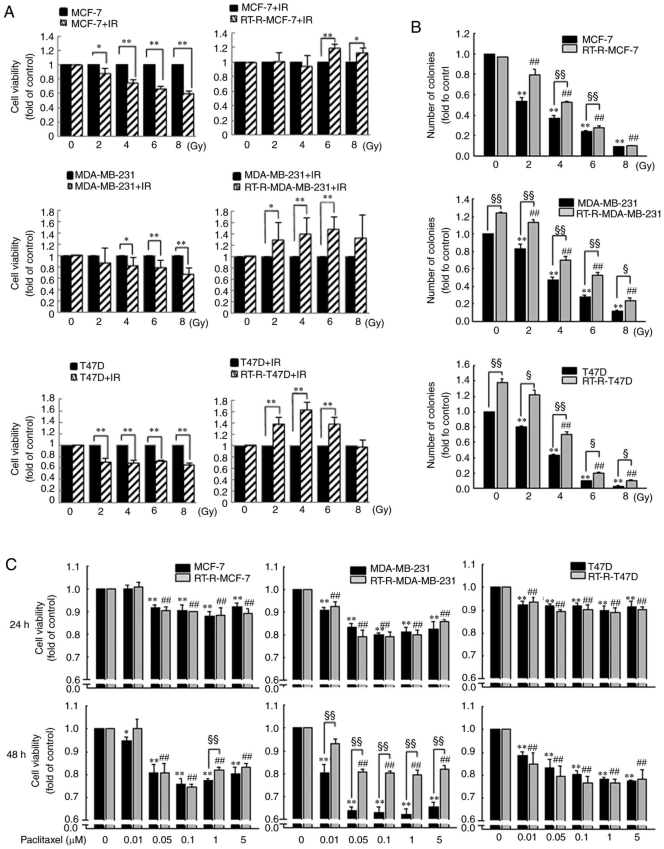 | Figure 1.Radioresistant breast cancer cells
established by repeated irradiation exhibit resistance to radiation
and chemotherapy. (A) The viabilities of MCF-7, RT-R-MCF-7,
MDA-MB-231, RT-R-MDA-MB-231, T47D and RT-R-T47D cells against
radiation treatment (2, 4, 6, 8 Gy) were examined as described in
the Materials and methods. *P<0.05, **P<0.01. (B) Parental
breast cancer cells (MCF-7, T47D and MDA-MB-231 cells) or
RT-R-breast cancer cells (RT-R-MCF-7, RT-R-T47D, RT-R-MDA-MB-231
cells) were irradiated (2, 4, 6, 8 Gy), and then colony formation
assays were performed. (C) Parental and RT-R-breast cancer cells
were treated with paclitaxel at the indicated doses (0.01–5 mM) for
24 and 48 h. Cell viability was determined and compared with the
untreated control groups. Data represent mean values ± standard
error of the mean of three independent experiments in triplicate.
**P<0.01 compared with parental breast cancer control cells;
##P<0.01 compared with RT-R-breast cancer control
cells; §P<0.05 and §§P<0.01 compared
between parental breast cancer cells and RT-R-breast cancer cells.
IR, irradiation; RT-R, radioresistant. |
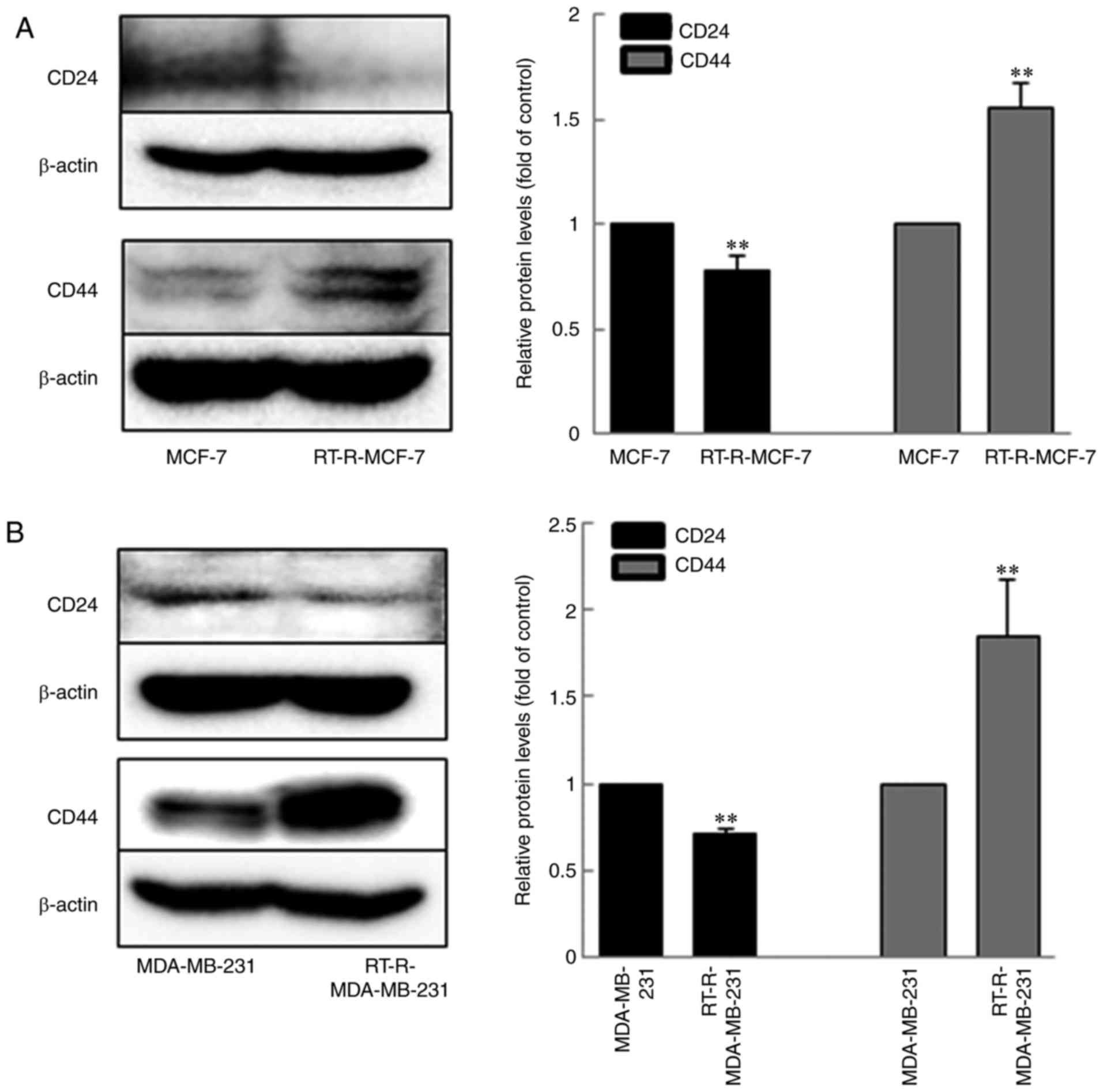
RT-R-MDA-MB-231 cells demonstrate
higher levels of CSCs markers CD44, but lower levels of CD24,
compared with MDA-MB-231 or RT-R-MCF-7 cells
It has been suggested that CSCs represent a possible
cause of tumor resistance to irradiation as well as chemotherapy
(15–17). Therefore, whether RT-R-MDA-MB-231
cells harbored more CSCs compared with MDA-MB-231 cells or
RT-R-MCF-7 cells derived from the low metastatic breast cancer
cells MCF-7 was investigated. When the expression levels of CD24
and CD44 were examined by western blot analysis, RT-R-MCF-7 and
RT-R-MDA-MB-231 cells revealed significantly higher expression
levels of CD44 and lower expression levels of CD24 (Fig. 2), compared with MCF-7 and MDA-MB-231
cells, respectively. RT-R-MDA-MB-231 and RT-R-MCF-7 cells expressed
a significantly higher level of CD44 compared with MDA-MB-231 cells
(~2-fold) and MCF-7 cells (~1.5-fold), respectively, indicating
that RT-R-MDA-MB-231 cells possessed a higher number of CSCs
compared with RT-R-MCF-7 cells. This result supports the idea that
RT-R-MDA-MB-231 cells derived from highly metastatic breast cancer
cells MDA-MB-231 are more resistant, compared with RT-R-MCF-7 cells
derived from low metastatic breast cancer cells MCF-7, due to the
level of CSCs present.
RT-R-MDA-MB-231 cells increase the
number of CD24low/CD44high cells and
expression levels of CSCs markers Notch-4, Oct-3/4 and ALDH1
compared with MDA-MB-231 cells
According to the aforementioned results,
RT-R-MDA-MB-231 cells were chosen to investigate the role of CSCs
in the increased invasiveness of RT-R-breast cancer cells and the
potential underlying mechanisms. First of all,
CD24low/CD44high cells were isolated from
MDA-MB-231 and RT-R-MDA-MB cells using isolation kit as
aforementioned, which confirmed that the number of the isolated
CD24low/CD44high cells from RT-R-MDA-MB-231
cells was significantly higher compared with MDA-MB-231 cells, as
indicated by flow cytometric analysis (Fig. 3A). Then, the expression levels of
CD24 or CD44 were analyzed by flow cytometry, and other CSC
markers, including Notch-4, Oct-3/4 and ALDH1 (18–22)
were determined using western blot analysis. Fig. 3B demonstrates that RT-R-MDA-MB-231
cells exhibited significantly higher CD44 and lower CD24 levels
compared with MDA-MB-231 cells. The CSC markers, Notch-4, Oct3/4
and ALDH1, were also significantly upregulated in RT-R-MDA-MB-231
cells compared withMDA-MB-231 cells (Fig. 3C).
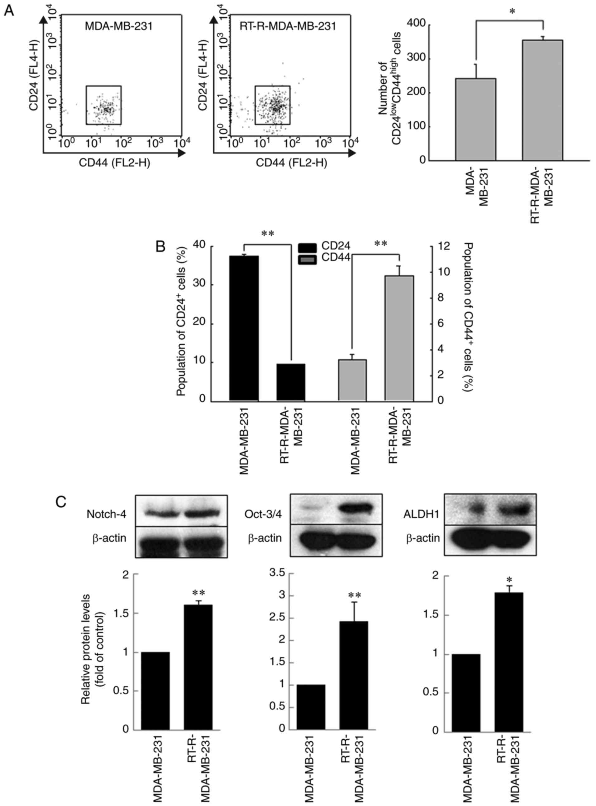 | Figure 3.RT-R-MDA-MB-231 cells increase the
populations of CD24low/CD44high cells, and
expression levels of other CSCs markers Notch-4, Oct-3/4 and ALDH1.
(A) CD24low/CD44high cells were isolated from
MDA-MB-231 and RT-R-MDA-MB cells, and then the number of
CD24low/CD44high breast cancer stem cells was
quantified by flow cytometry. (B) MDA-MB-231 and RT-R-MDA-MB-231
were labeled with anti-CD24 and anti-CD44 antibodies, and the
percentages of CD24 or CD44-expressed subpopulation in cells were
determined by flow cytometry. (C) Other cancer stem cell markers,
including Notch-4, Oct3/4 and ALDH1 were detected in the MDA-MB-231
and RT-R-MDA-MB-231 cells using the specific antibodies by western
blotting. Data represent mean values ± standard error of the mean
of three independent experiments in triplicate. *P<0.05 and
**P<0.01 compared with MDA-MB-231 cells. ALDH1, aldehyde
dehydrogenase 1; CD, cluster of differentiation; CSCs, cancer stem
cells; Oct, octamer-binding transcription factor; RT-R,
radioresistant. |
RT-R-MDA-MB-231 cells demonstrate
higher expression levels of adhesion molecules (AMs) and
epithelial-mesenchymal transition (EMT)-associated proteins,
resulting in enhanced migration, adhesion to ECs and invasion
through ECs compared with MDA-MB-231 cells
We previously reported that induction of AMs by
MDA-MB-231 serves an important role in cancer cell migration,
cancer cell adhesion to ECs and cancer cell invasion through ECs
(14). Thus, whether
RT-R-MDA-MB-231 cells exhibited higher expression of AMs, including
ICAM-1 and VCAM-1, was examined. ICAM-1 and VCAM-1 expression was
significantly increased in RT-R-MDA-MB-231 cells compared with
MDA-MB-231 cells (Fig. 4). Next,
adhesion and migration assays were performed. As expected,
RT-MDA-MB-231 cells exhibited significantly enhanced migration and
adhesion to ECs compared with MDA-MB-231 cells (Fig. 4B and C). Furthermore, the
invasiveness was significantly enhanced in RT-R-MDA-MB-231 cells
compared with MDA-MB-231 cells (Fig.
4D). Next, the expression levels of EMT markers were determined
in RT-R-MDA-MB-231 cells. Fig. 5A and
B demonstrates that RT-R-MDA-MB-231 cells significantly
upregulated the activity of MMP-9, and the expression of the
mesenchymal markers Snail and β-catenin, but downregulated the
expression of the epithelial marker E-cadherin. These results
suggest that RT-R-MDA-MB-231 cells increased the invasiveness of
cells by upregulating EMT-associated protein and AM expression.
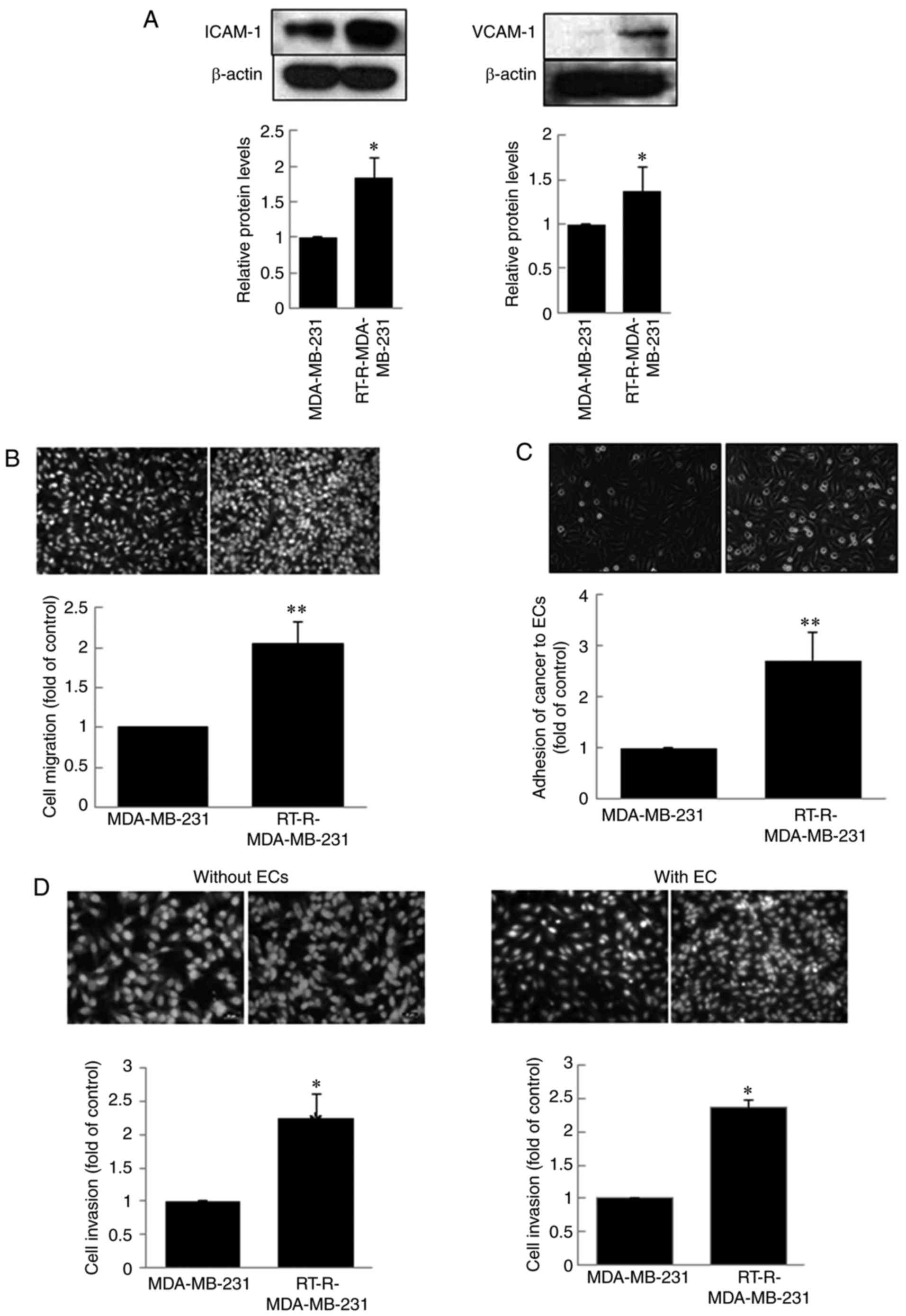 | Figure 4.RT-R-MDA-MB-231 cells exhibit higher
expression levels of ICAM-1 and VCAM-1, resulting in enhanced
migration, adhesion to ECs and invasion through ECs. (A) Protein
was extracted from the cells, and western blotting was performed to
detect ICAM-1 and VCAM-1 expression. (B) MDA-MB-231 and
RT-R-MDA-MB-231 cells were seeded into cell culture inserts. After
24 h, the cancer cells that had migrated across the insert well
membrane were stained with DAPI, counted under a fluorescence
microscope and quantified (×200 magnification). (C) MDA-MB-231 and
RT-R-MDA-MB-231 cells were cultured with ECs. After 30 min, the
remaining cell suspension was withdrawn, and the number of adherent
cells was counted under a light microscope and quantified (×200
magnification). (D) MDA-MB-231 and RT-R-MDA-MB-231 cells were
seeded onto the Matrigel-coated insert wells or EC-Matrigel-coated
insert wells in serum-free media, and the rest of the procedure was
performed as described for the Migration assay. Data represent mean
values ± standard error of the mean of three independent
experiments. *P<0.05 and **P<0.01 compared with MDA-MB-231
cells. EC, endothelial cell; ICAM-1, intercellular adhesion
molecule-1; RT-R, radioresistant; VCAM-1, vascular cell adhesion
molecule-1. |
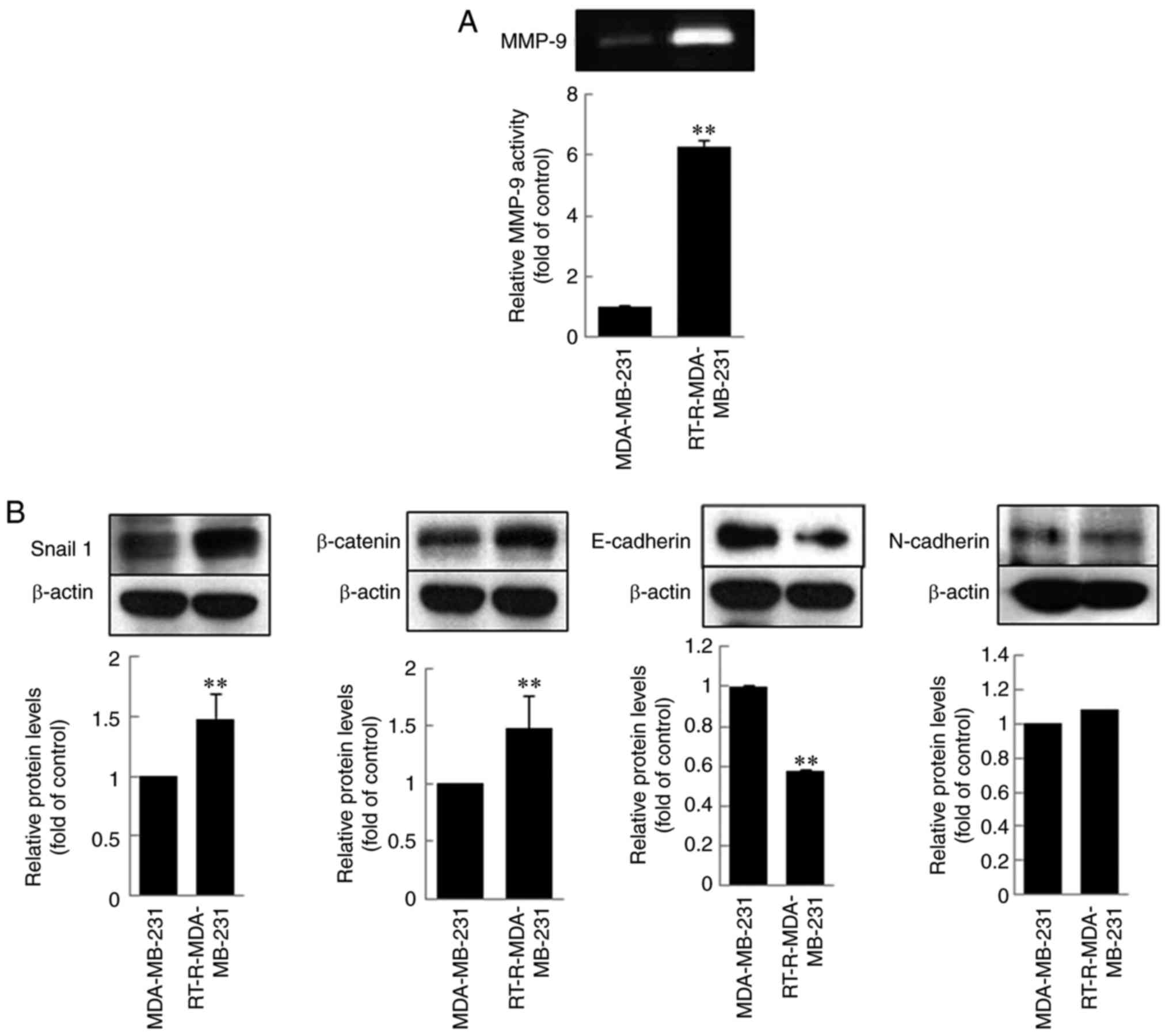 | Figure 5.RT-R-MDA-MB-231 cells exhibit higher
MMP-9 activity, and levels of EMT proteins, Snail-1 and β-catenin,
but decreased levels of E-cadherin. (A) Cells were serum-starved
overnight, and MMP gelatinase activities were determined from the
conditioned media by zymography (MMP-9; 92 kDa). (B) Cell lysates
were obtained from MDA-MB-231 and RT-R-MDA-MB-231 cells, and the
protein levels of Snail, N-cadherin, E-cadherin and β-catenin were
determined by western blot analysis. Data are presented as the mean
values ± standard error of the mean of three independent
experiments. **P<0.01 compared with MDA-MB-231 cells. EMT,
epithelial-mesenchymal transition; MMP-9, matrix
metalloproteinase-9; RT-R, radioresistant. |
Discussion
Radiotherapy is an important treatment option in
modern cancer therapy besides surgery and systemic therapy;
currently, >60% of all patients with cancer receive radiotherapy
(1,23). Unfortunately, tumor recurrence
following radiotherapy is common, with numerous causes for
radiotherapy failure and cancer recurrence, including metastasis
(23). One underlying cause of
tumor radioresistance is the existence of CSCs (13,15).
Indeed, preclinical data suggest that breast CSCs are enriched
following radiation and are particularly resistant to radiation
(10), as well as potentially to
other cancer therapies. Accordingly, the present study aimed to
determine whether RT-R-MDA-MB-231 breast cancer cells, which are
highly metastatic, harbor a larger CSC population, compared with
other low metastatic breast cancer cells. In addition, the current
study aimed to clarify whether CSCs, which exist among
RT-R-MDA-MB-231 cells, are responsible for the increased
invasiveness and resistance to cancer therapies, and if so, the
possible mechanisms for this behavior.
In the present study, it was demonstrated that
RT-R-MDA-MB-231 derived from highly metastatic breast cancer cells
expressed significantly higher levels of CD44 compared with low
metastatic breast cancer MDA-MB-231 or RT-R-MCF-7 cells, and
significantly increased the expression of other CSCs markers,
including Notch, Oct3/4 and ALDH1. These results suggested that
RT-R-MDA-MB-231 cells possessed more CSCs. In addition, the results
revealed that RT-R-breast cancer cells, derived through repeated
irradiation, exhibited significantly increased colony forming
abilities compared with the parental breast cancer cells. Notably,
RT-R-MDA-MB-231 cells were more resistant to paclitaxel treatment
compared with RT-R-MCF-7 or RT-R-T47D cells. There is controversy
regarding the use of ALDH1 as a breast CSCs marker. While Resetkova
et al (24) reported that no
significant increase in ALDH-1-positive cells following neoadjuvant
chemotherapy in surgical specimens, other researchers (25–27),
including Tanei et al (22)
reported that ALDH1 was a more significant predictive marker
compared with CD44+/CD24− for the
identification of breast CSCs in terms of chemotherapy resistance.
In the present study, ALDH1 levels were significantly increased in
RT-R-MDA-MB-231 cells compared with MDA-MB-231 cells, and ALDH1 was
not expressed in MCF-7 and T47D cells, even in RT-R-MCF-7 and
RT-R-T47D cells (data not shown), suggesting that ALDH1 may be a
potent marker of BCSCs, and ALDH1-positive breast CSCs may serve an
important role in radioresistance.
As aforementioned, AMs, including ICAM-1 and VCAM-1,
mediate cell migration and adhesion, resulting in cancer
recurrence, invasion and the development of distant metastases.
Thus, RT-R-MDA-MB-231 and MDA-MB-231 cells were compared in terms
of AM expression, cell migration, adhesion to ECs and invasion
through ECs. RT-R-MDA-MB-231 cells exhibited significantly
increased expression of ICAM-1 and VCAM-1, resulting in enhanced
migration and adhesion to ECs compared with MDA-MB-231 cells. In
addition, RT-R-MDA-MB-231 cells demonstrated significantly
increased invasion through ECs and expression of EMT-associated
proteins, including MMP-9, Snail-1 and β-catenin. According to the
reports, MMPs are well known factors that mediate invasion through
ECM remodeling (28,29), and induction of EMT promotes tumor
cell motility and invasion, potentially contributing to treatment
resistance (30–34). In addition, it has been proposed
that CSCs in primary tumors can metastasize to distant tissues or
organs and form metastatic colonies via EMT (13). Therefore, it is suggested that CSCs
in RT-R-MDA-MB-231 cells may promote invasion through AM expression
and EMT induction.
Taken together, the results of the current study
suggested that RT-R-breast cancer cells exhibited an increased
population of CSCs. In particular, RT-R-MDA-MB-231 cells derived
from highly metastatic breast cancer cells produced more CSCs,
which leads to the acquisition of resistance to other cancer
therapies besides radiotherapy. Furthermore, the results indicated
the possible mechanisms for the increase in invasiveness of
RT-R-MDA-MB-231 cells. CSCs that exist among RT-R-MDA-MB-231 cells
contribute to enhanced invasiveness by increasing cancer cell
migration, adhesion to ECs and invasion through ECs by promoting
EMT via the upregulation of the expression of AMs and
EMT-associated proteins (Fig. 6).
Therefore, it is suggested that a multi-targeted approach against
CSCs in combination with classic chemotherapy should be developed
to reduce breast cancer resistance and relapse rates.
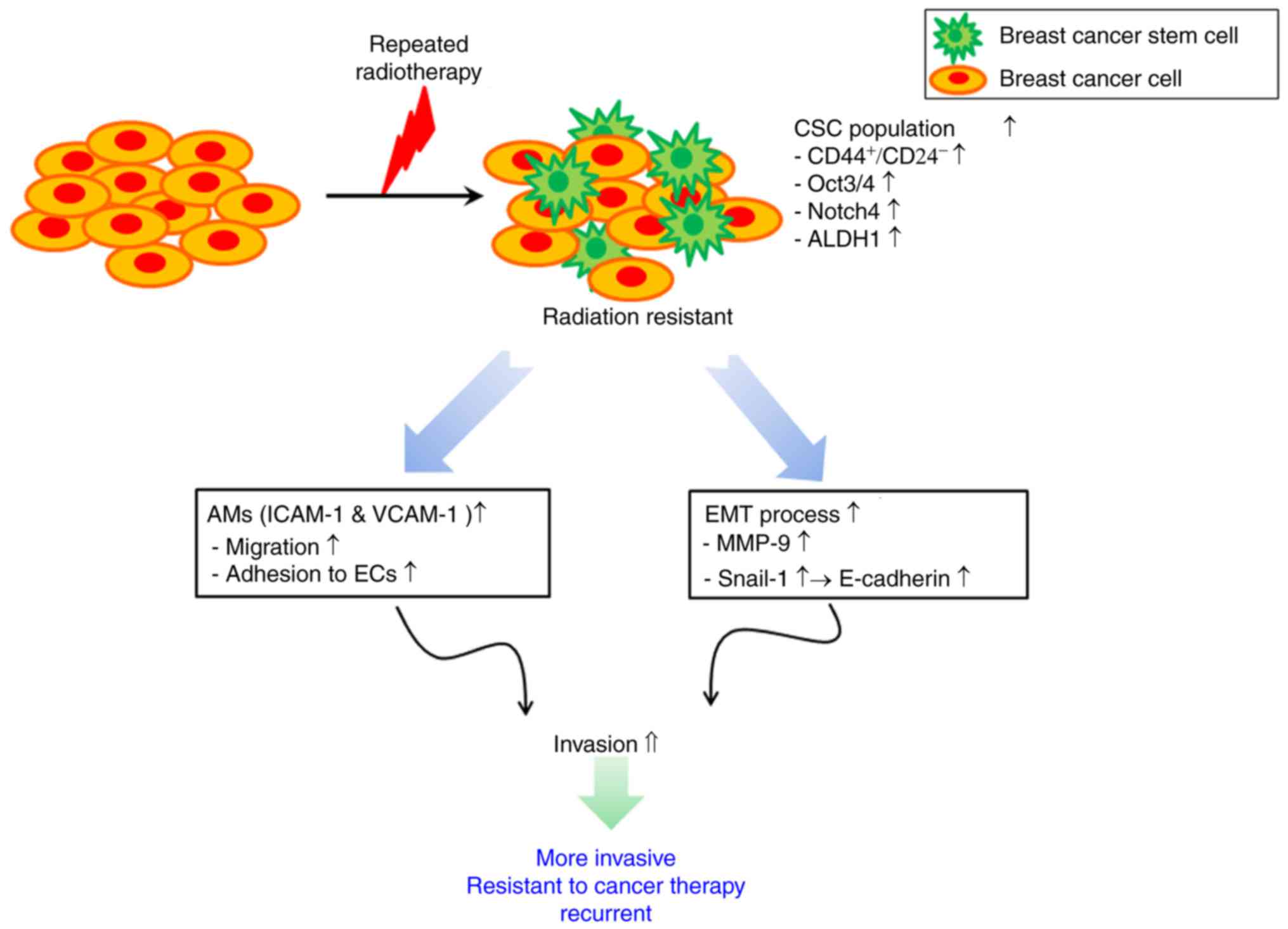 | Figure 6.Schematic representation of the
proposed role cancer stem cells on tumor growth and invasiveness in
the radioresistant breast cancer cells. AM, adhesion molecule;
ALDH1, aldehyde dehydrogenase 1; CD, cluster of differentiation;
CSC, cancer stem cell; ECs, endothelial cells; EMT,
epithelial-mesenchymal transition; ICAM-1, intercellular adhesion
molecule-1; MMP, matrix metalloproteinase; Notch, neurogenic locus
notch homolog; Oct, octamer-binding transcription factor; VCAM-1,
vascular cell adhesion molecule-1. |
Acknowledgements
Not applicable.
Funding
The present study was supported by Basic Science
Research Program through the National Research Foundation of Korea
funded by the Ministry of Education, Science and Technology (grant
no. NRF-2015R1A1A3A04001029) and by the Ministry of Science, ICT
and Future Planning (grant no. NRF-2015R1A5A2008833).
Availability of data and materials
The datasets used during the present study are
available from the corresponding author upon reasonable
request.
Authors' contributions
YSK performed the experiments and statistical
analyses; HJ performed the experiments and revised the manuscript;
JSL performed data analysis and helped with the interpretation of
data; SWP and KCC analysed the data and revised the manuscript
critically; KMK developed the methodology and discussed the data;
BKJ designed the study, developed the methodology and directed the
project; HJK designed the study, conceived the hypothesis, directed
the project and wrote the manuscript.
Ethics approval and consent to
participate
Not applicable.
Patient consent for publication
Not applicable.
Competing interests
The authors declare that they have no competing
interests.
Glossary
Abbreviations
Abbreviations:
|
ALDH1
|
aldehyde dehydrogenase 1
|
|
BCSC
|
breast cancer stem cell
|
|
BMDCs
|
bone marrow-derived dendritic
cells
|
|
CAM
|
cell adhesion molecules
|
|
DAPI
|
4′,6-diamidino-2-phenylindole
dihydrochloride
|
|
EC
|
endothelial cell
|
|
ECM
|
extracellular matrix
|
|
EMT
|
epithelial-mesenchymal transition
|
|
FBS
|
fetal bovine serum
|
|
SDS-PAGE
|
sodium dodecyl sulfate polyacrylamide
gel electrophoresis
|
|
SEM
|
standard error of the mean
|
|
RT-R
|
radioresistant
|
References
|
1
|
Torre LA, Bray F, Siegel RL, Ferlay J,
Lortet-Tieulent J and Jemal A: Global cancer statistics, 2012. CA
Cancer J Clin. 65:87–108. 2015. View Article : Google Scholar : PubMed/NCBI
|
|
2
|
Steeg PS: Tumor metastasis: Mechanistic
insights and clinical challenges. Nat Med. 12:895–904. 2006.
View Article : Google Scholar : PubMed/NCBI
|
|
3
|
Steeg PS: Cancer: Micromanagement of
metastasis. Nature. 449:671–673. 2007. View
Article : Google Scholar : PubMed/NCBI
|
|
4
|
Eccles SA and Welch DR: Metastasis: Recent
discoveries and novel treatment strategies. Lancet. 369:1742–1757.
2007. View Article : Google Scholar : PubMed/NCBI
|
|
5
|
Clevers H: The cancer stem cell: Premises,
promises and challenges. Nat Med. 17:313–319. 2011. View Article : Google Scholar : PubMed/NCBI
|
|
6
|
Sato R, Semba T, Saya H and Arima Y:
Concise review: Stem cells and epithelial-mesenchymal transition in
cancer: Biological implications and therapeutic targets. Stem
Cells. 34:1997–2007. 2016. View Article : Google Scholar : PubMed/NCBI
|
|
7
|
Clarke MF, Dick JE, Dirks PB, Eaves CJ,
Jamieson CH, Jones DL, Visvader J, Weissman IL and Wahl GM: Cancer
stem cells-perspectives on current status and future directions:
AACR workshop on cancer stem cells. Cancer Res. 66:9339–9344. 2006.
View Article : Google Scholar : PubMed/NCBI
|
|
8
|
Bertolini G, Roz L, Perego P, Tortoreto M,
Fontanella E, Gatti L, Pratesi G, Fabbri A, Andriani F, Tinelli S,
et al: Highly tumorigenic lung cancer CD133+ cells
display stem-like features and are spared by cisplatin treatment.
Proc Natl Acad Sci USA. 106:16281–16286. 2009. View Article : Google Scholar : PubMed/NCBI
|
|
9
|
Creighton CJ, Li X, Landis M, Dixon JM,
Neumeister VM, Sjolund A, Rimm DL, Wong H, Rodriguez A,
Herschkowitz JI, et al: Residual breast cancers after conventional
therapy display mesenchymal as well as tumor-initiating features.
Proc Natl Acad Sci USA. 106:13820–13825. 2009. View Article : Google Scholar : PubMed/NCBI
|
|
10
|
Li X, Lewis MT, Huang J, Gutierrez C,
Osborne CK, Wu MF, Hilsenbeck SG, Pavlick A, Zhang X, Chamness GC,
et al: Intrinsic resistance of tumorigenic breast cancer cells to
chemotherapy. J Natl Cancer Inst. 100:672–679. 2008. View Article : Google Scholar : PubMed/NCBI
|
|
11
|
Phillips TM, McBride WH and Pajonk F: The
response of CD24(−/low)/CD44+ breast cancer-initiating
cells to radiation. J Natl Cancer Inst. 98:1777–1785. 2006.
View Article : Google Scholar : PubMed/NCBI
|
|
12
|
Abraham BK, Fritz P, McClellan M,
Hauptvogel P, Athelogou M and Brauch H: Prevalence of
CD44+/CD24−/low cells in breast cancer may
not be associated with clinical outcome but may favor distant
metastasis. Clin Cancer Res. 11:1154–1159. 2005.PubMed/NCBI
|
|
13
|
Nizamutdinova IT, Lee GW, Lee JS, Cho MK,
Son KH, Jeon SJ, Kang SS, Kim YS, Lee JH, Seo HG, et al: Tanshinone
I suppreses growth and invasion of human breast cancer cells,
MDA-MB-231, through regulation of adhesion molecules.
Carcinogenesis. 29:1885–1892. 2008. View Article : Google Scholar : PubMed/NCBI
|
|
14
|
Jin H, Eun SY, Lee JS, Park SW, Lee JH,
Chang KC and Kim HJ: P2Y2 receptor activation by nucleotides
released from highly metastatic breast cancer cells increases tumor
growth and invasion via crosstalk with endothelial cells. Breast
Cancer Res. 16:R772014. View
Article : Google Scholar : PubMed/NCBI
|
|
15
|
Gupta PB, Chaffer CL and Weinberg RA:
Cancer stem cells: Mirage or reality? Nat Med. 15:1010–1012. 2009.
View Article : Google Scholar : PubMed/NCBI
|
|
16
|
Rosen JM and Jordan CT: The increasing
complexity of the cancer stem cell paradigm. Science.
324:1670–1673. 2009. View Article : Google Scholar : PubMed/NCBI
|
|
17
|
Rycaj K and Tang DG: Cancer stem cells and
radioresistance. Int J Radiat Biol. 90:615–621. 2014. View Article : Google Scholar : PubMed/NCBI
|
|
18
|
Al-Hajj M, Wicha MS, Benito-Hernandez A,
Morrison SJ and Clarke MF: Prospective identification of
tumorigenic breast cancer cells. Proc Natl Acad Sci USA.
100:3983–3988. 2003. View Article : Google Scholar : PubMed/NCBI
|
|
19
|
Ponti D, Costa A, Zaffaroni N, Pratesi G,
Petrangolini G, Coradini D, Pilotti S, Pierotti MA and Daidone MG:
Isolation and in vitro propagation of tumorigenic breast cancer
cells with stem/progenitor cell properties. Cancer Res.
65:5506–5511. 2005. View Article : Google Scholar : PubMed/NCBI
|
|
20
|
Koo BS, Lee SH, Kim JM, Huang S, Kim SH,
Rho YS, Bae WJ, Kang HJ, Kim YS, Moon JH and Lim YC: Oct4 is a
critical regulator of stemness in head and neck squamous carcinoma
cells. Oncogene. 34:2317–2324. 2015. View Article : Google Scholar : PubMed/NCBI
|
|
21
|
Tsai YH, VanDussen KL, Sawey ET, Wade AW,
Kasper C, Rakshit S, Bhatt RG, Stoeck A, Maillard I, Crawford HC,
et al: ADAM10 regulates Notch function in intestinal stem cells of
mice. Gastroenterology. 147:822–834.e13. 2014. View Article : Google Scholar : PubMed/NCBI
|
|
22
|
Tanei T, Morimoto K, Shimazu K, Kim SJ,
Tanji Y, Taguchi T, Tamaki Y and Noguchi S: Association of breast
cancer stem cells identified by aldehyde dehydrogenase 1 expression
with resistance to sequential Paclitaxel and epirubicin-based
chemotherapy for breast cancers. Clin Cancer Res. 15:4234–4241.
2009. View Article : Google Scholar : PubMed/NCBI
|
|
23
|
Begg A, Stewart F and Vens C: Strategies
to improve radiotherapy with targeted drugs. Nat Rev Cancer.
11:239–253. 2011. View
Article : Google Scholar : PubMed/NCBI
|
|
24
|
Resetkova E, Reis-Filho JS, Jain RK, Mehta
R, Thorat MA, Nakshatri H and Badve S: Prognostic impact of ALDH1
in breast cancer: A story of stem cells and tumor microenvironment.
Breast Cancer Res Treat. 123:97–108. 2010. View Article : Google Scholar : PubMed/NCBI
|
|
25
|
Ginestier C, Hur MH, Charafe-Jauffret E,
Monville F, Dutcher J, Brown M, Jacquemier J, Viens P, Kleer CG,
Liu S, et al: ALDH1 is a marker of normal and malignant human
mammary stem cells and a predictor of poor clinical outcome. Cell
Stem Cell. 1:555–567. 2007. View Article : Google Scholar : PubMed/NCBI
|
|
26
|
Morimoto K, Kim SJ, Tanei T, Shimazu K,
Tanji Y, Taguchi T, Tamaki Y, Terada N and Noguchi S: Stem cell
marker aldehyde dehydrogenase 1-positive breast cancers are
characterized by negative estrogen receptor, positive human
epidermal growth factor receptor type 2, and high Ki67 expression.
Cancer Sci. 100:1062–1068. 2009. View Article : Google Scholar : PubMed/NCBI
|
|
27
|
Charafe-Jauffret E, Ginestier C, Iovino F,
Tarpin C, Diebel M, Esterni B, Houvenaeghel G, Extra JM, Bertucci
F, Jacquemier J, et al: Aldehyde dehydrogenase 1-positive cancer
stem cells mediate metastasis and poor clinical outcome in
inflammatory breast cancer. Clin Cancer Res. 16:45–55. 2010.
View Article : Google Scholar : PubMed/NCBI
|
|
28
|
Blood CH and Zetter BR: Tumor interactions
with the vasculature: Angiogenesis and tumor metastasis. Biochim
Biophys Acta. 1032:89–118. 1990.PubMed/NCBI
|
|
29
|
Herren B, Levkau B, Raines EW and Ross R:
Cleavage of beta-catenin and plakoglobin and shedding of
VE-cadherin during endothelial apoptosis: Evidence for a role for
caspases and metalloproteinases. Mol Biol Cell. 9:1589–1601. 1998.
View Article : Google Scholar : PubMed/NCBI
|
|
30
|
Chang HY, Nuyten DS, Sneddon JB, Hastie T,
Tibshirani R, Sørlie T, Dai H, He YD, van't Veer LJ, Bartelink H,
et al: Robustness, scalability, and integration of a wound response
gene expression signature in predicting breast cancer survival.
Proc Natl Acad Sci USA. 102:3738–3743. 2005. View Article : Google Scholar : PubMed/NCBI
|
|
31
|
Mani SA, Guo W, Liao MJ, Eaton EN, Ayyanan
A, Zhou AY, Brooks M, Reinhard F, Zhang CC, Shipitsin M, et al: The
epithelial mesenchymal transition generates cells with properties
of stem cells. Cell. 133:704–715. 2008. View Article : Google Scholar : PubMed/NCBI
|
|
32
|
Morel AP, Lievre M, Thomas C, Hinkal G,
Ansieau S and Puisieux A: Generation of breast cancer stem cells
through epithelial-mesenchymal transition. PLoS One. 3:e28882008.
View Article : Google Scholar : PubMed/NCBI
|
|
33
|
Hennessy BT, Gonzalez-Angulo AM,
Stemke-Hale K, Gilcrease MZ, Krishnamurthy S, Lee JS, Fridlyand J,
Sahin A, Agarwal R, Joy C, et al: Characterization of a naturally
occurring breast cancer subset enriched in
epithelial-to-mesenchymal transition and stem cell characteristics.
Cancer Res. 69:4116–4124. 2009. View Article : Google Scholar : PubMed/NCBI
|
|
34
|
Aktas B, Tewes M, Fehm T, Hauch S, Kimmig
R and Kasimir-Bauer S: Stem cell and epithelial-mesenchymal
transition markers are frequently overexpressed in circulating
tumor cells of metastatic breast cancer patients. Breast Cancer
Res. 11:R462009. View Article : Google Scholar : PubMed/NCBI
|