Introduction
The 5-year survival rate for colorectal cancer is
undeniably increasing in many developed countries; for patients
diagnosed with colon cancer and rectal cancer in 22 countries
worldwide, it was 50% in the past (1995–1999) and has now reached
over 60%, according to data from 2005 to 2009 (1). Improvements in surgical techniques for
resectable cases of colorectal cancer and the development of
multidisciplinary treatments, particularly the introduction of
chemotherapy with molecular targeted therapeutic agents against
progressive or recurrent unresectable colorectal cancer, have
contributed to the improved survival rates. In recent years,
efforts have been actively made in the field of colorectal cancer
toward developing precision medicine to further improve the
prognosis, search for and identify novel biomarkers, and develop
new medicines including molecular targeted therapeutic drugs. In
particular, in recent years, elucidation of the genetic mutations
responsible for the development of colorectal cancer has been
achieved. However, with respect to RAS (KRAS/NRAS)/RAF mutation,
the positive effect of the anti-EGFR antibody could not be
determined (2).
Based on these findings, it is indispensable to
perform genetic analysis of RAS/RAF as a biomarker for predicting
the effect of anti-EGFR antibody drugs, when performing systemic
chemotherapy for unresectable and recurrent colorectal cancer.
Y-box-binding protein 1 (YB-1) is a transcription
factor that contains a cold shock domain highly conserved across
species, and binds to the Y-box (CCAAT) region in the promoter
domain of the major histocompatibility complex II gene HLA-DRα
(3) and the gene encoding the EGFR
enhancer (4). It has been reported
that YB-1 plays a crucial role in cell proliferation, invasion,
metastasis and drug resistance of cancer cells through the actions
of transcription, translational control and DNA repair (5).
Previous studies have reported that YB-1 expression
is a poor prognostic factor in gastric (6), cervical (7), non-small cell lung (8) and breast cancer (9). In a previous study, we also
demonstrated that the nuclear expression of YB-1 in colorectal
tumor cells led to a poor prognosis (10).
A significant correlation between HER2 expression
and YB-1 expression exists in estrogen receptor (ER)-positive
breast cancer, with the two elements being involved in tumor growth
and drug resistance. As a result, YB-1 expression in breast cancer
is reported to be a prognostic factor for a worse outcome (11). Furthermore, the co-expression of
EGFR and YB-1 can modulate the effect of the anti-EGFR antibody as
a treatment in non-small cell lung cancer (NSCLC) (8). Even in colorectal cancer, YB-1
expression has been correlated with poor prognosis (12,13);
however, the biological function and molecular correlation of YB-1
in colorectal cancer are not fully understood. The objective of the
present study was to elucidate the importance and biological role
of YB-1 in colorectal cancer at the molecular level.
Materials and methods
Cell culture
Five human colon cancer cell lines, T84, HT29,
HCT116, CaCo2 and SW480 were purchased from the American Type
Culture Collection (ATCC; Manassas, VA, USA). Next, the expression
of YB-1 and the mutational status of RAS/RAF were examined. Based
on these results, three cell lines, namely CaCo2, HCT116 and HT29
were selected and used for further experiments. All cells were
cultured in Roswell Park Memorial Institute (RPMI)-1640 medium
supplemented with 10% fetal bovine serum (FBS) (Fujifilm Wako Pure
Chemical Industries, Ltd., Osaka, Japan), 100 U/ml penicillin and
100 mg/l streptomycin. The cells were maintained in a humidified
incubator at 37°C with 5% CO2 (Sanyo Electric Biomedical
Co., Osaka, Japan).
Mutation analysis
For the detection of KRAS, NRAS and
BRAF mutations, genomic DNA was isolated from the cell
lines, using an AllPrep DNA/RNA/Protein Mini kit (Qiagen, Inc.,
Valencia, CA, USA) according to the manufacturer's protocol. The
genomic DNA was subjected to polymerase chain reaction (PCR)
amplification using the primers listed in Table I. The primer sets were designed to
amplify exons 2, 3 and 4 of KRAS and NRAS and exon 15
of BRAF. The PCR products were treated with ExoSap-IT PCR
Product Cleanup reagent (Thermo Fisher Scientific, Inc., Waltham,
MA, USA) according to the manufacturer's protocol to inactivate the
free primers and dNTPs, and then were subjected to sequencing using
the forward primers and BigDye® Terminator v3.1 (Thermo
Fisher Scientific, Inc.). Each 20-µl PCR reaction consisted of 2 µl
10X LA PCR buffer II, 2 µl 10 mmol/l dNTPs, 0.1 µl AmpliTaq Gold
(Thermo Fisher Scientific, Inc.), 2 µl of genomic DNA, 1 µl 100
pmol/µl forward primer, 1 µl 100 pmol/µl reverse primer and 12 µl
H2O. The cycling conditions were 95°C for 12 min; 10
cycles at 94°C for 15 sec, 55°C for 15 sec, and 72°C for 30 sec; 25
cycles at 89°C for 15 sec, 55°C for 15 sec, 72°C for 30 sec; and a
final extension at 72°C for 10 min. The PCR products were evaluated
by electrophoresis using 2% agarose gels. Sequencing was carried
out using an ABI 3100 Genetic Analyzer (Thermo Fisher Scientific,
Inc.) according to the manufacturer's protocol. The sequences
obtained were aligned to the consensus reference-sequences from the
University of California, Santa Cruz (UCSC) Genome Bioinformatics
website (http://genome.ucsc.edu/index.html) using the ClustalW
program (https://www.genome.jp/tools-bin/clustalw) to identify
nucleotide differences.
 | Table I.Primer sequences for the quantitative
polymerase chain reaction. |
Table I.
Primer sequences for the quantitative
polymerase chain reaction.
|
| Primers | Expected size
(bp) |
|---|
| KRAS-ex2-F |
5′-TTAACCTTATGTGTGACATGTTCTAA-3′ | 225 |
| KRAS-ex2-R |
5′-AGAATGGTCCTGCACCAGTAA-3′ |
|
| KRAS-ex3-F |
5′-CCAGACTGTGTTTCTCCCTTC-3′ | 286 |
| KRAS-ex3-R |
5′-TGCATGGCATTAGCAAAGAC-3′ |
|
| KRAS-ex4-F |
5′-AAGGACTCTGAAGATGTACCTATGG-3′ |
|
| KRAS-ex4-R |
5′-AAGAAGCAATGCCCTCTCAA-3′ | 294 |
| NRAS-ex2-F |
5′-GATGTGGCTCGCCAATTAAC-3′ | 220 |
| NRAS-ex2-R |
5′-CCGACAAGTGAGAGACAGGA-3′ |
|
| NRAS-ex3-F |
5′-CCCCTTACCCTCCACACC-3′ | 243 |
| NRAS-ex3-R |
5′-CACAAAGATCATCCTTTCAGAGAA-3′ |
|
| NRAS-ex4-F |
5′-AGCCACTGTACCCAGCCTAA-3′ | 250 |
| NRAS-ex4-R |
5′-TGCACAAATGCTGAAAGCTG-3′ |
|
| BRAF-ex15-F |
5′-TGCTTGCTCTGATAGGAAAATG-3′ | 228 |
| BRAF-ex15-R |
5′-AGCATCTCAGGGCCAAAAAT-3′ |
|
YB-1 knockdown
The small interfering RNAs (siRNAs) for YB-1
(siRNA-1: sense, 5′-GUAAAAUGGUUCAAUGUAAtt-3′ and antisense,
5′-UUACAUUGAACCAUUUUACtg-3′; and siRNA2: sense,
5′-CGAAGGUUUUGGGAACAGUtt-3′ and antisense,
5′-ACUGUUCCCAAAACCUUCGtt-3′) and the negative control siRNAs
(siCtr) were purchased from Life Technologies Corp. (Thermo Fisher
Scientific, Inc.). The siRNAs were transfected into the cells,
using Lipofectamine RNAiMAX Transfection reagent and Opti-MEM
medium (Invitrogen Life Technologies; Thermo Fisher Scientific,
Inc.) according to the manufacturer's recommendations. A total of
1×10⁴ cells/well were seeded in 96-well plates (Iwaki
Co., Ltd., Tokyo, Japan) and cultured for 24 h before transfection.
The adjusted amounts of siRNAs were added to each well. Reverse
transcription PCR (RT-PCR) and western blotting were performed 72 h
post-transfection, and MTT and migration assays were performed 48 h
post-transfection.
Quantitative real-time PCR
(RT-qPCR)
Total RNA was extracted using an AllPrep
DNA/RNA/Protein Mini kit (Qiagen, Inc.) according to the
manufacturer's protocol and reverse-transcribed into complementary
DNA (cDNA), using SuperScript IV Reverse Transcriptase (Promega
Corporation, Madison, WI, USA). RT-qPCR was performed with TaqMan
Gene Expression Assays (Applied Biosystems; Thermo Fisher
Scientific, Inc.) for YB-1 (Hs00358903_g1), GAPDH
(Hs02758991_g1), Bax (Hs00180269_m1) and Bcl-2
(Hs00608023_m1) using the TaqMan Gene Expression Master Mix
(Applied Biosystems; Thermo Fisher Scientific, Inc.) on a
StepOnePlus Real-Time PCR system (Applied Biosystems; Thermo Fisher
Scientific, Inc.). The levels of transcripts of the indicated genes
were standardized to the corresponding GAPDH transcript
levels. All values reported represent the average of at least three
independent experiments.
Western blotting
Total cell proteins were extracted using an AllPrep
DNA/RNA/Protein Mini kit (Qiagen, Inc.) according to the
manufacturer's protocol. Protein determination method is BCA. Mass
of protein loaded per lane is 20 µg. Proteins were separated by
sodium dodecyl sulfate-polyacrylamide gel electrophoresis SDS-PAGE
(4–12%) and transferred to polyvinylidene difluoride (PVDF)
membranes. After blocking with 5% milk in Tris-buffered saline
containing Tween-20 (TBST) buffer, the membranes were incubated at
4°C overnight with an anti-YB-1 (dilution 1:1,000; rabbit; cat. no.
ab12148; Abcam, Cambridge, MA, USA) or anti-EGFR (dilution 1:1,000;
rabbit; cat. no. 4267; Cell Signaling Technology, Inc., Danvers,
MA, USA) primary antibody. Subsequently, the membranes were
incubated for 1 h at room temperature with horseradish peroxidase
(HRP)-conjugated goat anti-rabbit IgG secondary antibody (dilution
1:5,000; cat. no. 7074; Cell Signaling Technology, Inc.). The
immunoreactive bands were visualized using Fusion-SL7-400 (Vilber
Lourmat, Marne Le-Vallée, France). Immunostaining using an
anti-β-actin primary antibody (dilution 1:5,000; mouse; cat. no.
ab6276; Abcam) was used as a loading control.
MTT assay
For the
3-(4,5-dimethylthiazol-2-yl)-2,5-diphenyltetrazolium bromide)
tetrazolium (MTT) reduction assays, the three colorectal cell lines
were used as either untreated (CaCo2, HT29 and HCT116), treated
with siYB-1 (CaCo2-siYB-1, HT29-siYB-1 and HCT116-siYB-1), or
treated with siCtr (CaCo2-siCtr, HT29-siCr and HCT116-siCtr). The
cells were added to 96-well plates (1×10⁴ cells/well)
and incubated for 2 days in a humidified incubator at 37°C with 5%
CO2. MTT Cell Proliferation Assay Reagent (Cell Biolabs,
Inc., San Diego, CA USA) was added (10 µl) to each well, and the
cells were incubated for 3 h at 37°C. Thereafter, the detergent
solution provided with the assay kit was added to each well (100
µl) and the cells were incubated for 3 h at room temperature while
being protected from the light. The absorbance of each sample for
each well was measured at 540 nm using a microplate reader
(VersaMax ELISA; Molecular Devices Japan K.K., Tokyo, Japan).
Migration assay
A total of 1×10⁴ CaCo2, CaCo2-siYB-1,
CaCo2-siCtr, HT29, HT29-siYB-1, HT29-siCtr, HCT116, HCT116-siYB-1,
or HCT116-siCtr cells/well were seeded into 96-well Collagen I
coated plates (Platypus Technologies, LCC, Fitchburg, WI, USA) and
incubated at 37°C to allow for cell attachment. After 24 h, all
silicon stoppers at the center of each well were removed, and any
unattached cells were removed by washing with 100 µl of sterile
phosphate-buffered saline (PBS). After 72 h, the number of migrated
cells was quantified using ImageJ software (ver. 1.51, NIH;
National Institutes of Health, Bethesda, MD, USA).
Statistical analysis
Each assay was independently performed three times.
The data were analyzed using JMP software (version 13.0.0; SAS
Institute Inc., Cary, NC, USA). The values were presented as the
means ± standard deviation (SD). The comparisons between groups
were analyzed using the Kruskal-Wallis test. P<0.05 was
considered to indicate a statistically significant difference.
Results
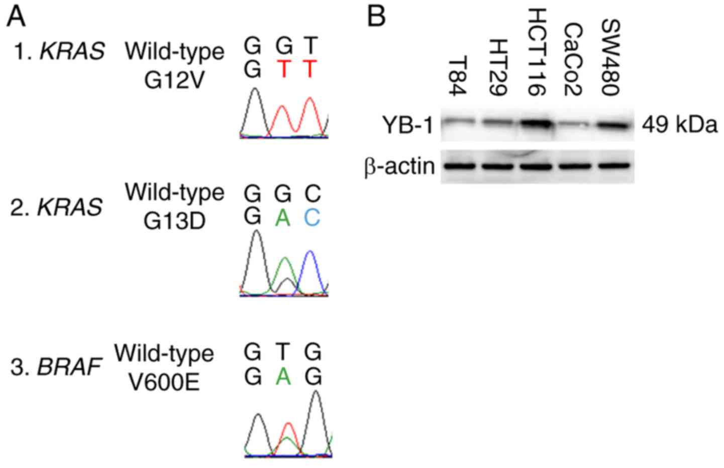
KRAS/NRAS/BRAF mutation profiles and
expression of YB-1 in human colon cancer cell lines
In colorectal cancer, the mutational status of
RAS markedly affects the characteristics of the cancer
cells. Therefore, in order to confirm the molecular background of
each of the five colon-cancer cell lines, we determined the genetic
mutation of RAS/RAF by direct sequence evaluation. A summary
of the KRAS/NRAS/BRAF gene mutations is presented in
Table II. The detected KRAS
mutations were G12V (GGT→GTT) and G13D (GGC→GAC), and the
BRAF mutation was V600E (GTG→GAG). The genotypes of the
mutant KRAS and mutant BRAF are displayed in Fig. 1A.
| Table II.KRAS/NRAS/BRAF mutation profiles of
cancer cell lines. |
Table II.
KRAS/NRAS/BRAF mutation profiles of
cancer cell lines.
|
| Cell line | Mutation |
|---|
| 1 | SW480 | G12V |
| 2 | HCT116 | G13D |
| 3 | T84 | G13D |
| 4 | CaCo2 | Wild-type |
| 5 | HT29 | V600E |
Subsequently, western blot analysis was performed to
investigate the levels of protein expression of YB-1 in the five
colon cancer cell lines. As revealed in Fig. 1B, the expression of YB-1 was
confirmed in all five cell lines. HCT116 demonstrated the highest
level of YB-1 expression among the cell lines. Notably, the
expression level of YB-1 tended to be higher in the cell lines
harboring the RAS/RAF mutation compared to that in the cell
line harboring wild-type RAS. To clarify the biological
function of YB-1 in colorectal cancer with respect to the
mutational status of the RAS, the CaCo2, HCT116 and HT29
cell lines were selected for further examination.
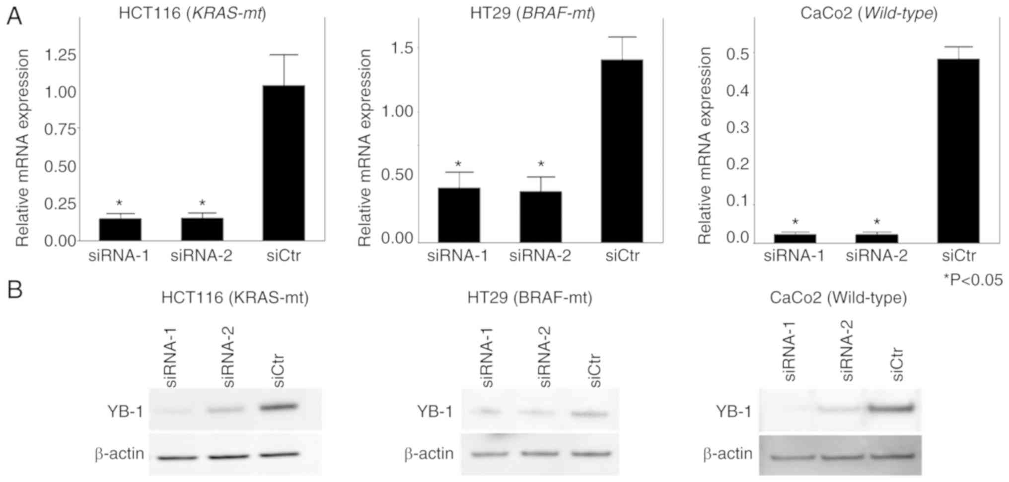
Knockdown of YB-1 in HCT16, HT29 and
CaCo2 cell lines inhibits cell proliferation and migration
Two siRNAs (siRNA-1 and siRNA-2) were transfected
into the cell lines HCT116, HT29 and CaCo2 in order to knockdown
YB-1. The suppression of YB-1 mRNA levels and protein levels were
confirmed by RT-qPCR and western blot analyses, respectively. As
revealed in Fig. 2A, the expression
level of YB-1 was significantly decreased in both
siRNA-1-transfected and siRNA-2-transfected cells compared to that
in siCtr-transfected cells (P<0.05). According to the RT-qPCR
analysis, the reduced level of YB-1 expression was not
significantly different between the siRNA-1-transfected and
siRNA-2-transfected groups. However, western blot analysis revealed
that the reduced level of YB-1 expression was higher in the
siRNA-1-transfected group compared to that in the
siRNA-2-transfected group (Fig.
2B). Based on these results, siRNA-1 was used in subsequent
in vitro experiments.
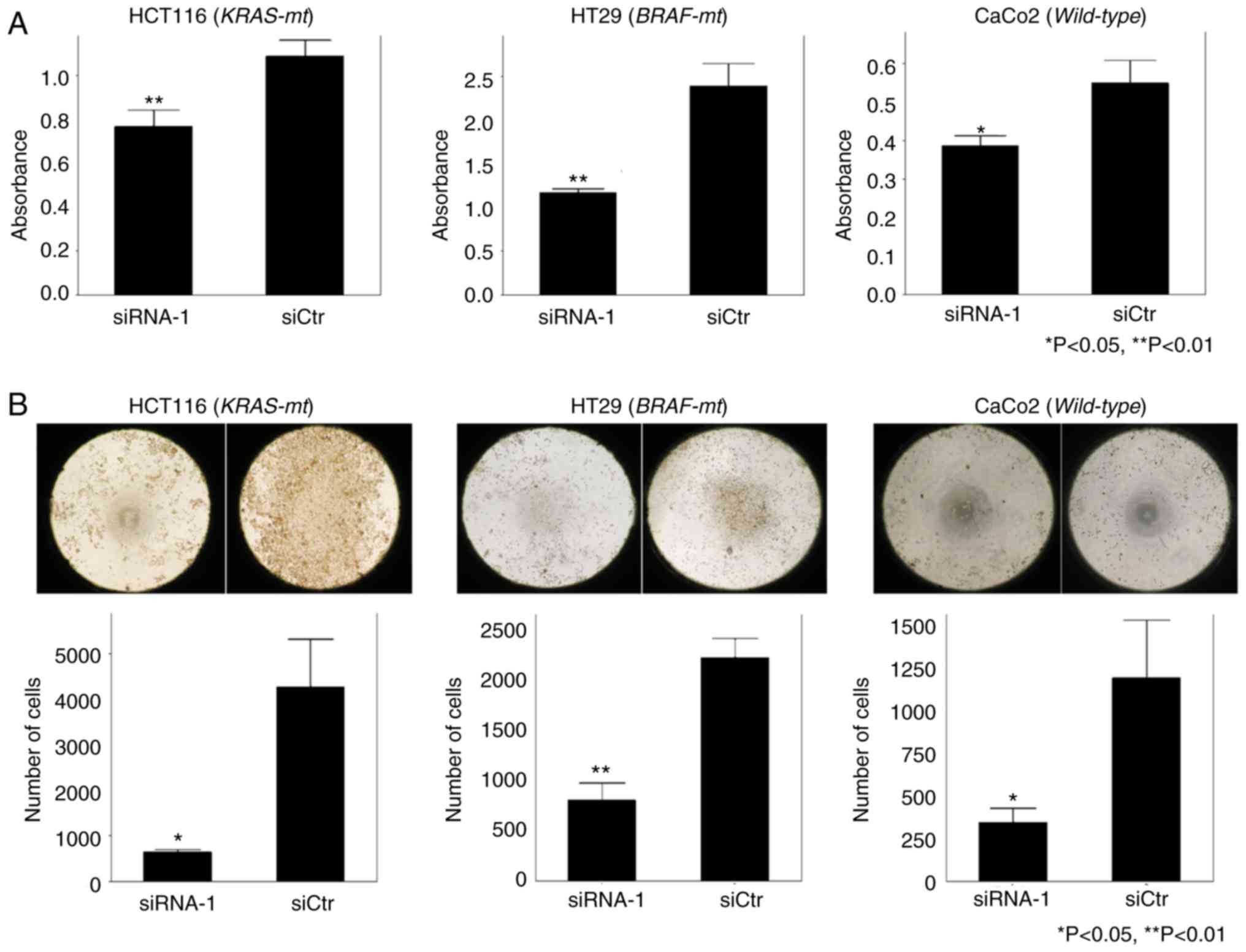
To investigate the role of YB-1 in cell
proliferation and migration, MTT assays and migration assays were
performed, respectively. In the MTT assays, proliferation potency
was significantly suppressed in all the cell lines transfected with
siRNA-1 compared to that in the siCtr-transfected group (Fig. 3A). Moreover, in the migration
assays, the number of cells transfected with si-RNA-1 that migrated
into the center of the well was significantly less than that of the
siCtr-transfected group (Fig. 3B).
Similar results were obtained from siRNA knockdown experiments
using the other cell lines. Overall, the effects of YB-1 on cell
proliferation and cell migration were not affected by the presence
or absence of mutations in KRAS or BRAF.
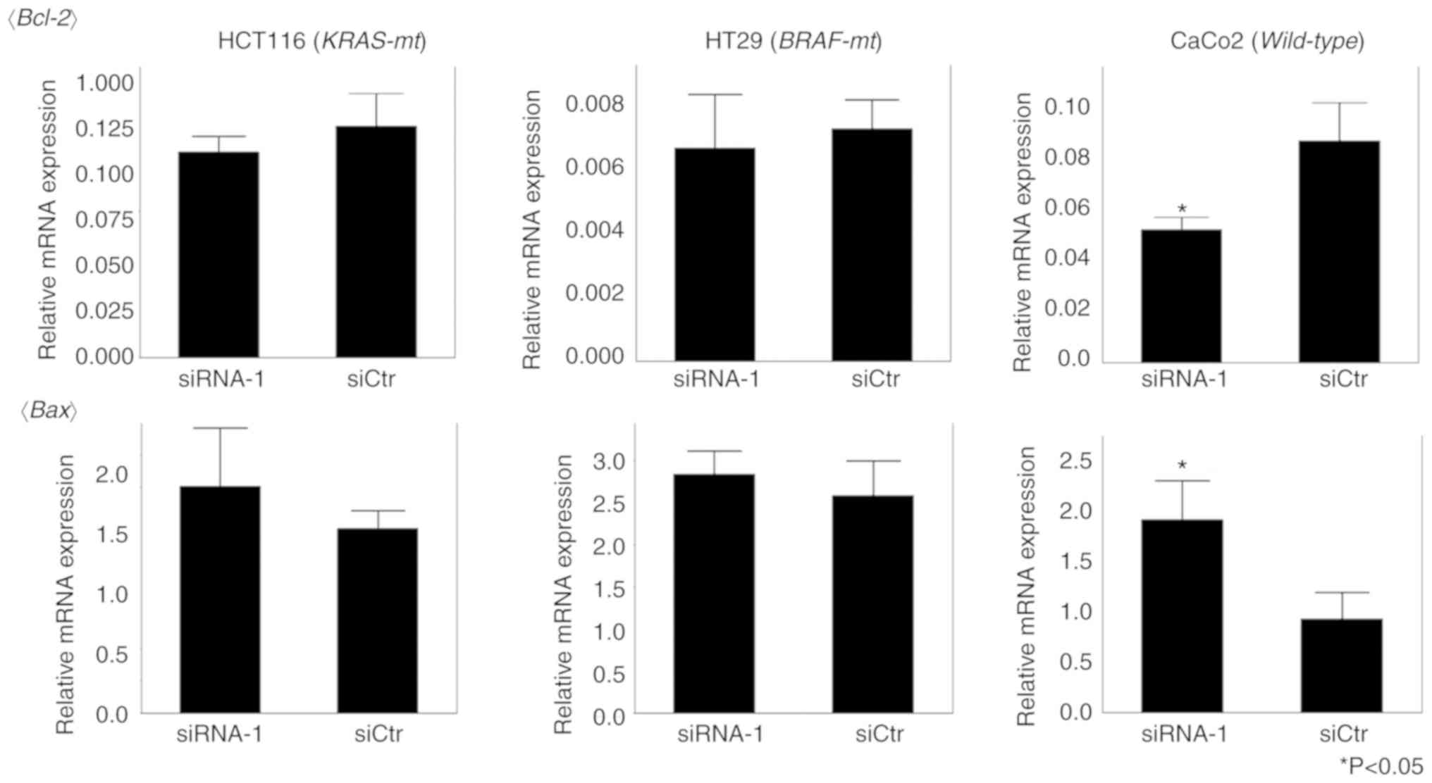
Knockdown of YB-1 induces
apoptosis-related genes in human colon cancer
Since there are few studies regarding the
relationship between YB-1 and apoptosis, the effect of YB-1 on
apoptosis was analyzed by evaluating alterations in the expression
of apoptosis-related genes Bax and Bcl-2 in
siRNA-1-treated cells compared to that in siCtr-treated cells, by
RT-qPCR analysis. There were no significant gene expression
differences between the siRNA-1-treated and siCtr-treated groups
for either HCT116 or HT29 cells. In contrast, Bcl-2
expression levels were significantly decreased (P=0.018) and
Bax levels were significantly increased (P=0.021) in
siRNA-1-treated CaCo2 cells compared to that in siCtr-treated CaCo2
cells (Fig. 4).
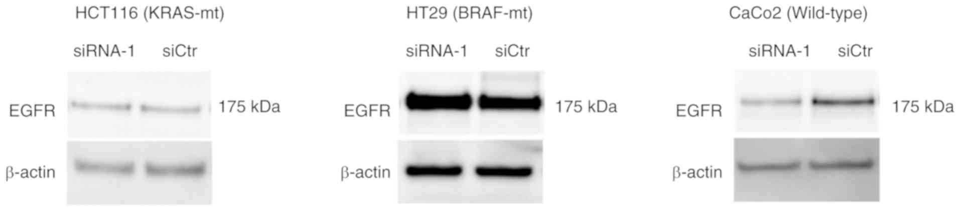
Knockdown of YB-1 reduces the
expression of EGFR in human colon cancer
EGFR protein expression was compared between the
siRNA-1-treated and siCtr-treated groups by western blot analysis.
There were no significant differences in EGFR expression between
the siRNA-1-treated and siCtr-treated groups for either HCT116 or
HT29 cells. However, EGFR expression was significantly decreased in
the siRNA-1-treated group of CaCo2 cells compared to that in
siCtr-treated CaCo2 cells (Fig.
5).
Discussion
Findings from the present study suggest that
Y-box-binding protein 1 (YB-1) was involved in promoting the
malignancy potential of colon cancer through the enhancement of
cell proliferation and -migration properties. Furthermore, YB-1
also contributed to the suppression of apoptosis and epidermal
growth factor receptor (EGFR) expression in colorectal cancer
cells. The alteration of apoptosis-related genes and EGFR
expression by YB-1 knockdown was confirmed by our results to occur
only in the cell lines expressing wild-type RAS/RAF and not
in those expressing mutated RAS/RAF. These results suggest
that the presence of the RAS/RAF mutation may affect the
suppression of apoptosis and promotion of EGFR expression by YB-1
in colorectal cancer.
Previous studies have reported that there is a clear
association between YB-1 expression and cell proliferation, and an
increase in the expression levels of YB-1 was correlated with the
expression of proliferation markers (14). In addition, there is a possible
relationship between YB-1 expression and hyperplasia (15).
Basaki et al (16) demonstrated that knockdown of YB-1
resulted in a reduction of the number of cells in the S phase for
multiple types of cancer, including breast, lung cancer and
leukemia. Furthermore, they indicated that downregulation of cyclin
D1 and upregulation of p21 occured as a result of YB-1 knockdown.
These investigators also reported that YB-1 was associated with the
cell-proliferation cycle (16). In
addition, Yan et al (17)
revealed that the downregulation of E-cadherin and the upregulation
of vimentin and N-cadherin occured in colon cancer cell lines
transfected with YB-1 and resulted in cancer cell
proliferation through the enhancement of epithelial-mesenchymal
transition (EMT).
The present study revealed that the knockdown of
YB-1 impaired cell proliferation and cell migration in colon cancer
cell lines, regardless of the status of RAS/RAF. While
additional studies are required, it is possible to conclude that
YB-1 is strongly involved in cancer cell proliferation and
mobility, independent of the type of cancer cell.
The knockdown of YB-1 in the colon cancer cell line
expressing wild-type RAS/RAF resulted in the upregulation of
Bax and downregulation of Bcl-2, which are both
associated with apoptosis. Similar results were observed in breast
(18), bladder cancer (19) and neuroblastoma (20). Our results may be explained by the
molecular relationship between YB-1 and p53, which selectively bind
with each other and cause the suppression of p53 function to induce
an anti-apoptotic effect. The enhancement of apoptosis by YB-1
knockdown was not observed in the colorectal cancer cell lines
HCT116 or HT29, which harbor mutated RAS/RAF.
In breast cancer, non-small cell lung and cervical
cancer, phosphorylation of YB-1 via the mitogen-activated protein
kinase (MAPK) pathway upregulated EGFR expression (7,21,22).
Previously, we reported that there is a positive correlation
between YB-1 expression and EGFR expression in colorectal cancer
tissue and high expression of both YB-1 and EGFR were associated
with poor prognosis (10).
In the present study, suppression of EGFR expression
by YB-1 knockdown was observed in the CaCo2 cell line, which
expresses wild-type RAS/RAF. It was considered that YB-1
regulated EGFR in colorectal cancer harboring wild-type
RAS/RAF, similar to that confirmed in other types of cancer.
However, the same phenomenon was not verified in the cell lines
HCT116 and HT29, which harbor the mutated RAS/RAF.
Our results were consistent with the promotion of
apoptosis and suppression of EGFR expression upon knockdown of YB-1
in colon cancer with wild-type RAS/RAF. In contrast, in
colorectal cancer cells harboring mutated RAS/RAF, there
were no significant differences in the promotion of apoptosis or
suppression of EGFR expression, despite the knockdown of YB-1.
In colorectal cancer, the expression of EGFR and
factors of the downstream MAPK pathway are involved in cancer
proliferation, invasion, metastasis and cell survival. In
RAS/RAF-mutated cell lines, the mutated proteins activate
the MAPK pathway independent of stimulation by EGFR, which help to
maintain cancer survival and proliferation (23). It has been reported that
extracellular signal-regulated kinases 1 and 2 (ERK 1/2) are
located downstream of the MAPK pathway and they suppress apoptosis
(24). Furthermore, in several
types of cancer, YB-1 functioned downstream of ERK 1/2 and promotes
proliferation, invasion and metastasis of cancer cells, resulting
in poor prognosis (25–27). Moreover, Chu et al (28) revealed that in colon cancer cells
with KRAS mutation, the expression of YB-1 was upregulated through
the MEK/Sp1/DNMT1/miR-137 pathway.
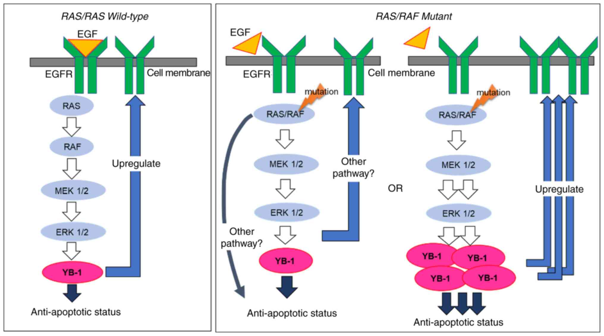
In colon cancer with wild-type RAS/RAF, it is
thought that YB-1 is activated via the MAPK pathway, which then
induces the suppression of apoptosis and upregulation of EGFR. On
the other hand, in RAS/RAF-mutated colorectal cancer, there
may be an alternative pathway not mediated by YB-1, or YB-1 may
exist in abundance due to constant expression activated by the MAPK
pathway. The regulation of YB-1 by siRNA may become insufficient
with the induction of apoptosis and the suppression of EGFR
expression not being achieved (Fig.
6). Based on these findings, YB-1 may be useful as a
therapeutic target in colon cancer harboring wild-type
RAS/RAF. Furthermore, there was a significant association
between the high expression of YB-1 and existence of mutated
RAS/RAF in colon cancer cell lines.
In conclusion, it is possible that YB-1 plays a
vital role in cell proliferation, cell migration, apoptosis and
EGFR expression in colon cancer. Furthermore, apoptosis and
expression of EGFR mediated through the control of YB-1 may be
affected by the mutational status of RAS/RAF.
Acknowledgments
We would like to thank Ms. Eriko Matsuo (Research
assistant at the Molecular Targeting Therapeutics Division, Kurume
University, Kurume, Japan) for research assistance in the present
study.
Funding
No funding was received.
Availability of data and materials
The datasets used during the present study are
available from the corresponding author upon reasonable
request.
Authors' contributions
SN designed the study and wrote the initial draft of
the manuscript. TSu contributed to the analysis and interpretation
of the data and assisted in the preparation of the manuscript. TK,
TY, KF, TSh and YA contributed to the collection and interpretation
of the data and critically reviewed the manuscript. All authors
approved the final version of the manuscript and agreed to be
accountable for all aspects of the work in an effort to ensure that
any questions related to the accuracy or integrity of any portion
of the work are appropriately addressed and resolved.
Ethics approval and consent to
participate
Since the present study used only
commercially-available human-derived cells, approval by the
institutional ethics committee was not necessary.
Patient consent for publication
Not applicable.
Competing interests
The authors declare that they have no competing
interests.
References
|
1
|
Allemani C, Weir HK, Carreira H, Harewood
R, Spika D, Wang XS, Bannon F, Ahn JV, Johnson CJ, Bonaventure A,
et al: Global surveillance of cancer survival 1995–2009: Analysis
of individual data for 25,676,887 patients from 279
population-based registries in 67 countries (CONCORD-2). Lancet.
385:977–1010. 2015. View Article : Google Scholar : PubMed/NCBI
|
|
2
|
Kawazoe A, Shitara K, Fukuoka S, Kuboki Y,
Bando H, Okamoto W, Kojima T, Fuse N, Yamanaka T, Doi T, et al: A
retrospective observational study of clinicopathological features
of KRAS, NRAS, BRAF and PIK3CA mutations in Japanese patients with
metastatic colorectal cancer. BMC Cancer. 15:2582015. View Article : Google Scholar : PubMed/NCBI
|
|
3
|
Didier DK, Schiffenbauer J, Woulfe SL,
Zacheis M and Schwartz BD: Characterization of the cDNA encoding a
protein binding to the major histocompatibility complex class II Y
box. Proc Natl Acad Sci USA. 85:7322–7326. 1988. View Article : Google Scholar : PubMed/NCBI
|
|
4
|
Sakura H, Maekawa T, Imamoto F, Yasuda K
and Ishii S: Two human genes isolated by a novel method encode
DNA-binding proteins containing a common region of homology. Gene.
73:499–507. 1988. View Article : Google Scholar : PubMed/NCBI
|
|
5
|
Lasham A, Print CG, Woolley AG, Dunn SE
and Braithwaite AW: YB-1: Oncoprotein, prognostic marker and
therapeutic target? Biochem J. 449:11–23. 2013. View Article : Google Scholar : PubMed/NCBI
|
|
6
|
Guo T, Yu Y, Yip GW, Baeg GH, Thike AA,
Lim TK, Tan PH, Matsumoto K and Bay BH: Y-box binding protein 1 is
correlated with lymph node metastasis in intestinal-type gastric
cancer. Histopathology. 66:491–499. 2015. View Article : Google Scholar : PubMed/NCBI
|
|
7
|
Nishio S, Ushijima K, Yamaguchi T,
Sasajima Y, Tsuda H, Kasamatsu T, Kage M, Ono M, Kuwano M and
Kamura T: Nuclear Y-box-binding protein-1 is a poor prognostic
marker and related to epidermal growth factor receptor in uterine
cervical cancer. Gynecol Oncol. 132:703–708. 2014. View Article : Google Scholar : PubMed/NCBI
|
|
8
|
Kashihara M, Azuma K, Kawahara A, Basaki
Y, Hattori S, Yanagawa T, Terazaki Y, Takamori S, Shirouzu K,
Aizawa H, et al: Nuclear Y-box binding protein-1, a predictive
marker of prognosis, is correlated with expression of HER2/ErbB2
and HER3/ErbB3 in non-small cell lung cancer. J Thorac Oncol.
4:1066–1074. 2009. View Article : Google Scholar : PubMed/NCBI
|
|
9
|
Janz M, Harbeck N, Dettmar P, Berger U,
Schmidt A, Jürchott K, Schmitt M and Royer HD: Y-box factor YB-1
predicts drug resistance and patient outcome in breast cancer
independent of clinically relevant tumor biologic factors HER2, uPA
and PAI-1. Int J Cancer. 97:278–282. 2002. View Article : Google Scholar : PubMed/NCBI
|
|
10
|
Shiraiwa S, Kinugasa T, Kawahara A, Mizobe
T, Ohchi T, Yuge K, Fujino S, Katagiri M, Shimomura S, Tajiri K, et
al: Nuclear Y-box-binding protein-1 expression predicts poor
clinical outcome in stage III colorectal cancer. Anticancer Res.
36:3781–3788. 2016.PubMed/NCBI
|
|
11
|
Fujii T, Kawahara A, Basaki Y, Hattori S,
Nakashima K, Nakano K, Shirouzu K, Kohno K, Yanagawa T, Yamana H,
et al: Expression of HER2 and estrogen receptor alpha depends upon
nuclear localization of Y-box binding protein-1 in human breast
cancers. Cancer Res. 68:1504–1512. 2008. View Article : Google Scholar : PubMed/NCBI
|
|
12
|
Yan X, Yan L, Zhou J, Liu S, Shan Z, Jiang
C, Tian Y and Jin Z: High expression of Y-box-binding protein 1 is
associated with local recurrence and predicts poor outcome in
patients with colorectal cancer. Int J Clin Exp Pathol.
7:8715–8723. 2014.PubMed/NCBI
|
|
13
|
Zhang Y, Zhao PW, Feng G, Xie G, Wang AQ,
Yang YH, Wang D and Du XB: The expression level and prognostic
value of Y-box binding protein-1 in rectal cancer. PLoS One.
10:e01193852015. View Article : Google Scholar : PubMed/NCBI
|
|
14
|
Feng Q, Huang S, Zhang A, Chen Q, Guo X,
Chen R and Yang T: Y-box protein 1 stimulates mesangial cell
proliferation via activation of ERK1/2. Nephron Exp Nephrol.
113:e16–e25. 2009. View Article : Google Scholar : PubMed/NCBI
|
|
15
|
Bergmann S, Royer-Pokora B, Fietze E,
Jürchott K, Hildebrandt B, Trost D, Leenders F, Claude JC, Theuring
F, Bargou R, et al: YB-1 provokes breast cancer through the
induction of chromosomal instability that emerges from mitotic
failure and centrosome amplification. Cancer Res. 65:4078–4087.
2005. View Article : Google Scholar : PubMed/NCBI
|
|
16
|
Basaki Y, Taguchi K, Izumi H, Murakami Y,
Kubo T, Hosoi F, Watari K, Nakano K, Kawaguchi H, Ohno S, et al:
Y-box binding protein-1 (YB-1) promotes cell cycle progression
through CDC6-dependent pathway in human cancer cells. Eur J Cancer.
46:954–965. 2010. View Article : Google Scholar : PubMed/NCBI
|
|
17
|
Yan XB, Zhu QC, Chen HQ, Peng JY, Chao HL,
Du HX, Wang ZG and Jin ZM: Knockdown of Y-box-binding protein-1
inhibits the malignant progression of HT-29 colorectal
adenocarcinoma cells by reversing epithelial-mesenchymal
transition. Mol Med Rep. 10:2720–2728. 2014. View Article : Google Scholar : PubMed/NCBI
|
|
18
|
Lee C, Dhillon J, Wang MY, Gao Y, Hu K,
Park E, Astanehe A, Hung MC, Eirew P, Eaves CJ and Dunn SE:
Targeting YB-1 in HER-2 overexpressing breast cancer cells induces
apoptosis via the mTOR/STAT3 pathway and suppresses tumor growth in
mice. Cancer Res. 68:8661–8666. 2008. View Article : Google Scholar : PubMed/NCBI
|
|
19
|
Shiota M, Yokomizo A, Itsumi M, Uchiumi T,
Tada Y, Song Y, Kashiwagi E, Masubuchi D and Naito S: Twist1 and
Y-box-binding protein-1 promote malignant potential in bladder
cancer cells. BJU Int. 108:E142–E149. 2011. View Article : Google Scholar : PubMed/NCBI
|
|
20
|
Wang H, Sun R, Gu M, Li S, Zhang B, Chi Z
and Hao L: shRNA-mediated silencing of Y-box binding protein-1
(YB-1) suppresses growth of neuroblastoma cell SH-SY5Y in vitro and
in vivo. PLoS One. 10:e01272242015. View Article : Google Scholar : PubMed/NCBI
|
|
21
|
Hyogotani A, Ito K, Yoshida K, Izumi H,
Kohno K and Amano J: Association of nuclear YB-1 localization with
lung resistance-related protein and epidermal growth factor
receptor expression in lung cancer. Clin Lung Cancer. 13:375–384.
2012. View Article : Google Scholar : PubMed/NCBI
|
|
22
|
Wu J, Lee C, Yokom D, Jiang H, Cheang MC,
Yorida E, Turbin D, Berquin IM, Mertens PR, Iftner T, et al:
Disruption of the Y-box binding protein-1 results in suppression of
the epidermal growth factor receptor and HER-2. Cancer Res.
66:4872–4879. 2006. View Article : Google Scholar : PubMed/NCBI
|
|
23
|
Hynes NE and Lane HA: ERBB receptors and
cancer: The complexity of targeted inhibitors. Nat Rev Cancer.
5:341–354. 2005. View
Article : Google Scholar : PubMed/NCBI
|
|
24
|
Bonni A, Brunet A, West AE, Datta SR,
Takasu MA and Greenberg ME: Cell survival promoted by the Ras-MAPK
signaling pathway by transcription-dependent and -independent
mechanisms. Science. 286:1358–1362. 1999. View Article : Google Scholar : PubMed/NCBI
|
|
25
|
Sutherland BW, Kucab J, Wu J, Lee C,
Cheang MC, Yorida E, Turbin D, Dedhar S, Nelson C, Pollak M, et al:
Akt phosphorylates the Y-box binding protein 1 at Ser102 located in
the cold shock domain and affects the anchorage-independent growth
of breast cancer cells. Oncogene. 24:4281–4292. 2005. View Article : Google Scholar : PubMed/NCBI
|
|
26
|
Basaki Y, Hosoi F, Oda Y, Fotovati A,
Maruyama Y, Oie S, Ono M, Izumi H, Kohno K, Sakai K, et al:
Akt-dependent nuclear localization of Y-box-binding protein 1 in
acquisition of malignant characteristics by human ovarian cancer
cells. Oncogene. 26:2736–2746. 2007. View Article : Google Scholar : PubMed/NCBI
|
|
27
|
Sinnberg T, Sauer B, Holm P, Spangler B,
Kuphal S, Bosserhoff A and Schittek B: MAPK and PI3K/AKT mediated
YB-1 activation promotes melanoma cell proliferation which is
counteracted by an autoregulatory loop. Exp Dermatol. 21:265–270.
2012. View Article : Google Scholar : PubMed/NCBI
|
|
28
|
Chu PC, Lin PC, Wu HY, Lin KT, Wu C,
Bekaii-Saab T, Lin YJ, Lee CT, Lee JC and Chen CS: Mutant KRAS
promotes liver metastasis of colorectal cancer, in part, by
upregulating the MEK-Sp1-DNMT1-miR-137-YB-1-IGF-IR signaling
pathway. Oncogene. 37:3440–3455. 2018. View Article : Google Scholar : PubMed/NCBI
|