Survival improvement in breast cancer has been
attained using surgical treatment, radiotherapy and targeted
therapy, however, patients experience discomfort related to
treatment related complications including breast cancer-related
lymphedema (BCRL) (1). The first
common consequence of trauma, infection, surgery or irradiation
injury is BCRL, especially in lymphadenectomy, and this manifests
as regionalized damage to the normal vasculature and an increase in
limb volume by ≥10% (2). BCRL
severely affects the quality of life of patients due to lifestyle
and occupational alterations, changes in functional status, as well
as changes in psychosocial and economic aspects (3–10).
Survivors of breast cancer suffer from a perpetual risk of BCRL
occurrence, with an average time of 14.4 months after treatment
(11,12) and an estimated risk of 14–40% after
treatment completion (13). Sentinel
node sampling techniques lower the risk estimation to 6–10%
(14).
Clinical manifestations of BCRL vary widely and
include swelling, pain, discomfort, reduced joint dexterity due to
fibrosis and hardening of affected tissues, as well as enhanced
infection risk caused by static protein-rich ambience fostering
bacteria. According to the National Comprehensive Cancer Network
Guidelines for Survivorship, Lymphedema 2018.1 (15), lymphedema can be categorized into 4
stages as presented in Table I. In
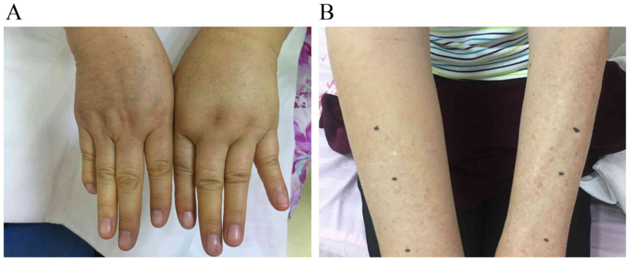
the earliest stage of BCRL, slight changes occur in the surface
architecture of arms or hands of patients accompanied by feelings
of limb heaviness, discomfort or both (Fig. 1A and B). The first common site of
swelling is the forearm, which is usually soft and the swelling
disappears by external compression. Initial swelling may also occur
in the axilla, scapular region or breast. In the
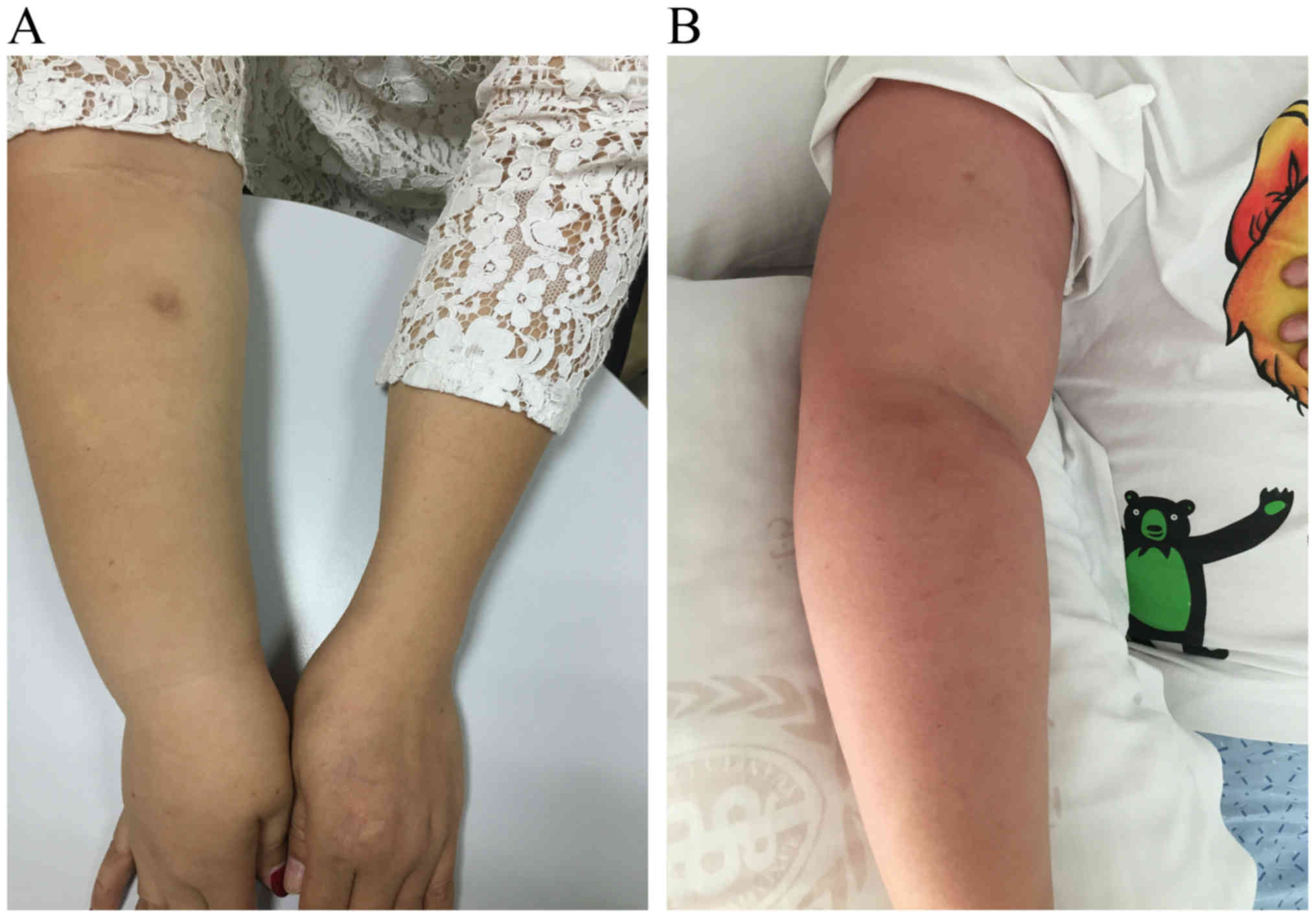
moderate-to-advanced stage, limb edema is no longer relieved by
lifting it or by external pressure; the affected area may become
larger and show a peau dorange appearance (Fig. 2A and B). Clinical symptoms vary
according to the severity and course of BCRL. BCRL is a natural
process that ranges from initial swelling to progressive structural
malformation, often occurring over a period of several weeks or
months (16).
The purpose of the present review is to discuss the
pathogenesis, risk factors, prevention, diagnosis and surveillance,
as well as the traditional and new therapeutic approaches for
BCRL.
The pathogenesis for lymphedema remains unclear,
however, the traditional view of lymphatic obstruction is
insufficient to explain the generation of lymphedema. There are
three linked newly-presented hypotheses about the pathogenesis of
BCRL.
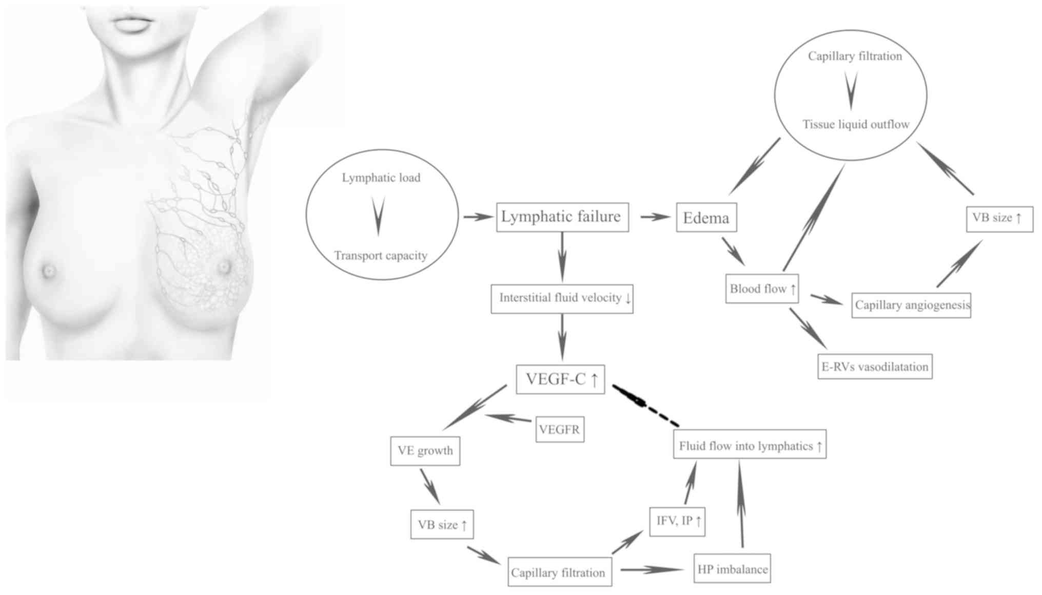
In a normal physiological state, there is a dynamic
equilibrium between lymphatic load and transport capacity that
makes the lymphatic system effective at absorbing and transporting
lymphatic fluid back to the venous system (17). Lymphatic load refers to the volume of
lymphatic fluid, which predominately includes interstitial water
and protein filtrate. Transport capacity is the maximum lymphatic
volume that can be transported by the lymphatics in a given period
of time. However, when the transport capacity is inadequate to meet
the needs of lymphatic load, lymphatic failure occurs and gives
rise to interstitial edema (Fig. 3)
(17).
Total arm blood flow (volume × blood flow/ml) is
increased in the swollen arm, but blood flow per unit volume is not
elevated, causing the vasodilatation of existing resistance vessels
and capillary angiogenesis near existing vessels. Capillary
angiogenesis can augment the surface area of the vascular bed size,
which, along with total arm blood flow, positively facilitates
capillary filtration in the whole limb. When the filtration load
exceeds the outflow of liquid from the tissue, edema of the limb is
further aggravated, forming a vicious circle (Fig. 3) (18).
Since Lymphatic failure leads to a decrease of the
interstitial fluid velocity, and subsequent lymphatic regeneration
and increased lymphatic vascular endothelial growth factor C
(VEGF-C) (19). Lymphatic growth
requires the binding of VEGF-C to the VEGF receptor (VEGFR)
(20). When blood flow is absent,
the increased VEGF-C diffuses to the VEGFR on blood vessels,
inducing vascular endothelial growth and increasing the vascular
bed size to promote capillary filtration (20). This contributes to an increased
interstitial fluid volume and interstitial pressure, and the
imbalance of hydrostatic pressure difference between the lymphatics
and the interstitium (21). These
factors in turn elicit the fluid flow towards those lymphatics,
thus curtailing the production of VEGF-C and finally reaching a
stable state (Fig. 3) (21). Due to the deterioration of lymphatic
failure, the fluid flow rate decreases again, which induces
production of VEGF-C and the cycle repeats until a new stable
equilibrium is reached.
Currently, there is no reliable way of
distinguishing patients who are likely to develop lymphedema, but a
consensus has been reached on some well-defined risk factors,
including axillary lymph node dissection (ALND), which is
associated with early-onset disease (22), regional lymph node radiotherapy,
which is related to late-onset disease (22), high body mass index (BMI) at the time
of breast cancer diagnosis (BMI ≥25 kg/m2) (23), a high number of positive lymph nodes
(>8) (24) and capsular invasion
of the tumor (24).
In order to reduce the incidence rate and avoid the
occurrence of BCRL, a position statement by the National Lymphedema
Network has outlined the recommended preventive measures, including
the avoidance of flight, trauma, skin infection, extreme
temperature, venipuncture (such as blood draws) and limb
compression such as blood pressure readings on the affected arm
(25). However, two clinical studies
demonstrated that injection, flight, blood draws and blood pressure
readings were not significantly associated with the increase in arm
volume, indicating that they are not risk factors for BCRL
(23,26). By contrast, Clark et al
(27) demonstrated that hospital
skin puncture was a high-risk factor for the development of
BCRL.
Similarly, it remains controversial whether age and
chemotherapy are risk factors for BCRL. Previous reports have
suggested that younger survivors are more likely to experience
lymphedema (28,29) because they tend to have more
aggressive tumors and more intensive therapy, but some studies have
indicated that older age is a high-risk factor (30,31).
Other studies have shown that age is not associated with BCRL
(32,33). It was demonstrated that women treated
with chemotherapy, specifically with taxane-based chemotherapy were
more likely to develop lymphedema (34,35), but
with inconsistent results (36).
Extreme temperature and hypertension are risk factors for BCRL that
have been confirmed in certain studies (37,38),
however, further investigation is required. A study attempted to
explore whether race affected the occurrence of BCRL, and it found
that black women may have a higher prevalence of BCRL than white
women (28 vs. 21%), although the results were not statistically
significant (39).
There may be a genetic predisposition for BRCL.
Studies have identified several single nucleotide polymorphisms
associated with the development of secondary lymphedema within the
genes for hepatocyte growth factor, Met protooncogene, gap junction
protein γ2, interleukin (IL)-1A, IL-4, IL-6, IL-10, IL-13, VEGF-C,
NF-κB, lymphocyte cytosolic protein 2, neutropilin 2,
spleen-associated tyrosine kinase, vascular cell adhesion molecule
1, forkhead box C2, VEGFR2, VEGFR3 and RAR-related orphan C
(40,41). By identifying patients with breast
cancer who harbor these molecular biomarkers, numerous
precautionary measures can be taken before surgery to reduce the
incidence of BCRL.
In the initial stage of breast cancer management,
especially before surgery, the assessment of risk factors and a
selection of appropriate surgical scenarios are available to
prevent the occurrence of BCRL (42). The BCRL rate is significantly reduced
in patients who receive lumpectomy compared with those who receive
total mastectomy or modified radical mastectomy (43). Historically, complete ALND is the
standard treatment of axillary intervention for certain patients,
including pregnant women, male patients, patients with inflammatory
breast tumor or those requiring mastectomy or receiving systemic
neo-adjuvant chemotherapy. In recent years, advances have been made
in identifying the population of patients who really need ALND.
Findings demonstrate that axillary lymph node biopsy (ALNB) is a
reliable and safe approach for predicting the status of residual
nodes following systematic neo-adjuvant chemotherapy (44–46).
Axillary intervention for patients undergoing mastectomy can be
downgraded to sentinel lymph node dissection (47), and those who are eligible for
lumpectomy can choose a feasible modality of ALNB (48), that does not increase the incidence
of lymphedema (49). Axillary
radiotherapy may effectively replace complete ALND to control
disease relapse and metastasis in patients who have had mastectomy
or y lumpectomy (50), and
potentially in elderly (>70 years) patients with node-negative
luminal breast cancer who have undergone lumpectomy and tamoxifen
treatment (51). The long-term
recurrence of early-stage breast cancer is associated with
biological characteristics instead of anatomical factors;
therefore, avoidance of axillary intervention purely for the
optimization of prognosis is suggested (52,53).
In 2007, two clinical studies introduced a
pioneering technique for mapping lymphatic drainage in the axillary
region called axillary reverse mapping (ARM) (54,55). ARM
can be used to identify the lymphatic drainage of the upper
extremities and the breast by injecting blue dye into the arm
during the ALND procedure, resulting in the exclusive removal of
lymphatics of the breast and the preservation of lymphatics of the
arm to avoid the incidence of lymphedema caused by resection of arm
lymphatics. This technique is underpinned by the assumption that
the lymphatic drainage of the arm and breast are separate in the
axillary region but are anatomically interconnected (56). An increasing body of clinical trials
has confirmed a significantly lower incidence rate of BCRL in women
undergoing ARM during ALND procedure compared with those receiving
ALND alone (57–59). However, it is necessary to consider
oncological safety when the arm nodes are conserved, particularly
for patients with sentinel lymph node-positivity, as the
co-localization of arm nodes and sentinel lymph nodes is as great
as 27%, which is a key factor in metastasis (60). Fortunately, the risk of metastasis
can be lowered if patients with sentinel lymph node receive
neo-adjuvant chemotherapy (61).
Accurate diagnosis of BCRL depends on a combination
of assessments that include risk evaluation, physical condition and
objective examination of patients (1). The common subjective clinical symptoms
are pain, swelling, numbness, arm heaviness, stiffness of affected
segments and impaired joint activity, but not all patients
experience these symptoms (1). Those
considered to be at high-risk should monitor their physical
condition by objective examination. An increasing number of
techniques and instruments are used for objective examination of
lymphedema, including limb measurements, bioimpedance spectroscopy
(BIS), dual-energy X-ray absorptiometry (DXA), magnetic resonance
imaging (MRI), computed tomography (CT), color Doppler imaging,
lymphoscintigraphy and indocyanine green (ICG) lymphography.
Clinically, BCRL is diagnosed in terms of the degree
of distortion of limb measurements. The standard methods for
obtaining limb measurements include perometry, limb circumference
and water displacement (1). However,
results vary widely because they depend on the subjective
estimation of operators, and the assessment of volume alone is
insufficient (62). Diagnosis tools
such as ultrasound and DXA, which allow assessment of arm tissue
composition, should be recommended for patients in which the
thickness of muscle in the affected arm is less than that in the
unaffected arm, and the muscle growth rates differ between the arms
(63). In 2018, Engin et al
(64) used a new volumeter called
‘easy volumeter’ to measure water displacement with regard to the
limb volume in patients with BCRL. The easy volumeter was designed
for home use and is more durable, lightweight and easier to clean
than a standard volumeter (64). In
this study, the measurements from easy volumeters were valid and
consistent with those of standard volumeters, suggesting that it
was a promising tool to investigate BCRL (64).
In 2000, a modality was added to the diagnosis tools
for lymphedema called BIS which measures the volume of
extracellular fluid via detecting a physical reaction to an
impressed electrical current (65).
In comparison to conventional methods, the measurement of BIS is
more objective and specific (80–99%) (66–69),
with a wide range of sensitivity (30–100%) (66–70), but
a higher false-negative rate (36%) (71). Notably, BIS permits identification of
lymphedema earlier, when it is in the subclinical stage, and tracks
disease progression persistently, making timely intervention of
lymphedema a reality (72). Timely
intervention dramatically reduces the incidence rate of lymphedema
from 36.4 to 4.4% (72).
DXA is effective and credible in quantifying the
soft-tissue masses of the upper and lower extremities and the
composition of arms, including fat, lean and bone mineral masses
(73). Compared with BIS and limb
circumference measurements, DXA has similar precision in detecting
the percentage differences between the affected and unaffected arms
(73). Moreover, DXA has superior
repeatability in volume measurement yields compared with the
measurements of limb circumference and water displacement,
particularly in the affected arm, but not in the unaffected arm
(74).
MRI has been used for decades to diagnose
lymphedema, especially when it is coupled with edema in fat tissue
(1). Compared to lymphoscintigraphy,
MRI has a higher specificity in detecting delayed lymphatic
drainage (85.7 vs. 66.7%) and greater sensitivity for delineating
the architecture of lymphatic vessels (100 vs. 83.3%) (75). In some instances, the excessive water
retention in subcutaneous tissue and reasons for lymphatic vessel
interruption or obstruction maybe determined by this technique
(1). Nevertheless, MRI is expensive
and cannot achieve real-time diagnosis (76). Commonly, the detection of lymphedema
is not recommended by CT or ultrasonography (US) due to their low
sensitivity, but CT can be used to assess the excessive growth of
fibrous tissue during the procedure of lymphedema (77). The low sensitivity of ultrasound can
be attributed to several factors, including excessive edema, tissue
fibrosis caused by irradiation injury and focal short-section vein
occlusion beneath the clavicle or in the deep pelvis (1). Venous obstruction occurs concurrently
with chronic lymphedema, with an incidence rate of 4.6%, and may be
falsely evaluated by US as a negative result owing to technical
difficulties (78). Fortunately,
this problem is solved by color Doppler imaging that can be used to
visualize vessels with a diameter of only 1–2 mm, thus enabling the
detection of the anatomy and function of damaged veins (78).
Provided that the aforementioned approaches cannot
affirm the diagnosis, the standard recommendation is to apply
radionuclide lymphoscintigraphy, a nuclear medicine imaging
technique that allows visualization of lymphatic drainage into the
axillary lymph nodes by subcutaneous injection of radiolabeled
sulfur colloid into the hand (79).
A tardive axillary visualization coupled with dermal
lymphangiectasia will occur if there is lymphedema (79). Early-stage lymphoscintigraphy is an
effective diagnostic tool; however, it has many disadvantages such
as radiation exposure, low resolution, high costs and increased
invasiveness (1). Lymphography is a
newer, systematic method to evaluate limb edema and lymph
circulation without radiation exposure (71). Lymphography has higher specificity
and sensitivity, and longer tracking capabilities, ranging from
subclinical to more advanced stages, compared with
lymphoscintigraphy (77,80–82). Of
note, the greatest advantage of lymphography is that it can be used
for the real-time monitoring of lymphatic vessels during surgery,
albeit not in a perfect way (82).
For example, if the lymph vessels beneath the subcutaneous level of
the skin are thicker than 2 cm, observation cannot be achieved by
ICG lymphography (82). The
advantages and disadvantages of all the diagnosis tools for
lymphedema are summarized in Table
II.
Lymphedema surveillance can be used to identify and
diagnose subclinical or early-stage disease, providing the
opportunity for early intervention and treatment of BCRL (83). Prospective interval surveillance
greatly optimizes the costs (84),
reduces the observed incidence (72)
and can reverse and prevent the progression of BCRL (85–87).
Data using direct provider costs of surveillance demonstrate that
it has potential to reduce direct treatment costs of BCRL
management (84); however, for
improving assessment, further data on the indirect costs must be
reported. At present, there are four main techniques for the
surveillance of BCRL: Water displacement, perometry, tonometry and
BIS. Water displacement is laborious, time-consuming and not
suitable for massive, continuous surveillance. Perometry, optical
assessment of limb volume, is less time-consuming but more
expensive. Tonometry is a noninvasive method that can detect
subclinical interstitial edema via continuously measuring the
dielectric constant in affected tissue and evaluating moisture
content (88). BIS is more specific,
but lacks sensitivity, and it may be the most frequently utilized
modality (71). However, no widely
adopted consensus has been reached regarding which technique is the
best for the surveillance of BCRL.
It is widely believed that the optimal management to
efficiently relieve lymphedema is complex decongestive treatment
(CDT), lymphatic physiotherapeutic intervention including manual
lymph drainage (MLD), skin care, physical exercise, long-term
education on self-management of lymphedema, compression bandages
and sleeve or stocking compression (1). Lymphoscintigraphy of upper limbs is a
valid tool to predict the prognosis of this combined strategy. CDT
can be provided with a commercialized product called
Linfadren®, which is a mixture of diosmin, coumarin and
arbutin, to further improve its efficacy without any adverse events
(89). Obesity reduces the
effectiveness of CDT (90). Every
treatment method for lymphedema has been gradually defined into
explicitness.
MLD, a universal treatment for lymphedema is a
massage technique that uses a special rhythmic pumping through
gentle, directed stretching of skin to massage the affected area
and stimulate lymphatic contractility, thus enhancing lymphatic
drainage (1). A meta-analysis found
that, compared with other treatments such as physical exercise,
skin care and compression therapy, additional MLD was unlikely to
achieve a significant reduction in the volume of the affected limb
(91). Of note, heterogeneity across
the analyzed studies was considerable and the sample size was
limited. Paradoxically, a subgroup analysis in a Cochrane
systematic review demonstrated that MLD was safe and, when used in
combination with compression bandages, may provide additional
benefits of swelling reduction for BCRL compared with the use of
compression bandages alone, particularly for patients with
mild-to-moderate disease (92).
Compression bandages used with a compression garment can
significantly reduce the volume of the edematous limb compared with
the usage of a compression garment alone (1). Generally, the bandaging method involves
a spiral-bandaging method and a figure-of-eight method (93). The figure-of-eight method is a more
effective approach in maintaining the correct position, is more
comfortable for the patient and has a replacement frequency of
either 5 times per week over a 4-week period or once per 2 days
over a 3-week period (93). Precast
adjustable compression systems, a novel technique that can be
easily used and removed by patients, may be an effective
alternative to compression bandages due to similar effects on
reducing excess limb volume (94).
When the affected limb reaches a minimum volume, self-care can be
accepted by the patient (1). A
myriad of advantages is attained by a CDT approach that reduces
edema volume, intensity of pain and arm heaviness, reinforces
lymphatic function, improves quality of life and lowers the
incidence of cellulitis (95–97).
The use of adjuvant tools such as a pneumatic
compression device (PCD) provides additional benefits in managing
lymphedema that is associated with reduction of outpatient services
and hospitalizations (1). The
initial volume reduction of a swollen limb can be maintained over a
long period of time by the use of PCD followed by the use of a
flexible and suitable compression garment. A meta-analysis of 7
randomized controlled trials (RCTs) showed that PCD can alleviate
lymphedema, however, there was no significant reduction in limb
volume between conventional management of lymphedema with or
without the usage of PCD (98).
These RCTs had limitations such as small sample sizes of 12–56
participants with considerable heterogeneity among them (98). Currently, the most advanced PCD uses
a calibrated, gradient compressor, and it is designed for home use
with multiple inflatable compartments to deliver external pneumatic
compression, more garment chambers and a higher level of
adjustability and programmability (99). A retrospective analysis demonstrated
that this adjunctive modality yields significant clinical and
economic effectiveness in treating cancer-related and
non-cancer-related lymphedema, demonstrated by a reduction in the
adjusted rate of cellulitis, the usage of lymphedema-related manual
therapy, outpatient visits and total lymphedema-related costs per
patient, excluding medical equipment costs (99).
It is widely known that a sedentary lifestyle leads
to being overweight or obese, factors that are associated with an
increased incidence of BCRL (14).
Participation in physical exercise during and after treatment for
breast cancer can ameliorate psychosocial and physical conditions,
resulting in active lifestyles with optimized survival (100). Traditionally, patients with
lymphedema or who are at risk for lymphedema tend to reduce
physical exercise due to concerns about disease exacerbation
(100). Some preliminary studies
have indicated that exercise neither causes lymphedema nor worsens
the disease (101–105). A slowly progressive weight-lifting
program does not increase the rate of lymphedema compared with no
exercise, and aerobic exercise, resistance training, stretching,
yoga, qigong and pilates are also safe (101,105).
Under specific circumstances, resistance training can even
substantially improve the lymphedema state and may prevent the
development of secondary lymphedema in patients (106).
There are a variety of surgical techniques for
lymphedema, including debulking resection, liposuction,
lymphatic-venous ‘end-to-end’ anastomoses (LVA) and vascularized
lymph node (VLN) transplantation with the advent of microsurgery
(1). Typically, LVA is only used in
the early-stage of the disease, but despite this limitation it
reduces limb volume or circumference effectively and improves
quality of life (107–109). LVA also has other advantages such
as reducing trauma, lowering the risk of complications and it can
be performed under local anesthesia (110,111).
After a 1-year follow-up of women undergoing LVA, >56.5% of
anastomoses are still patent (109).
A new program, called the ‘Lymphedema Microsurgical
Preventive Healing Approach (LYMPHA)’, combines the LVA technique
with the surgery of ALND, which anastomoses the collateral branch
of the axillary vein to the lymphatics of the arm, with a low
incidence rate of lymphedema of 4.05% (112,113).
An altered and simplified version of LYMPHA used during the surgery
of ALND dramatically decreases the lymphedema rate to 3% compared
with ALND alone, which has a higher rate of 13% (114). Two pilot studies proposed a new
technique called ‘dynamic-lymphaticovenular anastomosis,’ which
uses preoperative dynamic imaging of the forearm to determine the
incision points followed by microsurgery of LVA (115,116).
This technique achieves significant reduction of excess limb volume
compared with conventional LVA and results in no swelling rebound
after postoperative degradation or removal of compression garments
in a 12 month follow up period (115,116).
VLN transfer is a promising technique for treating
moderate-to-advanced stage lymphedema, and it has the ability to
lower the clinical grade, attenuate limb circumference, reduce the
incidence of cellulitis and improve the quality of life in patients
(117). However, it requires a
strict observation of the donor site because donor site lymphedema
is the most serious complication after this surgery (117). Patient selection and scrupulous
assessment of donor and recipient sites prior to VLN
transplantation are key factors for surgical success. This concern
may be removed by the technique of vascularized groin lymph node
(VGLN) flap transplantation (118).
Findings revealed VGLN flap harvesting does not cause iatrogenic
lymphedema at the donor site, but this surgery cannot be performed
in patients with a high risk of lower limb lymphedema due to
obesity, pre-existing lower limb edema or previous pelvic surgery
(118). Of note, the limitations of
LVA and VLN procedures have to be emphasized, as they are complex
and can only be provided by experts at tertiary care centers.
CDT is not an effective treatment for chronic
massive lymphedema with excess adipose tissue, as adipose tissue
cannot be eliminated through compression alone (1). Microsurgery often fails to attain
complete limb reduction because the newly formed adipose tissue
persisting under the skin of the patient with longstanding
non-pitting lymphedema is not resected. These perplexities are
solved by the use of liposuction, which can remove excess adipose
tissue, resulting in complete reduction of lymphedema (1). Liposuction is effective in removing
chronic non-pitting limb lymphedema with a large volume, which can
be completely attenuated in 1–3 months, with no recurrence of arm
swelling observed in long-term follow-up (119–123).
Ample evidence suggests that women with lymphedema who undergo
liposuction followed by compression bandages or compression
garments achieve significant benefits, as the mean reduction of
excess limb volume ranges from 101–118% (124–126)
and can be maintained at >100% during 21 years of follow-up
(127). In addition, liposuction
improves lymph flow (128),
increases blood flow to the skin so that it is approximately equal
to the flow in a normal arm (129),
and it does not injure the existing lymphatic vessels within the
affected limb (122,130). These characteristics significantly
reduce the incidence of erysipelas and cellulitis (131). The complications from liposuction
are limited, with a very low incidence rate; paresthesia of the
skin is the most typical complication and fades away within 3–6
months (132), and fibrous tissue
increases in some cases, specifically in women with a male
distribution of body fat (132).
The characteristics of current treatment scenarios for lymphedema
are summarized in Table III.
Previous findings illustrated that no medication has
the capacity to reduce lymphedema, as the lymphatic flow could not
be improved by any drugs, including diuretics that change
microvascular fluid filtration by increasing the excretion of
sodium chloride and water, (96).
With a greater understanding of the molecular mechanisms that
control lymphatic function, lymphedema may be reversed. The first
potential medication for the treatment of BCRL is reported to be in
phase I trials (133). An
increasing number of lymphedema therapy-related preclinical
investigations are performed in animal models of lymphatic disease
in which the genes encoding VEGF-C or VEFG-D are transferred into
the animal by adenoviruses or adeno-associated viruses (134,135).
This technique results in the development of many new lymphatic
capillaries and reduces edema following an initial promotion of
lymphatic extravasation (136).
Following VEGF-C therapy, the injured collecting lymphatic vessels
in mice undergo regeneration of lymphatic capillaries, which
subsequently remodel, differentiate and mature into functional
vessels (137). Similarly,
surgery-based damage of lymphatic vasculature in pigs can be
effectively repaired by VEGF-C therapy, which greatly enhances the
function and structural stability of transferred lymph nodes
(138). The combined program of the
microsurgery of VLN transfer followed by VEGF-C treatment may be
also equally beneficial in patients with lymphedema to foster
lymphatic microvascular anastomoses.
The success of stem cell therapy involving the
transplantation of autologous mesenchymal stromal cells derived
from adipose tissue, muscle and bone marrow to alleviate lymphedema
has been reported in certain preclinical studies (139–141),
and this has opened up a potential new field of treatment for this
disease. In two pilot studies, injection of adipose-derived
regenerative cells into the axillary region with fat grafting was
well-tolerated, and only a paucity of liposuction-related adverse
events occurred transiently (142,143).
After 6–12 months of follow-up, lymphedema was alleviated, without
rebound of the swelling limb (143). However, these promising results of
autologous stem cell therapy from the two studies must be
investigated in humans with RCTs. If the results in humans are
positive, increasing number of patients with lymphedema could
benefit from this surgery.
BCRL exerts a negative impact on the quality of life
of survivors of breast cancer. Precautionary measures and earlier
lymphedema surveillance combined with effective diagnostic tools,
such as BIS or ICG lymphography, are effective in reducing the
incidence of lymphedema and providing more opportunities for
intervention and treatment in subclinical and early-stages,
especially in high-risk patients. In a variety of treatment
strategies, the combination of CDT, PCD and appropriate physical
exercise can contribute to women having an apparent reduction of
excess limb volume and improvement of quality of life. Of the
surgical techniques, the usage of LVA in the early-stage of
disease, VLN transplantation in the moderate-to-advanced stage and
liposuction when lymphedema is coupled with excess adipose tissue
can reduce swollen limb volume to normal, and be maintained
long-term without rebound of swelling. Recently, molecular therapy
and autologous stem cell transplantation have been shown to
successfully alleviate lymphedema in preclinical studies, which may
lead to the development of novel targeted therapies for BCRL in the
future.
The authors would like to acknowledge Dr Han Qin
(Department of Sport Medicine, The Affiliated Hospital of Qingdao
University, Qingdao, China) for providing the map of lymphatics of
the breast.
No funding was received.
Not applicable.
LH made substantial contributions to the conception
of the review, HQ produced software that was used in the work, QW
drafted the manuscript and substantively revised it, and YS
contributed to the writing of the manuscript.
Not applicable.
Not applicable.
The authors declare that they have no competing
interests.
|
1
|
Rockson SG: Lymphedema after Breast Cancer
Treatment. N Eng J Med. 379:1937–1944. 2018. View Article : Google Scholar
|
|
2
|
Armer JM, Ballman KV, McCall L, Armer NC,
Sun Y, Udmuangpia T, Hunt KK, Mittendorf EA, Byrd DR, Julian TB and
Boughey JC: Lymphedema symptoms and limb measurement changes in
breast cancer survivors treated with neoadjuvant chemotherapy and
axillary dissection: Results of American college of surgeons
oncology group (ACOSOG) Z1071 (Alliance) substudy. Support Care
Cancer. 27:495–503. 2019. View Article : Google Scholar : PubMed/NCBI
|
|
3
|
Velanovich V and Szymanski W: Quality of
life of breast cancer patients with lymphedema. Am J Surg.
177:184–187. 1999. View Article : Google Scholar : PubMed/NCBI
|
|
4
|
Casley-Smith JR and Casley-Smith JR:
Modern treatment of lymphoedema II. The benzopyrones. Australas J
Dermatol. 33:69–74. 1992. View Article : Google Scholar : PubMed/NCBI
|
|
5
|
MM H: Functional and psychosocial aspects
of lymphedema in women treated for breast cancer. Innov Breast
Cancer Care. 3:97–100. 17–18.
|
|
6
|
Armer JM and Mallinckrodt BR: Post-breast
cancer treatment lymphedema. The secret epidemic Scope Phlebolog
Lympholog. 9:334–341. 2002.
|
|
7
|
Carter BJ: Womens experiences of
lymphedema. Oncol Nurs Forum. 24:875–882. 1997.PubMed/NCBI
|
|
8
|
Newman ML, Brennan M and Passik S:
Lymphedema complicated by pain and psychological distress: A case
with complex treatment needs. J Pain Symptom Manage. 12:376–379.
1996. View Article : Google Scholar : PubMed/NCBI
|
|
9
|
Passik SD and McDonald MV: Psychosocial
aspects of upper extremity lymphedema in women treated for breast
carcinoma. Cancer. 83 (12 Suppl American):2817–2820. 1998.
View Article : Google Scholar : PubMed/NCBI
|
|
10
|
Tobin MB, Lacey HJ, Meyer L and Mortimer
PS: The psychological morbidity of breast cancer-related arm
swelling. Psychological morbidity of lymphoedema. Cancer.
72:3248–3252. 1993. View Article : Google Scholar : PubMed/NCBI
|
|
11
|
DiSipio T, Rye S, Newman B and Hayes S:
Incidence of unilateral arm lymphoedema after breast cancer: A
systematic review and meta-analysis. Lancet Oncol. 14:500–515.
2013. View Article : Google Scholar : PubMed/NCBI
|
|
12
|
Specht MC, Miller CL, Russell TA, Horick
N, Skolny MN, OToole JA, Jammallo LS, Niemierko A, Sadek BT,
Shenouda MN, et al: Defining a threshold for intervention in breast
cancer-related lymphedema: What level of arm volume i ncrease
predicts progression? Breast cancer research and treatment.
140:485–494. 2013. View Article : Google Scholar : PubMed/NCBI
|
|
13
|
Rockson SG and Rivera KK: Estimating the
population burden of lymphedema. Ann N Y Acad Sci. 1131:147–154.
2008. View Article : Google Scholar : PubMed/NCBI
|
|
14
|
Hayes S, Cornish B and Newman B:
Comparison of methods to diagnose lymphoedema among breast cancer
survivors: 6-month follow-up. Breast Cancer Res Treat. 89:221–226.
2005. View Article : Google Scholar : PubMed/NCBI
|
|
15
|
Denlinger CS, Sanft T, Baker KS, Broderick
G, Demark-Wahnefried W, Friedman DL, Goldman M, Hudson M, Khakpour
N, King A, et al: Survivorship, version 2.2018, NCCN clinical
practice guidelines in oncology. J Natl Compr Cancer Netw.
16:1216–1247. 2018. View Article : Google Scholar
|
|
16
|
International Society of Lymphology: The
diagnosis and treatment of peripheral lymphedema. 2009 concensus
document of the international society of lymphology. Lymphology.
42:51–60. 2009.PubMed/NCBI
|
|
17
|
Lawenda BD, Mondry TE and Johnstone PA:
Lymphedema: A primer on the identification and management of a
chronic condition in oncologic treatment. CA Cancer J Clin.
59:8–24. 2009. View Article : Google Scholar : PubMed/NCBI
|
|
18
|
Mortimer PS: The pathophysiology of
lymphedema. Cancer. 83 (12 Suppl American):2798–2802. 1998.
View Article : Google Scholar : PubMed/NCBI
|
|
19
|
Goldman J, Conley KA, Raehl A, Bondy DM,
Pytowski B, Swartz MA, Rutkowski JM, Jaroch DB and Ongstad EL:
Regulation of lymphatic capillary regeneration by interstitial flow
in skin. Am J Physiol Heart Circ Physiol. 292:H2176–H2183. 2007.
View Article : Google Scholar : PubMed/NCBI
|
|
20
|
Joukov V, Pajusola K, Kaipainen A, Chilov
D, Lahtinen I, Kukk E, Saksela O, Kalkkinen N and Alitalo K: A
novel vascular endothelial growth factor, VEGF-C, is a ligand for
the Flt4 (VEGFR-3) and KDR (VEGFR-2) receptor tyrosine kinases.
EMBO J. 15:290–298. 1996. View Article : Google Scholar : PubMed/NCBI
|
|
21
|
Pajusola, Kaipainen, Chilov, Lahtinen,
Kukk, Saksela O, Kalkkinen N and Alitalo K: 2, 199621. Bates DO: An
interstitial hypothesis for breast cancer related lymphoedema.
Pathophysiology. 17:289–294. 2010. View Article : Google Scholar : PubMed/NCBI
|
|
22
|
McDuff SGR, Mina AI, Brunelle CL, Salama
L, Warren LEG, Abouegylah M, Swaroop M, Skolny MN, Asdourian M,
Gillespie T, et al: Timing of lymphedema after treatment for breast
cancer: When are patients most at risk? Int J Radiat Oncol Biol
Phys. 103:62–70. 2019. View Article : Google Scholar : PubMed/NCBI
|
|
23
|
Asdourian MS, Swaroop MN, Sayegh HE,
Brunelle CL, Mina AI, Zheng H, Skolny MN and Taghian AG:
Association between precautionary behaviors and breast
cancer-related lymphedema in patients undergoing bilateral surgery.
J Clin Oncol. 35:3934–3941. 2017. View Article : Google Scholar : PubMed/NCBI
|
|
24
|
Iyigun ZE, Duymaz T, Ilgun AS, Alco G,
Ordu C, Sarsenov D, Aydin AE, Celebi FE, Izci F, Eralp Y and Ozmen
V: Preoperative lymphedema-related risk factors in early-stage
breast cancer. Lymphat Res Biol. 16:28–35. 2018. View Article : Google Scholar : PubMed/NCBI
|
|
25
|
Network NL: Lymphedema risk reduction
practices. http://wwwlymphnetorg/pdfDocs/nlnriskreductionpdf2012
|
|
26
|
Ferguson CM, Swaroop MN, Horick N, Skolny
MN, Miller CL, Jammallo LS, Brunelle C, OToole JA, Salama L, Specht
MC and Taghian AG: Impact of ipsilateral blood draws, injections,
blood pressure measurements, and air travel on the risk of
lymphedema for patients treated for breast cancer. J Clin Oncol.
34:691–698. 2016. View Article : Google Scholar : PubMed/NCBI
|
|
27
|
Clark B, Sitzia J and Harlow W: Incidence
and risk of arm oedema following treatment for breast cancer: A
three-year follow-up study. QJM. 98:343–348. 2005. View Article : Google Scholar : PubMed/NCBI
|
|
28
|
Geller BM, Vacek PM, OBrien P and
Secker-Walker RH: Factors associated with arm swelling after breast
cancer surgery. J Womens Health (Larchmt). 12:921–930. 2003.
View Article : Google Scholar : PubMed/NCBI
|
|
29
|
Armer J and Fu MR: Age differences in
post-breast cancer lymphedema signs and symptoms. Cancer Nurs.
28:200–207; quiz208-209. 2005. View Article : Google Scholar : PubMed/NCBI
|
|
30
|
Larson D, Weinstein M, Goldberg I, Silver
B, Recht A, Cady B, Silen W and Harris JR: Edema of the arm as a
function of the extent of axillary surgery in patients with stage
I–II carcinom a of the breast treated with primary radiotherapy.
Int J Radiat Oncol Biol Phys. 12:1575–1582. 1986. View Article : Google Scholar : PubMed/NCBI
|
|
31
|
Engel J, Kerr J, Schlesinger-Raab A, Sauer
H and Holzel D: Axilla surgery severely affects quality of life:
Results of a 5-year prospective study in breast cancer patients.
Breast Cancer Res Treat. 79:47–57. 2003. View Article : Google Scholar : PubMed/NCBI
|
|
32
|
Paskett ED, Naughton MJ, McCoy TP, Case LD
and Abbott JM: The epidemiology of arm and hand swelling in
premenopausal breast cancer survivors. Cancer Epidemiol Biomarkers
Prev. 16:775–782. 2007. View Article : Google Scholar : PubMed/NCBI
|
|
33
|
Pezner RD, Patterson MP, Hill LR, Lipsett
JA, Desai KR, Vora N, Wong JY and Luk KH: Arm lymphedema in
patients treated conservatively for breast cancer: Relationship to
patient age and axillary node dissection technique. Int J Radiat
Oncol Biol Phys. 12:2079–2083. 1986. View Article : Google Scholar : PubMed/NCBI
|
|
34
|
Cariati M, Bains SK, Grootendorst MR,
Suyoi A, Peters AM, Mortimer P, Ellis P, Harries M, Van Hemelrijck
M and Purushotham AD: Adjuvant taxanes and the development of
breast cancer-related arm lymphoedema. Br J Surg. 102:1071–1078.
2015. View Article : Google Scholar : PubMed/NCBI
|
|
35
|
Penn IW, Chang YC, Chuang E, Chen CM,
Chung CF, Kuo CY and Chuang TY: Risk factors and prediction model
for persistent breast-cancer-related lymphedema: A 5-year cohort
study. Support Care Cancer. 27:991–1000. 2019. View Article : Google Scholar : PubMed/NCBI
|
|
36
|
Swaroop MN, Ferguson CM, Horick NK, Skolny
MN, Miller CL, Jammallo LS, Brunelle CL, OToole JA, Isakoff SJ,
Specht MC and Taghian AG: Impact of adjuvant taxane-based
chemotherapy on development of breast cancer-related lymphedema:
Results from a large prospective cohort. Breast Cancer Res Treat.
151:393–403. 2015. View Article : Google Scholar : PubMed/NCBI
|
|
37
|
Czerniec SA, Ward LC and Kilbreath SL:
Breast cancer-related arm lymphedema: Fluctuation over six months
and the effect of the weather. Lymphat Res Biol. 14:148–155. 2016.
View Article : Google Scholar : PubMed/NCBI
|
|
38
|
Rockson SG: Precipitating factors in
lymphedema: Myths and realities. Cancer. 83 (12 Suppl
American):2814–2846. 1998. View Article : Google Scholar : PubMed/NCBI
|
|
39
|
Meeske KA, Sullivan-Halley J, Smith AW,
McTiernan A, Baumgartner KB, Harlan LC and Bernstein L: Risk
factors for arm lymphedema following breast cancer diagnosis in
Black women and White women. Breast Cancer Res Treat. 113:383–391.
2009. View Article : Google Scholar : PubMed/NCBI
|
|
40
|
Visser J, van Geel M, Cornelissen AJM, van
der Hulst RRWJ and Qiu SS: Breast cancer-related lymphedema and
genetic predisposition: A systematic review of the literature.
Lymphat Res Biol. 17:288–293. 2019. View Article : Google Scholar : PubMed/NCBI
|
|
41
|
Newman B, Lose F, Kedda MA, Francois M,
Ferguson K, Janda M, Yates P, Spurdle AB and Hayes SC: Possible
genetic predisposition to lymphedema after breast cancer. Lymphat
Res Biol. 10:2–13. 2012. View Article : Google Scholar : PubMed/NCBI
|
|
42
|
Tandra P, Kallam A and Krishnamurthy J:
Identification and management of lymphedema in patients with breast
cancer. J Oncol pract. 15:255–262. 2019. View Article : Google Scholar : PubMed/NCBI
|
|
43
|
Tsai RJ, Dennis LK, Lynch CF, Snetselaar
LG, Zamba GK and Scott-Conner C: The risk of developing arm
lymphedema among breast cancer survivors: A meta-analysis of
treatment factors. Ann Surg Oncol. 16:1959–1972. 2009. View Article : Google Scholar : PubMed/NCBI
|
|
44
|
Gentilini O and Veronesi U: Abandoning
sentinel lymph node biopsy in early breast cancer? A new trial in
progress at the European institute of oncology of milan (SOUND:
Sentinel node vs observation after axillary UltraSouND). Breast.
21:678–681. 2012. View Article : Google Scholar : PubMed/NCBI
|
|
45
|
Cardoso F, vant Veer LJ, Bogaerts J,
Slaets L, Viale G, Delaloge S, Pierga JY, Brain E, Causeret S,
DeLorenzi M, et al: 70-Gene signature as an aid to treatment
decisions in early-stage breast cancer. N Engl J Med. 375:717–729.
2016. View Article : Google Scholar : PubMed/NCBI
|
|
46
|
Sparano JA, Gray RJ, Makower DF, Pritchard
KI, Albain KS, Hayes DF, Geyer CE Jr, Dees EC, Perez EA, Olson JA
Jr, et al: Prospective validation of a 21-gene expression assay in
breast cancer. N Engl J Med. 373:2005–2014. 2015. View Article : Google Scholar : PubMed/NCBI
|
|
47
|
Gradishar WJ, Anderson BO, Balassanian R,
Blair SL, Burstein HJ, Cyr A, Elias AD, Farrar WB, Forero A,
Giordano SH, et al: NCCN guidelines insights: Breast cancer,
version 1.2017. J Natl Compr Canc Netw. 15:433–451. 2017.
View Article : Google Scholar : PubMed/NCBI
|
|
48
|
Levenhagen K, Davies C, Perdomo M, Ryans K
and Gilchrist L: Diagnosis of upper quadrant lymphedema secondary
to cancer: Clinical practice guideline from the oncology section of
the American physical therapy association. Phys Ther. 97:729–745.
2017. View Article : Google Scholar : PubMed/NCBI
|
|
49
|
Lyman GH, Somerfield MR, Bosserman LD,
Perkins CL, Weaver DL and Giuliano AE: Sentinel lymph node biopsy
for patients with early-stage breast cancer: American society of
clinical oncology clinical practice guideline update. J Clin Oncol.
35:561–564. 2017. View Article : Google Scholar : PubMed/NCBI
|
|
50
|
International Society of Lymphology: The
diagnosis and treatment of peripheral lymphedema: 2013 Consensus
document of the international society of lymphology. Lymphology.
46:1–11. 2013.PubMed/NCBI
|
|
51
|
Hughes KS, Schnaper LA, Bellon JR,
Cirrincione CT, Berry DA, McCormick B, Muss HB, Smith BL, Hudis CA,
Winer EP and Wood WC: Lumpectomy plus tamoxifen with or without
irradiation in women age 70 years or older with early breast
cancer: Long-term follow-up of CALGB 9343. J Clin Oncol.
31:2382–2387. 2013. View Article : Google Scholar : PubMed/NCBI
|
|
52
|
Czerniec SA, Ward LC, Refshauge KM, Beith
J, Lee MJ, York S and Kilbreath SL: Assessment of breast
cancer-related arm lymphedema--comparison of physical measurement
methods and self-report. Cancer Invest. 28:54–62. 2010. View Article : Google Scholar : PubMed/NCBI
|
|
53
|
Cormier JN, Xing Y, Zaniletti I, Askew RL,
Stewart BR and Armer JM: Minimal limb volume change has a
significant impact on breast cancer survivors. Lymphology.
42:161–175. 2009.PubMed/NCBI
|
|
54
|
Thompson M, Korourian S, Henry-Tillman R,
Adkins L, Mumford S, Westbrook KC and Klimberg VS: Axillary reverse
mapping (ARM): A new concept to identify and enhance lymphatic
preservation. Ann Surg Oncol. 14:1890–1895. 2007. View Article : Google Scholar : PubMed/NCBI
|
|
55
|
Nos C, Lesieur B, Clough KB and Lecuru F:
Blue dye injection in the arm in order to conserve the lymphatic
drainage of the arm in breast cancer patients requiring an axillary
dissection. Ann Surg Oncol. 14:2490–2496. 2007. View Article : Google Scholar : PubMed/NCBI
|
|
56
|
Pavlista D and Eliska O: Analysis of
direct oil contrast lymphography of upper limb lymphatics
traversing the axilla - a lesson from the past - contribution to
the concept of axillary reverse mapping. Eur J Surg Oncol.
38:390–394. 2012. View Article : Google Scholar : PubMed/NCBI
|
|
57
|
Gennaro M, Maccauro M, Sigari C, Casalini
P, Bedodi L, Conti AR, Caraceni A and Bombardieri E: Selective
axillary dissection after axillary reverse mapping to prevent
breast-cancer-related lymphoedema. Eur J Surg Oncol. 39:1341–1345.
2013. View Article : Google Scholar : PubMed/NCBI
|
|
58
|
Pasko JL, Garreau J, Carl A, Ansteth M,
Glissmeyer M and Johnson N: Axillary reverse lymphatic mapping
reduces patient perceived incidence of lymphedema after axillary
dissection in breast cancer. Am J Surg. 209:890–895. 2015.
View Article : Google Scholar : PubMed/NCBI
|
|
59
|
Yue T, Zhuang D, Zhou P, Zheng L, Fan Z,
Zhu J, Hou L, Yu F, Dong X, Xiao L and He Q: A prospective study to
assess the feasibility of axillary reverse mapping and evaluate its
effect on preventing lymphedema in breast cancer patients. Clin
Breast Cancer. 15:301–306. 2015. View Article : Google Scholar : PubMed/NCBI
|
|
60
|
Noguchi M, Noguchi M, Ohno Y, Morioka E,
Nakano Y, Kosaka T, Kurose N and Minato H: Feasibility study of
axillary reverse mapping for patients with clinically node-negative
breast cancer. Eur J Surg Oncol. 42:650–656. 2016. View Article : Google Scholar : PubMed/NCBI
|
|
61
|
Beek MA, Gobardhan PD, Klompenhouwer EG,
Rutten HJ, Voogd AC and Luiten EJ: Axillary reverse mapping (ARM)
in clinically node positive breast cancer patients. Eur J Surg
Oncol. 41:59–63. 2015. View Article : Google Scholar : PubMed/NCBI
|
|
62
|
McLaughlin SA, Staley AC, Vicini F,
Thiruchelvam P, Hutchison NA, Mendez J, MacNeill F, Rockson SG,
DeSnyder SM, Klimberg S, et al: Considerations for clinicians in
the diagnosis, prevention, and treatment of breast cancer-related
lymphedema: Recommendations from a multidisciplinary expert ASBrS
panel: Part 1: Definitions, assessments, education, and future
directions. Ann Surg Oncol. 24:2818–2826. 2017. View Article : Google Scholar : PubMed/NCBI
|
|
63
|
Bok SK, Jeon Y and Hwang PS:
Ultrasonographic evaluation of the effects of progressive resistive
exercise in breast cancer-related lymphedema. Lymphat Res Biol.
14:18–24. 2016. View Article : Google Scholar : PubMed/NCBI
|
|
64
|
Engin O, Akalin E, Saribay E, Aslan C,
Sahin E and Alper S: Easy volumeter in detection of breast
cancer-related lymphedema: A validity study. Lymphat Res Biol.
17:543–549. 2019. View Article : Google Scholar : PubMed/NCBI
|
|
65
|
Cornish BH, Chapman M, Thomas BJ, Ward LC,
Bunce IH and Hirst C: Early diagnosis of lymphedema in postsurgery
breast cancer patients. Ann N Y Acad Sci. 904:571–575. 2000.
View Article : Google Scholar : PubMed/NCBI
|
|
66
|
Cornish BH, Chapman M, Hirst C, Mirolo B,
Bunce IH, Ward LC and Thomas BJ: Early diagnosis of lymphedema
using multiple frequency bioimpedance. Lymphology. 34:2–11.
2001.PubMed/NCBI
|
|
67
|
Bundred NJ, Stockton C, Keeley V, Riches
K, Ashcroft L, Evans A, Skene A, Purushotham A, Bramley M and
Hodgkiss T; Investigators of BEA/PLACE studies, : Comparison of
multi-frequency bioimpedance with perometry for the early detection
and intervention of lymphoedema after axillary node clearance for
breast cancer. Breast Cancer Res Treat. 151:121–129. 2015.
View Article : Google Scholar : PubMed/NCBI
|
|
68
|
Fu MR, Cleland CM, Guth AA, Kayal M, Haber
J, Cartwright F, Kleinman R, Kang Y, Scagliola J and Axelrod D:
L-dex ratio in detecting breast cancer-related lymphedema:
Reliability, sensitivity, and specificity. Lymphology. 46:85–96.
2013.PubMed/NCBI
|
|
69
|
Barrio AV, Eaton A and Frazier TG: A
prospective validation study of bioimpedance with volume
displacement in early-stage breast cancer patients at risk for
lymphedema. Ann Surg Oncol. 22 (Suppl 3):S370–S375. 2015.
View Article : Google Scholar : PubMed/NCBI
|
|
70
|
Czerniec SA, Ward LC, Lee MJ, Refshauge
KM, Beith J and Kilbreath SL: Segmental measurement of breast
cancer-related arm lymphoedema using perometry and bioimpedance
spectroscopy. Support Care Cancer. 19:703–710. 2011. View Article : Google Scholar : PubMed/NCBI
|
|
71
|
Qin ES, Bowen MJ and Chen WF: Diagnostic
accuracy of bioimpedance spectroscopy in patients with lymphedema:
A retrospective cohort analysis. J Plast Reconstr Aesthet Surg.
71:1041–1050. 2018. View Article : Google Scholar : PubMed/NCBI
|
|
72
|
Soran A, Ozmen T, McGuire KP, Diego EJ,
McAuliffe PF, Bonaventura M, Ahrendt GM, DeGore L and Johnson R:
The importance of detection of subclinical lymphedema for the
prevention of breast cancer-related clinical lymphedema after
axillary lymph node dissection; A prospective observational study.
Lymphat Res Biol. 12:289–294. 2014. View Article : Google Scholar : PubMed/NCBI
|
|
73
|
Newman AL, Rosenthall L, Towers A, Hodgson
P, Shay CA, Tidhar D, Vigano A and Kilgour RD: Determining the
precision of dual energy x-ray absorptiometry and bioelectric
impedance spectroscopy in the assessment of breast cancer-related
lymphedema. Lymphat Res Biol. 11:104–109. 2013. View Article : Google Scholar : PubMed/NCBI
|
|
74
|
Gjorup C, Zerahn B and Hendel HW:
Assessment of volume measurement of breast cancer-related
lymphedema by three methods: Circumference measurement, water
displacement, and dual energy X-ray absorptiometry. Lymphat Res
Biol. 8:111–119. 2010. View Article : Google Scholar : PubMed/NCBI
|
|
75
|
Bae JS, Yoo RE, Choi SH, Park SO, Chang H,
Suh M and Cheon GJ: Evaluation of lymphedema in upper extremities
by MR lymphangiography: Comparison with lymphoscintigraphy. Magn
Reson Imaging. 49:63–70. 2018. View Article : Google Scholar : PubMed/NCBI
|
|
76
|
Partsch H: Practical aspects of indirect
lymphography and lymphoscintigraphy. Lymphat Res Biol. 1:71–73;
discussion 73–74. 2003. View Article : Google Scholar : PubMed/NCBI
|
|
77
|
Mihara M, Hara H, Araki J, Kikuchi K,
Narushima M, Yamamoto T, Iida T, Yoshimatsu H, Murai N, Mitsui K,
et al: Indocyanine green (ICG) lymphography is superior to
lymphoscintigraphy for diagnostic imaging of early lymphedema of
the upper limbs. PloS One. 7:e381822012. View Article : Google Scholar : PubMed/NCBI
|
|
78
|
Szuba A, Razavi M and Rockson SG:
Diagnosis and treatment of concomitant venous obstruction in
patients with secondary lymphedema. J Vasc Interv Radiol.
13:799–803. 2002. View Article : Google Scholar : PubMed/NCBI
|
|
79
|
Rockson SG: Lymphedema after breast cancer
treatment. N Engl J Med. 380:6942019. View Article : Google Scholar : PubMed/NCBI
|
|
80
|
Chen WF, Zhao H, Yamamoto T, Hara H and
Ding J: Indocyanine green lymphographic evidence of surgical
efficacy following microsurgical and supermicrosurgical lymphedema
reconstructions. J Reconstr Microsurg. 32:688–698. 2016. View Article : Google Scholar : PubMed/NCBI
|
|
81
|
Yamamoto T, Matsuda N, Doi K, Oshima A,
Yoshimatsu H, Todokoro T, Ogata F, Mihara M, Narushima M, Iida T
and Koshima I: The earliest finding of indocyanine green
lymphography in asymptomatic limbs of lower extremity lymphedema
patients secondary to cancer treatment: The modified dermal
backflow stage and concept of subclinical lymphedema. Plast
Reconstr Surg. 128:314e–321e. 2011. View Article : Google Scholar : PubMed/NCBI
|
|
82
|
Yamamoto T, Yoshimatsu H, Narushima M,
Yamamoto N, Hayashi A and Koshima I: Indocyanine green lymphography
findings in primary leg lymphedema. Eur J Vasc Endovasc Surg.
49:95–102. 2015. View Article : Google Scholar : PubMed/NCBI
|
|
83
|
Torres Lacomba M, Yuste Sánchez MJ, Zapico
Goñi A, Prieto Merino D, Mayoral del Moral O, Cerezo Téllez E and
Minayo Mogollón E: Effectiveness of early physiotherapy to prevent
lymphoedema after surgery for breast cancer: Randomised, single
blinded, clinical trial. BMJ. 340:b53962010. View Article : Google Scholar : PubMed/NCBI
|
|
84
|
Stout NL, Pfalzer LA, Springer B, Levy E,
McGarvey CL, Danoff JV, Gerber LH and Soballe PW: Breast
cancer-related lymphedema: Comparing direct costs of a prospective
surveillance model and a traditional model of care. Phys Ther.
92:152–163. 2012. View Article : Google Scholar : PubMed/NCBI
|
|
85
|
Stout Gergich NL, Pfalzer LA, McGarvey C,
Springer B, Gerber LH and Soballe P: Preoperative assessment
enables the early diagnosis and successful treatment of lymphedema.
Cancer. 112:2809–2819. 2008. View Article : Google Scholar : PubMed/NCBI
|
|
86
|
Boccardo FM, Ansaldi F, Bellini C, Accogli
S, Taddei G, Murdaca G, Campisi CC, Villa G, Icardi G, Durando P,
et al: Prospective evaluation of a prevention protocol for
lymphedema following surgery for breast cancer. Lymphology. 42:1–9.
2009.PubMed/NCBI
|
|
87
|
Box RC, Reul-Hirche HM, Bullock-Saxton JE
and Furnival CM: Physiotherapy after breast cancer surgery: Results
of a randomised controlled study to minimise lymphoedema. Breast
Cancer Res Treat. 75:51–64. 2002. View Article : Google Scholar : PubMed/NCBI
|
|
88
|
Czerniec SA, Ward LC and Kilbreath SL:
Assessment of breast cancer-related lymphedema: A comparison of
moisture meter and spot bioimpedance measurement. Lymphat Res Biol.
13:10–19. 2015. View Article : Google Scholar : PubMed/NCBI
|
|
89
|
Cacchio A, Prencipe R, Bertone M, De
Benedictis L, Taglieri L, DElia E, Centoletti C and Di Carlo G:
Effectiveness and safety of a product containing diosmin, coumarin,
and arbutin (Linfadren®) in addit ion to complex
decongestive therapy on management of breast cancer-related
lymphedema. Support Care Cancer. 27:1471–1480. 2019. View Article : Google Scholar : PubMed/NCBI
|
|
90
|
Duyur Cakit B, Pervane Vural S and Ayhan
FF: Complex decongestive therapy in breast cancer-related
lymphedema: Does obesity affect the outcome negatively? Lymphat Res
Biol. 17:45–50. 2019. View Article : Google Scholar : PubMed/NCBI
|
|
91
|
Huang TW, Tseng SH, Lin CC, Bai CH, Chen
CS, Hung CS, Wu CH and Tam KW: Effects of manual lymphatic drainage
on breast cancer-related lymphedema: A systematic review and
meta-analysis of randomized controlled trials. World J Surg Oncol.
11:152013. View Article : Google Scholar : PubMed/NCBI
|
|
92
|
Ezzo J, Manheimer E, McNeely ML, Howell
DM, Weiss R, Johansson KI, Bao T, Bily L, Tuppo CM, Williams AF and
Karadibak D: Manual lymphatic drainage for lymphedema following
breast cancer treatment. Cochrane Database Syst Rev.
21:CD0034752015.
|
|
93
|
Johansson K, Albertsson M, Ingvar C and
Ekdahl C: Effects of compression bandaging with or without manual
lymph drainage treatment in patients with postoperative arm
lymphedema. Lymphology. 32:103–110. 1999.PubMed/NCBI
|
|
94
|
Pujol-Blaya V, Salinas-Huertas S, Catasus
ML, Pascual T and Belmonte R: Effectiveness of a precast adjustable
compression system compared to multilayered compression bandages in
the treatment of breast cancer-related lymphoedema: A randomized,
single-blind clinical trial. Clin Rehabil. 33:631–641. 2019.
View Article : Google Scholar : PubMed/NCBI
|
|
95
|
Mobarakeh ZS, Mokhtari-Hesari P,
Lotfi-Tokaldany M, Montazeri A, Heidari M and Zekri F: Combined
decongestive therapy and reduction of pain and heaviness in
patients with breast cancer-related lymphedema. Support Care
Cancer. 27:3805–3811. 2019. View Article : Google Scholar : PubMed/NCBI
|
|
96
|
Moffatt CJ, Doherty DC, Franks PJ and
Mortimer PS: Community-based treatment for chronic edema: An
effective service model. Lymphat Res Biol. 16:92–99. 2018.
View Article : Google Scholar : PubMed/NCBI
|
|
97
|
Ochalek K, Partsch H, Gradalski T and
Szygula Z: Do compression sleeves reduce the incidence of arm
lymphedema and improve quality of life? two-year results from a
prospective randomized trial in breast cancer survivors. Lymphat
Res Biol. 17:70–77. 2019. View Article : Google Scholar : PubMed/NCBI
|
|
98
|
Shao Y, Qi K, Zhou QH and Zhong DS:
Intermittent pneumatic compression pump for breast cancer-related
lymphedema: A systematic review and meta-analysis of randomized
controlled trials. Oncol Res Treat. 37:170–174. 2014. View Article : Google Scholar : PubMed/NCBI
|
|
99
|
Karaca-Mandic P, Hirsch AT, Rockson SG and
Ridner SH: The cutaneous, net clinical, and health economic
benefits of advanced pneumatic compression devices in patients with
lymphedema. JAMA Dermatol. 151:1187–1193. 2015. View Article : Google Scholar : PubMed/NCBI
|
|
100
|
Hayes SC, Reul-Hirche H and Turner J:
Exercise and secondary lymphedema: Safety, potential benefits, and
research issues. Med Sci Sports Exerc. 41:483–489. 2009. View Article : Google Scholar : PubMed/NCBI
|
|
101
|
Ahmed RL, Thomas W, Yee D and Schmitz KH:
Randomized controlled trial of weight training and lymphedema in
breast cancer survivors. J Clin Oncol. 24:2765–2772. 2006.
View Article : Google Scholar : PubMed/NCBI
|
|
102
|
Harris SR and Niesen-Vertommen SL:
Challenging the myth of exercise-induced lymphedema following
breast cancer: A series of case reports. J Surg Oncol. 74:95–98,
Discussion 98–99. 2000. View Article : Google Scholar : PubMed/NCBI
|
|
103
|
Lane K, Jespersen D and McKenzie DC: The
effect of a whole body exercise programme and dragon boat training
on arm volume and arm circumference in women treated for breast
cancer. Eur J Cancer Care (Engl). 14:353–358. 2005. View Article : Google Scholar : PubMed/NCBI
|
|
104
|
McKenzie DC and Kalda AL: Effect of upper
extremity exercise on secondary lymphedema in breast cancer
patients: A pilot study. J Clin Oncol. 21:463–466. 2003. View Article : Google Scholar : PubMed/NCBI
|
|
105
|
Panchik D, Masco S, Zinnikas P, Hillriegel
B, Lauder T, Suttmann E, Chinchilli V, McBeth M and Hermann W:
Effect of exercise on breast cancer-related lymphedema: What the
lymphatic surgeon needs to know. J Reconstr Microsurg. 35:37–45.
2019. View Article : Google Scholar : PubMed/NCBI
|
|
106
|
Keilani M, Hasenoehrl T, Neubauer M and
Crevenna R: Resistance exercise and secondary lymphedema in breast
cancer survivors-a systematic review. Support Care Cancer.
24:1907–1916. 2016. View Article : Google Scholar : PubMed/NCBI
|
|
107
|
Markkula SP, Leung N, Allen VB and Furniss
D: Surgical interventions for the prevention or treatment of
lymphoedema after breast cancer treatment. Cochrane Database Syst
Rev. 2:CD0114332019.PubMed/NCBI
|
|
108
|
Cornelissen AJM, Beugels J, Ewalds L,
Heuts EM, Keuter XHA, Piatkowski A, van der Hulst RRWJ and Qiu Shao
SS: Effect of lymphaticovenous anastomosis in breast cancer-related
lymphedema: A review of the literature. Lymphat Res Biol.
16:426–434. 2018. View Article : Google Scholar : PubMed/NCBI
|
|
109
|
Winters H, Tielemans HJP, Verhulst AC,
Paulus VAA, Slater NJ and Ulrich DJO: The long-term patency of
lymphaticovenular anastomosis in breast cancer-related lymphedema.
Ann Plast Surg. 82:196–200. 2019. View Article : Google Scholar : PubMed/NCBI
|
|
110
|
Granzow JW, Soderberg JM, Kaji AH and
Dauphine C: Review of current surgical treatments for lymphedema.
Ann Surg Oncol. 21:1195–1201. 2014. View Article : Google Scholar : PubMed/NCBI
|
|
111
|
Chan VS, Narushima M, Hara H, Yamamoto T,
Mihara M, Iida T and Koshima I: Local anesthesia for
lymphaticovenular anastomosis. Ann Plast Surg. 72:180–183. 2014.
View Article : Google Scholar : PubMed/NCBI
|
|
112
|
Boccardo F, Casabona F, De Cian F,
Friedman D, Murelli F, Puglisi M, Campisi CC, Molinari L, Spinaci
S, Dessalvi S and Campisi C: Lymphatic microsurgical preventing
healing approach (LYMPHA) for primary surgical prevention of breast
cancer-related lymphedema: Over 4 years follow-up. Microsurgery.
34:421–424. 2014. View Article : Google Scholar : PubMed/NCBI
|
|
113
|
Boccardo F, Casabona F, De Cian F,
Friedman D, Villa G, Bogliolo S, Ferrero S, Murelli F and Campisi
C: Lymphedema microsurgical preventive healing approach: A new
technique for primary prevention of arm lymphedema after
mastectomy. Ann Surg Oncol. 16:703–708. 2009. View Article : Google Scholar : PubMed/NCBI
|
|
114
|
Ozmen T, Lazaro M, Zhou Y, Vinyard A and
Avisar E: Evaluation of simplified lymphatic microsurgical
preventing healing approach (S-LYMPHA) for the prevention of breast
cancer-related clinical lymphedema after axillary lymph node
dissection. Ann Surg. 270:1156–1160. 2019. View Article : Google Scholar : PubMed/NCBI
|
|
115
|
Khan AA, Hernan I, Adamthwaite JA and
Ramsey KWD: Feasibility study of combined dynamic imaging and
lymphaticovenous anastomosis surgery for breast cancer-related
lymphoedema. Br J Surg. 106:100–110. 2019. View Article : Google Scholar : PubMed/NCBI
|
|
116
|
Seki Y, Kajikawa A, Yamamoto T, Takeuchi
T, Terashima T and Kurogi N: The dynamic-lymphaticovenular
anastomosis method for breast cancer treatment-related lymphedema:
Creation of functional lymphaticovenular anastomoses with use of
preoperative dynamic ultrasonography. J Plast Reconstr Aesthet
Surg. 72:62–70. 2019. View Article : Google Scholar : PubMed/NCBI
|
|
117
|
Pappalardo M, Patel K and Cheng MH:
Vascularized lymph node transfer for treatment of extremity
lymphedema: An overview of current controversies regarding donor
sites, recipient sites and outcomes. J Surg Oncol. 117:1420–1431.
2018. View Article : Google Scholar : PubMed/NCBI
|
|
118
|
Liu HL, Pang SY and Lee CC: Donor limb
assessment after vascularized groin lymph node transfer for the
treatment of breast cancer-related lymphedema: Clinical and
lymphoscintigraphy findings. J Plast Reconstr Aesthet Surg.
72:216–224. 2019. View Article : Google Scholar : PubMed/NCBI
|
|
119
|
Brorson H and Svensson H: Liposuction
combined with controlled compression therapy reduces arm lymphedema
more effectively than controlled compression therapy alone. Plast
Reconstr Surg. 102:1058–1067; discussion 1068. 1998. View Article : Google Scholar : PubMed/NCBI
|
|
120
|
Brorson H and Svensson H: Complete
reduction of lymphoedema of the arm by liposuction after breast
cancer. Scand J Plast Reconstr Surg Hand Surg. 31:137–143. 1997.
View Article : Google Scholar : PubMed/NCBI
|
|
121
|
Brorson H OK, Olsson G and Svensson B:
Liposuction of postmastectomy arm lymphedema completely removes
excess volume: A thirteen-year study (Quad erat demonstrandum). Eur
J Lymphol. 17:92007.
|
|
122
|
Brorson H, Svensson H, Norrgren K and
Thorsson O: Liposuction reduces arm lymphedema without
significantly altering the already impaired lymph transport.
Lymphology. 31:156–172. 1998.PubMed/NCBI
|
|
123
|
Brorson H: Liposuction in lymphedema
treatment. J Reconstr Microsurg. 32:56–65. 2016.PubMed/NCBI
|
|
124
|
Lamprou DA, Voesten HG, Damstra RJ and
Wikkeling OR: Circumferential suction-assisted lipectomy in the
treatment of primary and secondary end-stage lymphoedema of the
leg. Br J Surg. 104:84–89. 2017. View Article : Google Scholar : PubMed/NCBI
|
|
125
|
Damstra RJ, Voesten HG, Klinkert P and
Brorson H: Circumferential suction-assisted lipectomy for
lymphoedema after surgery for breast cancer. Br J Surg. 96:859–864.
2009. View Article : Google Scholar : PubMed/NCBI
|
|
126
|
Schaverien MV, Munro KJ, Baker PA and
Munnoch DA: Liposuction for chronic lymphoedema of the upper limb:
5 Years of experience. J Plast Reconstr Aesthet Surg. 65:935–942.
2012. View Article : Google Scholar : PubMed/NCBI
|
|
127
|
Brorson H: Complete reduction of arm
lymphedema following breast cancer - A prospective twenty-one years
study. Plast Reconstructive Surg. 136 (4 Suppl):S134–S135. 2015.
View Article : Google Scholar
|
|
128
|
Boyages J, Kastanias K, Koelmeyer LA,
Winch CJ, Lam TC, Sherman KA, Munnoch DA, Brorson H, Ngo QD,
Heydon-White A, et al: Liposuction for advanced lymphedema: A
multidisciplinary approach for complete reduction of arm and leg
swelling. Ann Surg Oncol. 22 (Suppl 3):S1263–S1270. 2015.
View Article : Google Scholar : PubMed/NCBI
|
|
129
|
Brorson H and Svensson H: Skin blood flow
of the lymphedematous arm before and after liposuction. Lymphology.
30:165–172. 1997.PubMed/NCBI
|
|
130
|
Frick A, Hoffmann JN, Baumeister RG and
Putz R: Liposuction technique and lymphatic lesions in lower legs:
Anatomic study to reduce risks. Plast Reconstr Surg. 103:1868–1873;
discussion 1874–1875. 1999. View Article : Google Scholar : PubMed/NCBI
|
|
131
|
Lee D, Piller N, Hoffner M, Manjer J and
Brorson H: Liposuction of postmastectomy arm lymphedema decreases
the incidence of erysipelas. Lymphology. 49:85–92. 2016.PubMed/NCBI
|
|
132
|
Schaverien MV, Munnoch DA and Brorson H:
Liposuction treatment of lymphedema. Semin Plast Surg. 32:42–47.
2018. View Article : Google Scholar : PubMed/NCBI
|
|
133
|
Alitalo K: The lymphatic vasculature in
disease. Nat Med. 17:1371–1380. 2011. View Article : Google Scholar : PubMed/NCBI
|
|
134
|
Karkkainen MJ, Saaristo A, Jussila L,
Karila KA, Lawrence EC, Pajusola K, Bueler H, Eichmann A, Kauppinen
R, Kettunen MI, et al: A model for gene therapy of human hereditary
lymphedema. Proc Natl Acad Sci U S A. 98:12677–12682. 2001.
View Article : Google Scholar : PubMed/NCBI
|
|
135
|
Kriederman BM, Myloyde TL, Witte MH,
Dagenais SL, Witte CL, Rennels M, Bernas MJ, Lynch MT, Erickson RP,
Caulder MS, et al: FOXC2 haploinsufficient mice are a model for
human autosomal dominant lymphedema-distichiasis syndrome. Hum Mol
Genet. 12:1179–1185. 2003. View Article : Google Scholar : PubMed/NCBI
|
|
136
|
Norrmen C, Tammela T, Petrova TV and
Alitalo K: Biological basis of therapeutic lymphangiogenesis.
Circulation. 123:1335–1351. 2011. View Article : Google Scholar : PubMed/NCBI
|
|
137
|
Tammela T, Saaristo A, Holopainen T,
Lyytikkä J, Kotronen A, Pitkonen M, Abo-Ramadan U, Ylä-Herttuala S,
Petrova TV and Alitalo K: Therapeutic differentiation and
maturation of lymphatic vessels after lymph node dissection and
transplantation. Nat Med. 13:1458–1466. 2007. View Article : Google Scholar : PubMed/NCBI
|
|
138
|
Lahteenvuo M, Honkonen K, Tervala T,
Tammela T, Suominen E, Lähteenvuo J, Kholová I, Alitalo K,
Ylä-Herttuala S and Saaristo A: Growth factor therapy and
autologous lymph node transfer in lymphedema. Circulation.
123:613–620. 2011. View Article : Google Scholar : PubMed/NCBI
|
|
139
|
Shimizu Y, Shibata R, i Shintani S, Ishii
M and Murohara T: Therapeutic lymphangiogenesis with implantation
of adipose-derived regenerative cells. J Am Heart Assoc.
1:e0008772012. View Article : Google Scholar : PubMed/NCBI
|
|
140
|
Conrad C, Niess H, Huss R, Huber S, von
Luettichau I, Nelson PJ, Ott HC, Jauch KW and Bruns CJ: Multipotent
mesenchymal stem cells acquire a lymphendothelial phenotype and
enhance lymphatic regeneration in vivo. Circulation. 119:281–289.
2009. View Article : Google Scholar : PubMed/NCBI
|
|
141
|
Hwang JH, Kim IG and Lee JY, Piao S, Lee
DS, Lee TS, Ra JC and Lee JY: Therapeutic lymphangiogenesis using
stem cell and VEGF-C hydrogel. Biomaterials. 32:4415–4423. 2011.
View Article : Google Scholar : PubMed/NCBI
|
|
142
|
Toyserkani NM, Jensen CH, Andersen DC,
Sheikh SP and Sørensen JA: Treatment of breast cancer-related
lymphedema with adipose-derived regenerative cells and fat grafts:
A feasibility and safety study. Stem Cells Transl Med. 6:1666–1672.
2017. View Article : Google Scholar : PubMed/NCBI
|
|
143
|
Toyserkani NM, Jensen CH, Tabatabaeifar S,
Jørgensen MG, Hvidsten S, Simonsen JA, Andersen DC, Sheikh SP and
Sørensen JA: Adipose-derived regenerative cells and fat grafting
for treating breast cancer-related lymphedema: Lymphoscintigraphic
evaluation with 1 year of follow-up. J Plast Reconstr Aesthet Surg.
72:71–77. 2019. View Article : Google Scholar : PubMed/NCBI
|