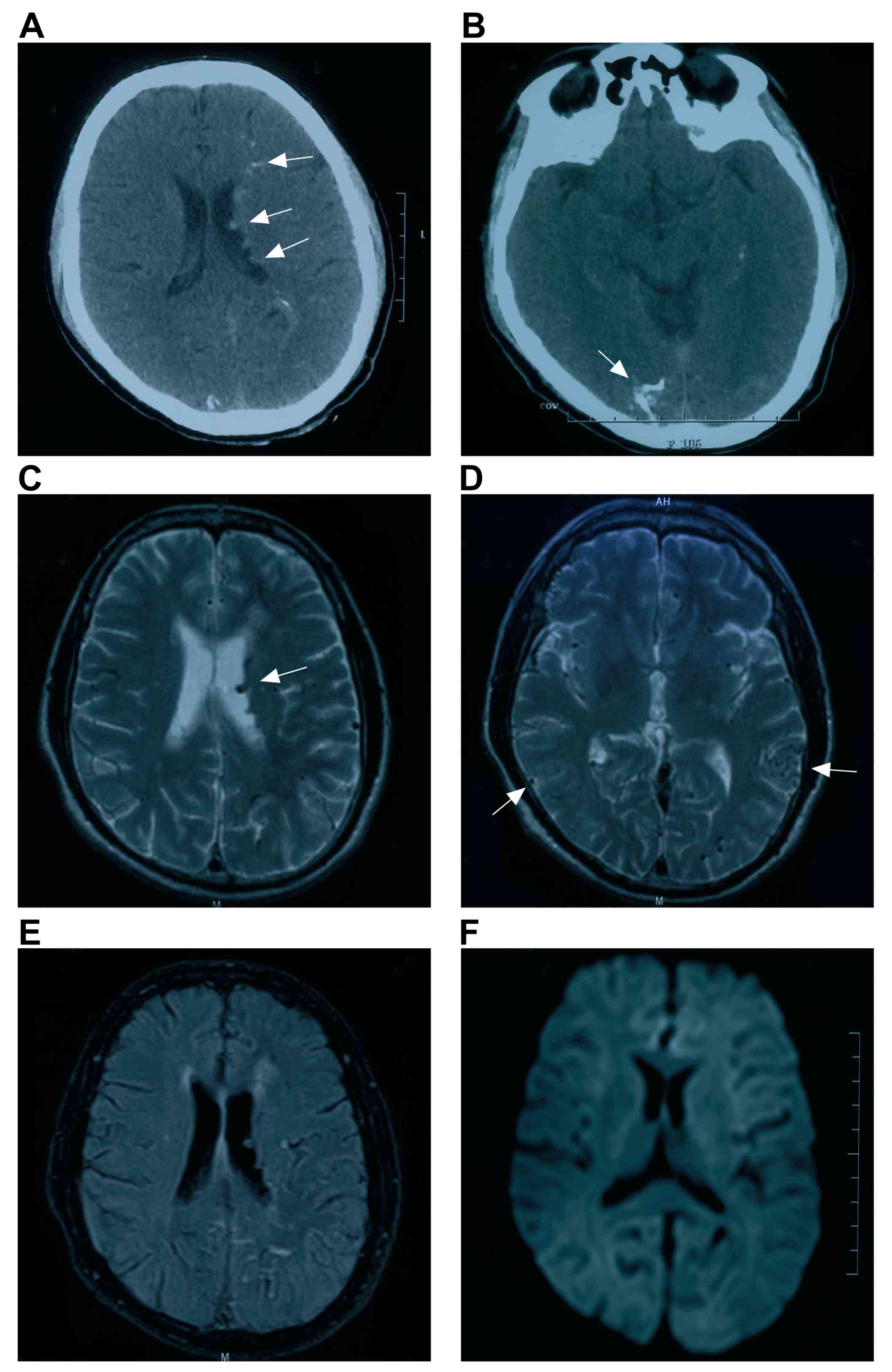
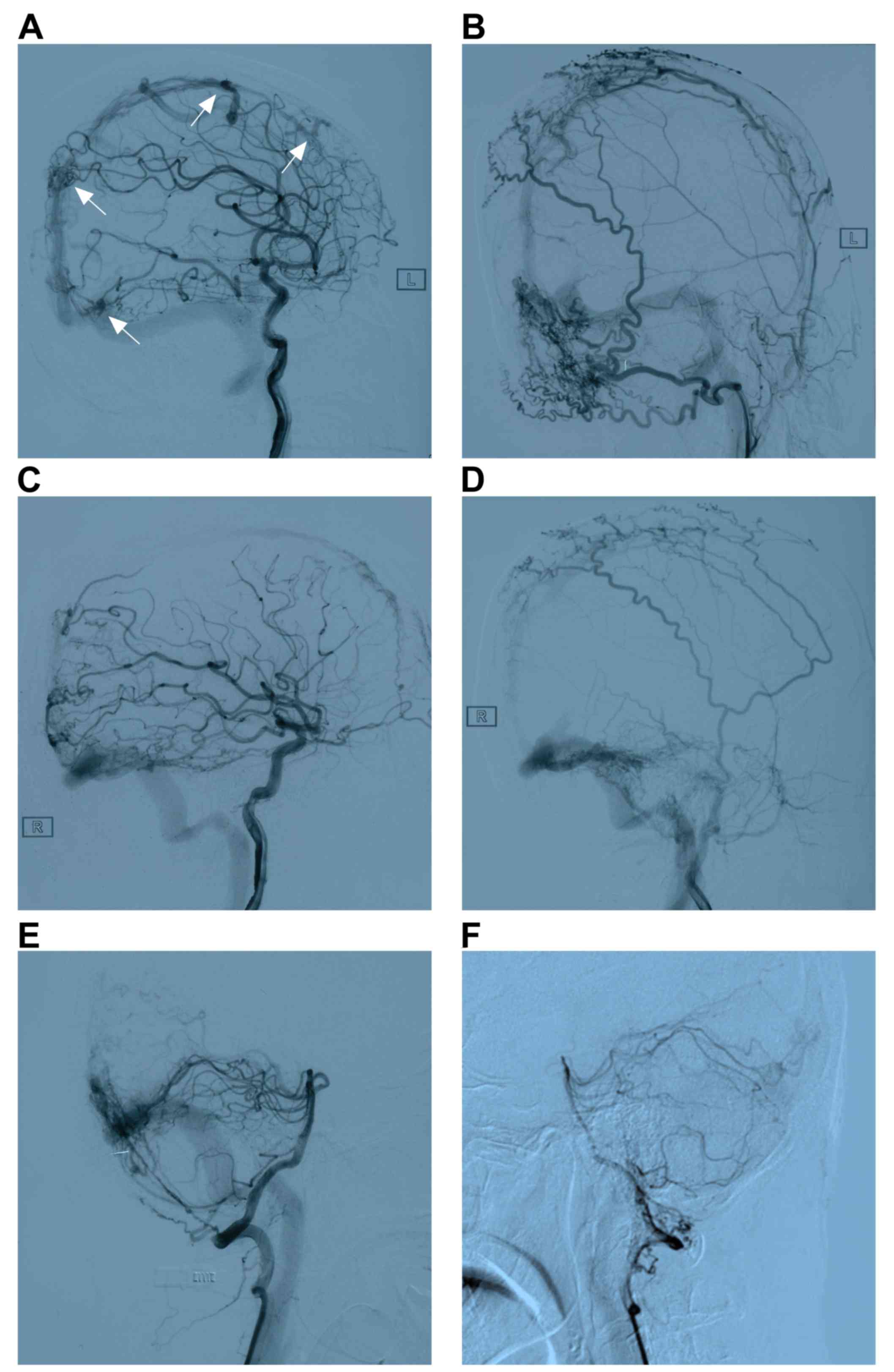
|
1
|
Barnwell SL, Halbach VV, Dowd CF,
Higashida RT, Hieshima GB and Wilson CB: Multiple dural
arteriovenous fistulas of the cranium and spine. AJNR Am J
Neuroradiol. 12:441–445. 1991.PubMed/NCBI
|
|
2
|
Ha SY, Kwon YS, Kim BM, Kim DI and Kim DJ:
Clinical and angiographic characteristics of multiple dural
arteriovenous shunts. AJNR Am J Neuroradiol. 33:1691–1695. 2012.
View Article : Google Scholar : PubMed/NCBI
|
|
3
|
van Dijk JM, TerBrugge KG, Willinsky RA
and Wallace MC: Multiplicity of dural arteriovenous fistulas. J
Neurosurg. 96:76–78. 2002. View Article : Google Scholar : PubMed/NCBI
|
|
4
|
Fujita A, Nakamura M and Tamaki N:
Multiple dural arteriovenous fistulas involving both the cavernous
sinus and the posterior fossa: Report of two cases and review of
the literature. No Shinkei Geka. 29:1065–1072. 2001.PubMed/NCBI
|
|
5
|
Folkman J: Successful treatment of an
angiogenic disease. N Engl J Med. 320:1211–1212. 1989. View Article : Google Scholar : PubMed/NCBI
|
|
6
|
Yu J, Lv X, Li Y and Wu Z: Therapeutic
progress in pediatric intracranial dural arteriovenous shunts: A
review. Interv Neuroradiol. 22:548–556. 2016. View Article : Google Scholar : PubMed/NCBI
|
|
7
|
Fudaba H, Kubo T, Goda M, Sugita K,
Morishige M, Onishi K, Ishii K, Anan M, Nagai Y and Fujiki M: The
Potentiality for development of multiple dural arteriovenous
fistulas after ligation of the internal jugular vein. A case
report. NMC Case Rep J. 4:71–73. 2017. View Article : Google Scholar : PubMed/NCBI
|
|
8
|
Oh SJ, Chon YI, Kong SK and Goh EK:
Multiple dural arteriovenous fistulas presenting as pulsatile
tinnitus treated with external manual compression. J Audiol Otol.
21:156–159. 2017. View Article : Google Scholar : PubMed/NCBI
|
|
9
|
Martinez-Burbano B, Correa Diaz EP and
Jácome Sánchez C: Evolutionary history of multiple dural fistula. J
Investig Med High Impact Case Rep. 4: View Article : Google Scholar : 2016.PubMed/NCBI
|
|
10
|
Mitsuhara T, Ikawa F, Ohbayashi N, Shirozu
H, Abiko M and Ichinose N: A case of multiple dural arteriovenous
fistulas treated by multiple modalities. No Shinkei Geka.
39:575–580. 2011.PubMed/NCBI
|
|
11
|
Shankar JJ, Terbrugge Karel and Krings T:
Multiple spinal and cranial dural arteriovenous fistulas. J
Neurosurg Spine. 15:113–116. 2011. View Article : Google Scholar : PubMed/NCBI
|
|
12
|
Spittau B, Millán DS, El-Sherifi S, Hader
C, Singh TP, Motschall E, Vach W, Urbach H and Meckel S: Dural
arteriovenous fistulas of the hypoglossal canal: Systematic review
on imaging anatomy, clinical findings, and endovascular management.
J Neurosurg. 122:883–903. 2015. View Article : Google Scholar : PubMed/NCBI
|
|
13
|
Kubota Y, Ueda T, Kaku Y and Sakai N:
Development of a dural arteriovenous fistula around the jugular
valve after transvenous embolization of cavernous dural
arteriovenous fistula. Surg Neurol. 51:174–176. 1999. View Article : Google Scholar : PubMed/NCBI
|
|
14
|
Rahmanian A, Farrokhi MR, Alibai EA and
Masoudi MS: Multiple intracranial dural arteriovenous fistula. J
Res Med Sci. 18:360–362. 2013.PubMed/NCBI
|
|
15
|
Minamide H, Hayashi Y and Uchiyama N:
Multiple tentorial dural arteriovenous fistulas with acquired pial
arteriovenous fistula presented with unilateral eye symptoms
successfully treated by transarterial embolization and direct
surgery. A case report. No Shinkei Geka. 45:239–245.
2017.PubMed/NCBI
|
|
16
|
Kusaka N, Sugiu K, Katsumata A, Nakashima
H, Tamiya T and Ohmoto T: The importance of venous hypertension in
the formation of dural arteriovenous fistulas: A case report of
multiple fistulas remote from sinus thrombosis. Neuroradiology.
43:980–984. 2001. View Article : Google Scholar : PubMed/NCBI
|
|
17
|
Mirza FA and Fraser JF: Multiple dural and
pial arteriovenous fistulae in a twenty-four-year-old woman in the
setting of superior sagittal sinus thrombosis: Case report and
review of literature. J Stroke Cerebrovasc Dis. 25:e192–e199. 2016.
View Article : Google Scholar : PubMed/NCBI
|
|
18
|
Nishijima M, Takaku A, Endo S, Kuwayama N,
Koizumi F, Sato H and Owada K: Etiological evaluation of dural
arteriovenous malformations of the lateral and sigmoid sinuses
based on histopathological examinations. J Neurosurg. 76:600–606.
1992. View Article : Google Scholar : PubMed/NCBI
|
|
19
|
Saito A, Takahashi N, Furuno Y, Kamiyama
H, Nishimura S, Midorikawa H and Nishijima M: Multiple isolated
sinus dural arteriovenous fistulas associated with antithrombin III
deficiency. A case report. Neurol Med Chir (Tokyo). 48:455–459.
2008. View Article : Google Scholar : PubMed/NCBI
|
|
20
|
Desal HA, Lee SK, Kim BS, Raoul S,
Tymianski M and TerBrugge KG: Multiple de novo vascular
malformations in relation to diffuse venous occlusive disease. A
case report. Neuroradiology. 47:38–42. 2005. View Article : Google Scholar : PubMed/NCBI
|
|
21
|
Matsubara S, Satoh K, Satomi J, Shigekiyo
T, Kinouchi T, Miyake H and Nagahiro S: Acquired pial and dural
arteriovenous fistulae following superior sagittal sinus thrombosis
in patients with protein S deficiency: A report of two cases.
Neurol Med Chir (Tokyo). 54:245–252. 2014. View Article : Google Scholar : PubMed/NCBI
|
|
22
|
Sugiura Y, Miyamoto T, Takehara S, Sumiya
K and Nozaki T: Multiple dural arteriovenous fistulas following
extensive sinus thrombosis. A case report. No Shinkei Geka.
24:379–383. 1996.PubMed/NCBI
|
|
23
|
Aoun SG, Bendok BR and Batjer HH: Acute
management of ruptured arteriovenous malformations and dural
arteriovenous fistulas. Neurosurg Clin N Am. 23:87–103. 2012.
View Article : Google Scholar : PubMed/NCBI
|
|
24
|
Russell SM, Woo HH and Nelson PK:
Transarterial wedged-catheter, flow-arrest, N-butyl cyanoacrylate
embolization of three dural arteriovenous fistulae in a single
patient. Interv Neuroradiol. 9:283–290. 2003. View Article : Google Scholar : PubMed/NCBI
|
|
25
|
Chaloupka JC, Marx WF and Kallmes DF:
Dural arteriovenous fistulas. J Neurosurg. 94:858–861.
2001.PubMed/NCBI
|
|
26
|
Bai Y, He C, Zhang H and Ling F: De novo
multiple dural arteriovenous fistulas and arteriovenous
malformation after embolization of cerebral arteriovenous fistula.
A case report. Childs Nerv Syst. 28:1981–1983. 2012. View Article : Google Scholar : PubMed/NCBI
|
|
27
|
Vilela P, Willinsky R and Terbrugge K:
Treatment of intracranial venous occlusive disease with sigmoid
sinus angioplasty and stent placement in a case of infantile
multifocal dural arteriovenous shunts. Interv Neuroradiol. 7:51–60.
2001. View Article : Google Scholar : PubMed/NCBI
|
|
28
|
Ushikoshi S, Kikuchi Y and Miyasaka K:
Multiple dural arteriovenous shunts in a 5-year-old boy. AJNR Am J
Neuroradiol. 20:728–730. 1999.PubMed/NCBI
|
|
29
|
Prats-Sánchez LA, Hervás-García JV,
Becerra JL, Lozano M, Castaño C, Munuera J, Escudero D and
García-Esperón C: Multiple intracranial arteriovenous fistulas in
Cowden syndrome. J Stroke Cerebrovasc Dis. 25:e93–e94. 2016.
View Article : Google Scholar : PubMed/NCBI
|
|
30
|
Gist TL, Rangel-Castilla L, Krishna C,
Roman GC, Cech DA and Diaz O: Endovascular management of six
simultaneous intracranial dural arteriovenous fistulas in a single
patient. J Neurointerv Surg. 6:e162014. View Article : Google Scholar : PubMed/NCBI
|
|
31
|
Li M, Lin N, Wu J, Liang J and He W:
Multiple intracranial aneurysms associated with multiple dural
arteriovenous fistulas and cerebral arteriovenous malformation.
World Neurosurg. 77:3982012. View Article : Google Scholar : PubMed/NCBI
|
|
32
|
Pascoe HM, Lui EH, Mitchell P and Gaillard
F: Progressive subcortical calcifications secondary to venous
hypertension in an intracranial dural arteriovenous fistula. J Clin
Neurosci. 39:98–101. 2017. View Article : Google Scholar : PubMed/NCBI
|
|
33
|
Borden JA, Wu JK and Shucart WA: A
proposed classification for spinal and cranial dural arteriovenous
fistulous malformations and implications for treatment. J
Neurosurg. 82:166–179. 1995. View Article : Google Scholar : PubMed/NCBI
|
|
34
|
Cognard C, Gobin YP, Pierot L, Bailly AL,
Houdart E, Casasco A, Chiras J and Merland JJ: Cerebral dural
arteriovenous fistulas: Clinical and angiographic correlation with
a revised classification of venous drainage. Radiology.
194:671–680. 1995. View Article : Google Scholar : PubMed/NCBI
|
|
35
|
Dogan M, Kahraman AS, Firat C, Ak M,
Yildirim O and Dogan DG: Multiple dural arteriovenous fistulas
involving the cavernous sinus, transverse sinus, sigmoid sinus and
spinal drainage: CT angiography findings in 14-year-old boy. Eur
Rev Med Pharmacol Sci. 16:1305–1306. 2012.PubMed/NCBI
|
|
36
|
Zeidman SM, Monsein LH, Arosarena O,
Aletich V, Biafore JA, Dawson RC, Debrun GM and Hurko O:
Reversibility of white matter changes and dementia after treatment
of dural fistulas. AJNR Am J Neuroradiol. 16:1080–1083.
1995.PubMed/NCBI
|
|
37
|
Iizuka Y, Rodesch G, Garcia-Monaco R,
Alvarez H, Burrows P, Hui F and Lasjaunias P: Multiple cerebral
arteriovenous shunts in children: Report of 13 cases. Childs Nerv
Syst. 8:437–444. 1992. View Article : Google Scholar : PubMed/NCBI
|
|
38
|
Signorelli F, Gory B, Maduri R, Guyotat J,
Pelissou-Guyotat I, Chirchiglia D, Riva R and Turjman F:
Intracranial dural arteriovenous fistulas: A review of their
current management based on emerging knowledge. J Neurosurg Sci.
61:193–206. 2017.PubMed/NCBI
|
|
39
|
Serulle Y, Miller TR and Gandhi D: Dural
arteriovenous fistulae: Imaging and management. Neuroimaging Clin N
Am. 26:247–258. 2016. View Article : Google Scholar : PubMed/NCBI
|
|
40
|
Tsai LK, Liu HM and Jeng JS: Diagnosis and
management of intracranial dural arteriovenous fistulas. Expert Rev
Neurother. 16:307–318. 2016. View Article : Google Scholar : PubMed/NCBI
|
|
41
|
Awad IA, Little JR, Akarawi WP and Ahl J:
Intracranial dural arteriovenous malformations: Factors
predisposing to an aggressive neurological course. J Neurosurg.
72:839–850. 1990. View Article : Google Scholar : PubMed/NCBI
|
|
42
|
Mendonça N, Santos G, Duro D, Machado E,
Goulão A and Santana I: Multiple dural arteriovenous fistulas
presenting as rapidly progressive dementia. Neurologist.
18:130–132. 2012. View Article : Google Scholar : PubMed/NCBI
|
|
43
|
Netravathi M, Pal PK, Bharath RD and
Ravishankar S: Intracranial dural arteriovenous fistula presenting
as parkinsonism and cognitive dysfunction. J Clin Neurosci.
18:138–140. 2011. View Article : Google Scholar : PubMed/NCBI
|
|
44
|
Abe K, Okuda O, Ohishi H, Sonobe M and
Arai H: Multiple dural arteriovenous fistulas causing rapid
progressive dementia successfully treated by endovascular surgery.
A case report. Neurol Med Chir (Tokyo). 54:145–149. 2014.
View Article : Google Scholar : PubMed/NCBI
|
|
45
|
Mejia P, Piedra LM and Merchan-Del Hierro
X: Rapidly progressive dementia and parkinsonism associated to
multiple dural arteriovenous fistulas. Rev Neurol. 64:214–218.
2017.PubMed/NCBI
|
|
46
|
Takai K, Komori T and Taniguchi M:
Microvascular anatomy of spinal dural arteriovenous fistulas:
Arteriovenous connections and their relationships with the dura
mater. J Neurosurg Spine. 23:526–533. 2015. View Article : Google Scholar : PubMed/NCBI
|
|
47
|
Hetts SW, Moftakhar P, Maluste N,
Fullerton HJ, Cooke DL, Amans MR, Dowd CF, Higashida RT and Halbach
VV: Pediatric intracranial dural arteriovenous fistulas:
Age-related differences in clinical features, angioarchitecture,
and treatment outcomes. J Neurosurg Pediatr. 18:602–610. 2016.
View Article : Google Scholar : PubMed/NCBI
|
|
48
|
Alerhand S and Lay C: Spontaneous
iIntracerebral hemorrhage. Emerg Med Clin North Am. 35:825–845.
2017. View Article : Google Scholar : PubMed/NCBI
|
|
49
|
Kunz WG, Schuler F, Sommer WH, Fabritius
MP, Havla L, Meinel FG, Reiser MF, Ertl-Wagner B and Thierfelder
KM: Wavelet-based angiographic reconstruction of computed
tomography perfusion data: Diagnostic value in cerebral venous
sinus thrombosis. Invest Radiol. 52:302–309. 2017. View Article : Google Scholar : PubMed/NCBI
|
|
50
|
Okamura A, Nakaoka M, Ohbayashi N, Yahara
K and Nabika S: Intraoperative cone-beam computed tomography
contributes to avoiding hypoglossal nerve palsy during transvenous
embolization for dural arteriovenous fistula of the anterior
condylar confluence. Interv Neuroradiol. 22:584–589. 2016.
View Article : Google Scholar : PubMed/NCBI
|
|
51
|
Lescher S, Gehrisch S, Klein S and
Berkefeld J: Time-resolved 3D rotational angiography: Display of
detailed neurovascular anatomy in patients with intracranial
vascular malformations. J Neurointerv Surg. 9:887–894. 2017.
View Article : Google Scholar : PubMed/NCBI
|
|
52
|
Azuma M, Hirai T, Shigematsu Y, Kitajima
M, Kai Y, Yano S, Nakamura H, Makino K, Iryo Y and Yamashita Y:
Evaluation of intracranial dural arteriovenous fistulas: Comparison
of unenhanced 3T 3D time-of-flight MR angiography with digital
subtraction angiography. Magn Reson Med Sci. 14:285–293. 2015.
View Article : Google Scholar : PubMed/NCBI
|
|
53
|
Willinsky R, Terbrugge K, Montanera W,
Mikulis D and Wallace MC: Venous congestion: An MR finding in dural
arteriovenous malformations with cortical venous drainage. AJNR Am
J Neuroradiol. 15:1501–1507. 1994.PubMed/NCBI
|
|
54
|
Ushikoshi S, Kikuchi Y, Houkin K, Saito H
and Abe H: Multiple dural arteriovenous fistulas. Neurol Med Chir
(Tokyo). 38:478–484. 1998. View Article : Google Scholar : PubMed/NCBI
|
|
55
|
Fiumara E, Tumbiolo S, Bellomonte ML,
Savatteri P, Finazzo F and La Gattuta F: Resection of the
transverse sinuses and confluence of sinuses for treatment of
multiple dural arteriovenous fistulas. A case report. J Neurosurg.
100:348–352. 2004. View Article : Google Scholar : PubMed/NCBI
|
|
56
|
Sato K, Matsumoto Y, Endo H and Tominaga
T: A hemorrhagic complication after Onyx embolization of a
tentorial dural arteriovenous fistula: A caution about subdural
extension with pial arterial supply. Interv Neuroradiol.
23:307–312. 2017. View Article : Google Scholar : PubMed/NCBI
|
|
57
|
Luo CB, Chang FC and Teng MM: Update of
embolization of intracranial dural arteriovenous fistula. J Chin
Med Assoc. 77:610–617. 2014. View Article : Google Scholar : PubMed/NCBI
|
|
58
|
Watanabe T, Matsumaru Y, Sonobe M, Asahi
T, Onitsuka K, Sugita K, Takahashi S and Nose T: Multiple dural
arteriovenous fistulae involving the cavernous and sphenoparietal
sinuses. Neuroradiology. 42:771–774. 2000. View Article : Google Scholar : PubMed/NCBI
|
|
59
|
Nakamura M, Tamaki N, Hara Y and Nagashima
T: Two unusual cases of multiple dural arteriovenous fistulas.
Neurosurgery. 41:288–292; discussion 292–283. 1997. View Article : Google Scholar : PubMed/NCBI
|
|
60
|
Lin N, Brouillard AM, Mokin M, Natarajan
SK, Snyder KV, Levy EI and Siddiqui AH: Direct access to the middle
meningeal artery for embolization of complex dural arteriovenous
fistula: A hybrid treatment approach. J Neurointerv Surg.
7:e242015. View Article : Google Scholar : PubMed/NCBI
|
|
61
|
Vougioukas VI, Coulin CJ, Shah M, Berlis
A, Hubbe U and Van Velthoven V: Benefits and limitations of image
guidance in the surgical treatment of intracranial dural
arteriovenous fistulas. Acta Neurochir (Wien). 148:145–153,
discussion 153. 2006. View Article : Google Scholar : PubMed/NCBI
|
|
62
|
Al-Mefty O, Jinkins JR and Fox JL:
Extensive dural arteriovenous malformation. A case report. J
Neurosurg. 65:417–420. 1986. View Article : Google Scholar : PubMed/NCBI
|
|
63
|
Yen CP, Lanzino G and Sheehan JP:
Stereotactic radiosurgery of intracranial dural arteriovenous
fistulas. Neurosurg Clin N Am. 24:591–596. 2013. View Article : Google Scholar : PubMed/NCBI
|
|
64
|
Dmytriw AA, Schwartz ML, Cusimano MD,
Mendes Pereira V, Krings T, Tymianski M, Radovanovic I and Agid R:
Gamma Knife radiosurgery for the treatment of intracranial dural
arteriovenous fistulas. Interv Neuroradiol. 23:211–220. 2017.
View Article : Google Scholar : PubMed/NCBI
|
|
65
|
Bertalanffy A, Dietrich W, Kitz K and
Bavinzski G: Treatment of dural arteriovenous fistulae (dAVF's) at
the superior sagittal sinus (SSS) using embolisation combined with
micro- or radiosurgery. Minim Invasive Neurosurg. 44:205–210. 2001.
View Article : Google Scholar : PubMed/NCBI
|
|
66
|
Kim DJ, terBrugge K, Krings T, Willinsky R
and Wallace C: Spontaneous angiographic conversion of intracranial
dural arteriovenous shunt: Long-term follow-up in nontreated
patients. Stroke. 41:1489–1494. 2010. View Article : Google Scholar : PubMed/NCBI
|
|
67
|
Friedman JA, Meyer FB, Nichols DA, Coffey
RJ, Hopkins LN, Maher CO, Meissner ID and Pollock BE: Fatal
progression of posttraumatic dural arteriovenous fistulas
refractory to multimodal therapy. A case report. J Neurosurg.
94:831–835. 2001. View Article : Google Scholar : PubMed/NCBI
|
|
68
|
Torok CM, Nogueira RG, Yoo AJ,
Leslie-Mazwi TM, Hirsch JA, Stapleton CJ, Patel AB and Rabinov JD:
Transarterial venous sinus occlusion of dural arteriovenous
fistulas using ONYX. Interv Neuroradiol. 22:711–716. 2016.
View Article : Google Scholar : PubMed/NCBI
|