Introduction
Idiopathic achalasia is a motor disease of the
esophagus characterized by the absence of peristalsis and
incomplete relaxation of the lower esophageal sphincter. The most
common clinical symptoms of achalasia include dysphagia,
regurgitation, chest pain and weight loss. Currently available
therapeutic options include medical treatment (such as calcium
channel blockers and nitrates), botulinum toxin injection,
pneumatic dilation, Heller myotomy and peroral endoscopic myotomy
(POEM) (1). In particular, POEM is
emerging as the treatment of choice for a significant proportion of
patients (1). The etiology and
pathophysiology of achalasia remain poorly understood, although it
has been associated with a variety of putative mechanisms,
including infection, autoimmune reactions, inflammatory processes
and genetic factors (2,3). In addition, a possible association
between achalasia and infection has been suggested. Several types
of causal viruses, including herpes simplex virus, bornavirus,
varicella zoster, measles and human papilloma virus, have all been
documented to be involved (4). The
contribution of human leukocyte antigen class II genes to the
susceptibility of achalasia has also been demonstrated previously
(3,5). Achalasia has been associated with the
functional loss of myenteric plexus ganglion cells and neurons in
the distal esophagus and with substantial immune cell infiltration
that consists predominantly of T lymphocytes, which appears to
decrease alongside disease progression (6). Moreover, the detection of circulating
anti-myenteric plexus autoantibodies in patients with achalasia and
its occurrence in association with other autoimmune diseases
(7) supports the role of an
inflammatory component mediated by the immune system within this
disorder. Characterization of infiltrating cells in the tissues
(muscle specimens) has largely been performed using histological or
immunohistochemical analyses, which produced conflicting reports
(7,8). Conversely, studies investigating the
involvement of circulating levels of inflammatory mediators,
cytokines, chemokines and growth factors in patients with achalasia
remain insufficient (9,10).
Therefore, the present study aimed to characterize
the systemic inflammatory patterns in the serum of patients with
achalasia and healthy individuals. The aim of the present study was
to explore the role of cytokines, chemokines and growth factors in
the pathogenesis of achalasia, to gain some insight into the
disease mechanisms and etiology of the disease.
Patients and methods
Patient enrolment and serum
collection
A total of 68 patients (35 male and 33 female; age
range, 27-84 years; median age, 59 years) diagnosed clinically with
achalasia and confirmed by manometry were enrolled between October
2014 and March 2018 at the Division of Gastroenterology of ‘Casa
Sollievo della Sofferenza’ Hospital (San Giovanni Rotondo, Italy).
The study protocol followed the ethical guidelines of the
Declaration of Helsinki (11), was
approved by the Institutional Ethics Committee of the ‘Casa
Sollievo della Sofferenza’ hospital (approval no. 139/CE on
28/10/2014) and all patients provided written informed consent.
Esophageal manometry was performed using a low compliance perfusion
system (12), where the manometric
criteria used for achalasia diagnosis included esophageal
aperistalsis and poor lower esophageal sphincter relaxation
(13).
The medical records were also reviewed using a
validated questionnaire. Clinical data regarding symptoms of
regurgitation, dysphagia, chest pain and weight loss were collected
and classified by following the scoring system previously described
by Eckardt et al (14). Concomitant autoimmune
diagnosis, including uveitis, type I diabetes, rheumatoid arthritis
and thyroid diseases, pneumatic dilatation of the esophagus and/or
surgical therapy were reported. The healthy control group was
comprised of 39 age- and sex-matched healthy blood donors (20 male
and 19 female; age range, 30-79 years; median age, 59 years), who
were recruited in the same time period and volunteered to donate to
the blood bank of ‘Casa Sollievo della Sofferenza’ Hospital. The
exclusion criteria for these samples were diagnoses of
comorbidities, such as diabetes, autoimmune disease or any
associated inflammatory or infectious diseases and administration
of treatment with anti-inflammatory drugs. Written informed consent
was obtained from all subjects. Blood samples from the patients and
controls were collected from the Laboratory of Research, Division
of Gastroenterology of ‘Casa Sollievo della Sofferenza’ Hospital
and placed into Vacutainer® Plus Plastic SST™ tubes with
Gel for Serum Separation (BD Biosciences). The samples were then
centrifuged at 1,620 x g for 15 min at room temperature before the
sera were aliquoted into NUNC-cryovial tubes (Thermo Fisher
Scientific Inc.) and stored at -80˚C until the time of analysis to
prevent protein degradation. None of the serum samples had been
previously thawed prior to thawing for the assay.
Cytokines, chemokines and growth
factor measurement
A panel of 27 cytokines and chemokines were measured
in the serum samples in duplicate using the Bio-Plex Pro Human
Cytokine assay in 96-well plates (Bio-Rad Laboratories, Inc.),
according to the protocols of the manufacturer. The Bio-Plex Pro
Human Cytokine assay panel integrates a network of biologically
relevant cytokines and chemokines in a single assay. It applies a
standard sandwich ELISA method, which enables the simultaneous
quantification and analysis of the following 27 cytokines,
chemokines and growth factors: IL-1β, IL-1 receptor antagonist,
IL-2, IL-4, IL-5, IL-6, IL-7, IL-8, IL-9, IL-10, IL-12p70, IL-13,
IL-15, IL-17, Hu eotaxin, basic fibroblast growth factor (bFGF),
granulocyte colony-stimulating factor (GCSF),
granulocyte-macrophage colony-stimulating factor (GM-CSF),
interferon (IFN)-γ, IFN-γ-induced protein 10 (IP-10), monocyte
chemotactic protein (MCP)-1, macrophage inflammatory protein
(MIP)-1α, platelet-derived growth factor (PDGF), MIP-1β, regulated
upon activation normal T cell expressed and presumably secreted
(RANTES), TNF-α and vascular endothelial growth factor (VEGF).
The appropriate analyte standards and samples were
diluted in standard diluent and sample diluent, respectively. The
median coefficient of variation was set at <25% for all
cytokines analyzed. Bio-Plex Manager version 6.1 (Bio-Rad
Laboratories, Inc.) was used for data analyses. Protein
concentrations were calculated using the appropriate standard
curves.
Statistical analysis
Clinical characteristics of patients with achalasia
and healthy individuals were presented as the mean ± standard
deviation, and as observed frequencies (and percentages) for
continuous and categorical variables. Distribution of protein
concentration in sera was evaluated for each considered analyte
using quantile-quantile plots and the distribution of data was
assessed using a Shapiro-Wilk test. Due to log-normal
distributions, comparisons of mean protein concentrations between
the patient and control groups were performed using an unpaired
two-samples t-test of the log-transformed values (log-pg/ml).
P-values were also adjusted for multiple comparisons using
Bonferroni correction. Two-sided P<0.05 was considered to
indicate a statistically significant difference. For the
concentration of those cytokines, chemokines and growth factors
that could be detected but not necessarily quantified as an exact
value, a visual evaluation approach proposed by the Expert Working
Group of the International Conference on Harmonisation of Technical
Requirements for Registration of Pharmaceuticals for Human Use was
performed (15). This is because
the Bio-Plex Pro Human Cytokine assay has limits of quantification,
which is defined as the lowest and the highest amount of analyte in
a sample that can be quantitatively determined with suitable
precision and accuracy. These unquantified concentrations were
replaced by suitable values, which were estimated using the
following protocol: i) For each analyte, the quantification limit
was determined by observing the minimum and maximum concentration
values observed in the data sample; ii) in cases where the
unquantified concentration was below the limit of quantification,
this was replaced by one that is 10% lower than the minimum
detected value; and iii) otherwise, this would be replaced by one
that is 10% higher than the maximum detected value. All statistical
analyses were performed using R Core Team (16) with the following packages: Tableone
(github.com/kaz-yos/tableone), tidyr
(github.com/tidyverse/tidyr), ggplot2
(github.com/tidyverse/ggplot2).
Results
Clinical characteristics of the
patients
The general and clinical characteristics of patients
with achalasia are summarized in Table
I. In total, 35 patients (51.5%) underwent surgical myotomy and
18 patients (26.5%) were successfully managed only by pneumatic
dilatation. In addition, 15 patients (22%) underwent both
esophagomyotomy and pneumatic dilatation, whilst 21 patients
(30.9%) developed megaesophagus.
 | Table IGeneral characteristics of the
patients and controls. |
Table I
General characteristics of the
patients and controls.
| Characteristics | Patients | Controls |
|---|
| Sample size, n | 68 | 39 |
| Sex, n,
male/female | 35/33 | 20/19 |
| Age, mean ± standard
deviation | 59±13 | 60±14 |
| Dysphagia, n (%) | 63 (92.6) | - |
| Esophageal
regurgitation, n (%) | 57 (83.8) | - |
| Chest pain, n
(%) | 11 (16.2) | - |
| Weight loss, n
(%) | | |
|
<5 | 48 (70.6) | - |
|
5-10 | 13 (19.1) | - |
|
>10 | 7 (10.3) | - |
| Autoimmune condition,
n (%) | 18 (26.5) | - |
| Megaesophagus, n
(%) | 21(30) | - |
| Pneumatic dilatation,
n (%) | 18 (26.5) | - |
| Surgical myotomy, n
(%) | 35 (51.5) | - |
| Pneumatic dilatation
and surgical myotomy, n (%) | 15(22) | - |
Comparison of cytokines, chemokines
and growth factor concentrations in the sera of patients with
achalasia and healthy individuals
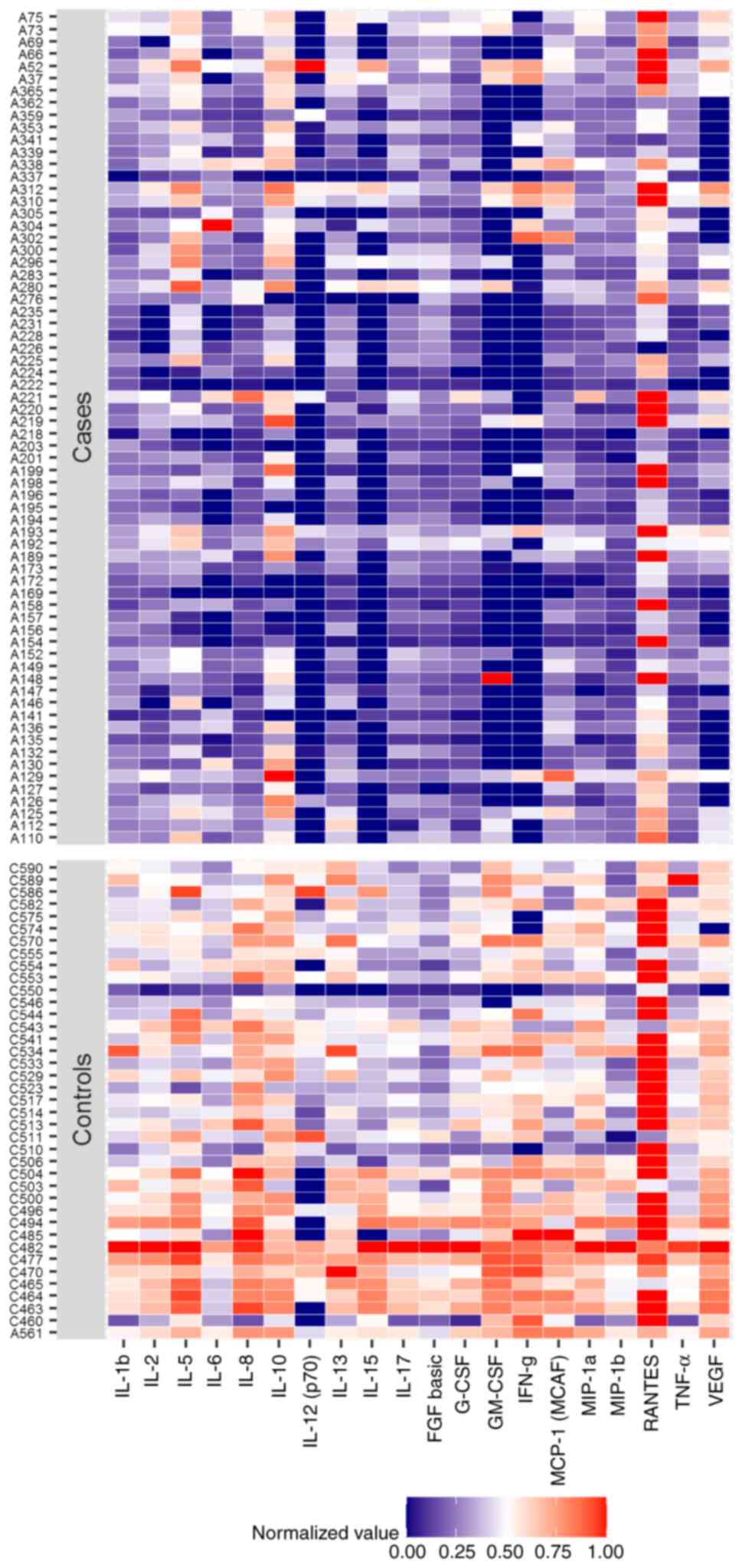
Analysis of the concentrations of markers between
the two study groups detected reduced levels of several cytokines,
namely IL-1β, IL-2, IL-5, IL-6, IL-8, IL-10, IL-12p70, IL-13,
IL-15, IL-17, bFGF, G-CSF, GM-CSF, IFN-γ, MCP-1, MIP-1α, MIP-1β,
RANTES, TNF-α and VEGF, in the patients with achalasia compared
with those in the control group (all P<0.001; Table II). Additionally, limits of
quantification were detected for IL-6, IL-12p70, IL-15, GM-CSF,
IFN-γ, VEGF and RANTES in >15% of the observations (Table SI). A heatmap of the normalized
protein concentrations (that is the values from 0 to 1), which
resulted in significantly different between patients and controls,
is depicted in Fig. 1. In addition,
to evaluate if the inflammatory profile differed between the
cohorts, the 27 markers were re-grouped into the ‘chemokines’,
‘anti-inflammatory’, ‘pro-inflammatory’ and ‘growth factors’
categories. However, no significant changes were observed (data not
shown).
| Table IIConcentration of proteins in the sera
of patients with achalasia and controls. |
Table II
Concentration of proteins in the sera
of patients with achalasia and controls.
| Analyte | Controls,
n=39f | Patients,
n=68f | Raw
P-valuea | Bonferroni adjusted
P-valueb |
|---|
| IL-1β | 2.49±0.97 | 1.07±0.52 |
<0.001e |
<0.001e |
| IL-1Ra | 5.16±0.83 | 4.70±0.76 | 0.005d | 0.127 |
| IL-2 | 2.77±0.51 | 1.91±0.49 |
<0.001e |
<0.001e |
| IL-4 | 2.78±0.45 | 2.52±0.41 | 0.003d | 0.082 |
| IL-5 | 3.67±0.49 | 3.20±0.48 |
<0.001e |
<0.001e |
| IL-6 | 2.72±0.71 | 1.58±1.09 |
<0.001e |
<0.001e |
| IL-7 | 2.98±0.38 | 2.83±0.40 | 0.060 | 1.000 |
| IL-8 | 5.52±1.39 | 2.53±0.91 |
<0.001e |
<0.001e |
| IL-9 | 5.79±0.18 | 5.76±0.15 | 0.259 | 1.000 |
| IL-10 | 2.31±0.51 | 1.64±1.02 |
<0.001e | 0.006d |
| IL-12p70 | 1.37±0.93 | 0.38±0.64 |
<0.001e |
<0.001e |
| IL-13 | 2.83±0.78 | 1.74±0.58 |
<0.001e |
<0.001e |
| IL-15 | 5.09±0.58 | 4.11±0.46 |
<0.001e |
<0.001e |
| IL-17 | 3.87±0.51 | 3.31±0.36 |
<0.001e |
<0.001e |
| Eotaxin | 5.71±0.48 | 5.68±0.47 | 0.769 | 1.000 |
| FGF basic | 3.73±0.31 | 3.52±0.18 |
<0.001e |
<0.001e |
| G-CSF | 7.30±0.68 | 6.17±0.43 |
<0.001e |
<0.001e |
| GM-CSF | 2.08±0.92 | 0.06±1.00 |
<0.001e |
<0.001e |
| IFN-γ | 2.75±1.15 | 0.67±1.14 |
<0.001e |
<0.001e |
| IP-10 | 6.32±0.54 | 6.07±0.54 | 0.022c | 0.602 |
| MCP-1 | 4.58±0.62 | 3.94±0.57 |
<0.001e |
<0.001e |
| MIP-1α | 4.16±0.96 | 2.00±0.76 |
<0.001e |
<0.001e |
| PDGF-BB | 8.46±0.61 | 8.19±0.47 | 0.012c | 0.323 |
| MIP-1β | 5.16±0.33 | 4.79±0.14 |
<0.001e |
<0.001e |
| RANTES | 9.95±0.50 | 9.41±0.59 |
<0.001e |
<0.001e |
| TNF-α | 4.10±0.37 | 3.58±0.27 |
<0.001e |
<0.001e |
| VEGF | 5.57±0.51 | 4.50±0.64 |
<0.001e |
<0.001e |
Association between serum levels of
cytokines, chemokines and growth factors and clinical data in
achalasia
To assess the relationship between the tested serum
levels and the clinical parameters of the patients (sex, dysphagia,
esophageal regurgitation, chest pain, weight loss, autoimmune
conditions, pneumatic dilatation, surgical myotomy and
megaesophagus), an association analysis was performed. After the
classification of the patients with achalasia were classified
according to sex, the serum levels of bFGF were found to be higher
in male patients compared with those in female patients (P=0.001).
Significant changes in the evaluated proteins after
re-categorization of the patients into the other remaining clinical
characteristics were not found (P>0.05; data not shown).
Discussion
A number of hypotheses point toward a multifactorial
etiopathogenesis of achalasia (2,17,18).
One of those was suggested to be viral and
autoimmune factors triggering inflammatory changes within the
tissue with ensuing damage to the myenteric plexus (2). It has been hypothesized that various
pathogens, including herpes simplex virus, varicella zoster,
measles and human papilloma virus, may play a causative role in
triggering tissue inflammation (4);
however, none have been categorically proven to be a causal agent,
since not all infected patients develop the disease (4). Chronic viral infection may trigger
aberrant immune responses, where under appropriate genetic and
environmental settings, loss of inhibitory neurons in the myenteric
plexus occurs in the lower esophageal sphincter (LES), which
releases vasoactive intestinal peptide and nitric oxide synthase
(NOS), resulting in failure of LES muscle relaxation and loss of
esophageal peristalsis (19).
Although the pathophysiological factors responsible for alterations
in enteric innervation remain unknown, the presence of lymphocytic
infiltration of the myenteric plexus, occurrence of circulating
antimyenteric neuronal antibodies and association with antigens of
the class II major histocompatibility complex all support the
existence of an immuno-inflammatory mechanism underlying neuronal
cell loss. Involvement of cytokines and inflammatory mediators may
be responsible for changes in the myenteric neuronal phenotype
(6). Cytokines and chemokines
released into the surrounding myenteric area can affect gene
expression in the enteric neurons, such as NOS, consequently
affecting LES function (20). In
addition, they may serve an important role in nerve regeneration
(21) and are able to activate
microglia, leading to neuronal excitation or neuronal loss
(22,23). The significant decrease in the
number of myenteric neurons in the distal esophagus and the LES, in
addition to the association of this reduction with inflammatory
cell infiltration, has been previously described through
histological examination of the resected specimens of patients with
achalasia, and was hypothesized to represent a secondary phenomenon
(24). At the early stages of the
disease, the inflammatory infiltrates in the esophageal myenteric
plexus are predominantly composed of the Th1, Th2 and regulatory
cell subsets (6). In addition, Th17
and Th22 cells have also been reported to be the main cellular
component in the LES muscle (25).
Recently, a CD4+-dominant inflammatory T-cell population
in the infiltrate of histological specimens at all stages of the
disease and at all levels of the esophagus was found (8).
Few studies have measured the levels of circulating
inflammatory biomarkers, namely cytokines, chemokines and growth
factors, and deduced their differences between patients with
achalasia and controls. Wang et al (9) previously measured the expression of
IL-17 and IL-22 in the serum, which was collected before and during
peroral endoscopic myotomy. It was found that the levels of IL-17
and IL-22 were significantly increased in the 14 patients who were
diagnosed with achalasia compared with those in 14 healthy
individuals. By contrast, Clayton et al (10) found no differences in the levels of
TNF-α receptor, IL-6, IFN-γ, IL-12, IL-17, IL-22 and IL-23 in the
serum of patients with achalasia compared with those with
gastroesophageal reflux disease, in addition to those amongst the
three subtypes of achalasia. However, Clayton et al
(10) did not include a control
group in their study. Due to these scarce and controversial
findings, the present study detected and quantified the differences
in the serum levels of a large panel of 27 biomarkers in 68
patients with achalasia, which were compared with those in the 39
healthy controls, with the aim of investigating the possibility of
an inflammatory basis for the etiology of the achalasia. The serum
protein levels of all markers in the patients with achalasia were
found to be significantly lower compared with those in the control
group, with the exception of IL-1 receptor antagonist, IL-4, IL-7,
IL-9, eotaxin, IP-10 and PDGF, which did not show statistical
significance. However, when the association between biomarker
levels and clinical parameters of the patients with achalasia was
analyzed, no differences were observed. At present, no explanation
may be provided as to why higher inflammatory biomarker levels were
found in the serum of the control cohort. Our working hypothesis
was that the inflammatory profile observed in the inflammatory
infiltrates in the tissue of patients with achalasia, which is
considered to be the cause of reduction or loss of neurons, could
also have been detected at systemic levels. The lack of elevations
in the levels of inflammatory biomarkers tested in the circulation
of patients, including cytokines, chemokines and growth factors,
might indicate that achalasia is a disease that is caused by local
inflammation. Specifically, the plasma biomarker levels may not
accurately reflect the extent of tissue inflammation in one organ.
Although the serum cytokine profiles of healthy subjects were
previously investigated using magnetic bead-based multiplex
immunoassays (26,27), the results from these previous
studies are not completely comparable with those from the present
study. Therefore, further studies with larger cohorts are required
to corroborate these findings. Furthermore, variations in marker
profiles identified amongst studies emphasizes the importance of
considering differences in study designs and clinicopathological
parameters of the participants, including inflammatory indices and
inflammatory diseases that develop during data interpretation. In
the present study, the control group included samples from healthy
subjects collected from the Blood Bank of the Transfusion Service
of the ‘Casa Sollievo della Sofferenza’ Hospital. As mentioned
above, information on the serum cytokine profiles in achalasia is
limited and therefore, the results of previous studies may not be
entirely comparable with those from the present study. The apparent
differences reported between the present and previous studies
(9,10) may also be explained by the
heterogeneity in the characteristics of the patients, the controls
and the methodology used. In effect, although all authors applied
immunoenzymatic assays for marker detection, the kits utilized were
different. The present study utilized a large panel that integrated
relevant cytokines and chemokines into a single assay, enabling the
simultaneous interrogation of 27 biomarkers in a single well. This
multiplex technology has been used to determine the presence and
quantify the levels of underrepresented cytokines in an accurate,
sensitive and reproducible manner (28). By contrast, in the study by
Clayton et al (10), each
kit varied in sample amount and dilution, assay incubation times,
volume of conjugates and substrates for all cytokines and receptors
analyzed. They measured the levels of 7 markers, including IL-22
and IL-23, which were not tested in the present study and no
control group was analyzed. The Human ELISA kit utilized by Wang
et al (9) determined IL-17
and IL-22 levels, where it was only possible to compare IL-17
levels. Finally, since the present study was designed to
characterize the systemic inflammation patterns in achalasia
compared with those in controls, it did not analyze the
immunohistochemical features of the inflammatory infiltrate within
the myenteric plexus of the patients, which constitutes a
limitation. Moreover, further validation using a larger sample size
of sera from patients with achalasia is necessary to confirm the
results of the present study.
In conclusion, the present results suggest that the
inflammatory processes and the presence of an inflammatory
infiltrate within the myenteric plexus of patients with achalasia
observed previously appears to be a local event and is not
reflected in the circulation. Therefore, further investigations
should be undertaken to identify specific inflammatory biomarkers
in serum samples that can contribute to the early diagnosis of the
disease and to improve our understanding of the etiopathogenesis of
achalasia.
Supplementary Material
Frequency of observations with
unquantified analytes concentration below and above the LOQ along
with the corresponding replaced values.
Acknowledgements
Not applicable.
Funding
This study was supported by a grant from the Italian Ministry of
Health.
Availability of data and materials
The datasets used and/or analyzed during the present
study are available from the corresponding author on reasonable
request.
Authors' contributions
AL conceived and designed the study, analyzed and
interpreted the data, and drafted the manuscript. AF and MC
performed the statistical analysis. AM, AC and GB provided the
clinical data and recruited the patients. AG contributed to serum
processing and performed the experiments. OP, AP and AA contributed
to study design and revision of the manuscript. All authors revised
the manuscript and accepted the final version to be published. AL
and AP confirm the authenticity of all the raw data.
Ethics approval and consent to
participate
Written informed consent was obtained from all
patients. The study protocol followed the ethical guidelines of the
Declaration of Helsinki and was approved by the Institutional
Ethics Committee (approval no. 139/CE).
Patient consent for publication
Not applicable.
Competing interests
The authors declare that they have no competing
interests.
References
|
1
|
Vaezi MF, Pandolfino JE, Yadlapati RH,
Greer KB and Kavitt RT: ACG clinical guidelines: Diagnosis and
management of achalasia. Am J Gastroenterol. 115:1393–1411.
2020.PubMed/NCBI View Article : Google Scholar
|
|
2
|
Boeckxstaens GE, Zaninotto G and Richter
JE: Achalasia. Lancet. 383:83–93. 2014.PubMed/NCBI View Article : Google Scholar
|
|
3
|
Gockel I, Becker J, Wouters MM, Niebisch
S, Gockel HR, Hess T, Ramonet D, Zimmermann J, Vigo AG, Trynka G,
et al: Common variants in the HLA-DQ region confer susceptibility
to idiopathic achalasia. Nat Genet. 46:901–904. 2014.PubMed/NCBI View
Article : Google Scholar
|
|
4
|
Boeckxstaens GE: Achalasia: Virus-induced
euthanasia of neurons? Am J Gastroenterol. 103:1610–1612.
2008.PubMed/NCBI View Article : Google Scholar
|
|
5
|
Latiano A, De Giorgio R, Volta U, Palmieri
O, Zagaria C, Stanghellini V, Barbara G, Mangia A, Andriulli A,
Corinaldesi R and Annese V: HLA and enteric antineuronal antibodies
in patients with achalasia. Neurogastroenterol. Motil. 18:520–525.
2006.PubMed/NCBI View Article : Google Scholar
|
|
6
|
Clark SB, Rice TW, Tubbs RR, Richter JE
and Goldblum JR: The nature of the myenteric infiltrate in
achalasia: An immunohistochemical analysis. Am J Surg Pathol.
24:1153–1158. 2000.PubMed/NCBI View Article : Google Scholar
|
|
7
|
Furuzawa-Carballeda J, Aguilar-León D,
Gamboa-Domínguez A, Valdovinos MA, Nuñez-Álvarez C,
Martín-del-Campo LA, Enríquez AB, Coss-Adame E, Svarch AE,
Flores-Nájera A, et al: Achalasia-an autoimmune inflammatory
disease: A cross-sectional study. J Immunol Res.
2015(729217)2015.PubMed/NCBI View Article : Google Scholar
|
|
8
|
Döhla M, Leichauer K, Gockel I, Niebisch
S, Thieme R, Lundell L, Schumacher J, Becker J, Rieker RJ, Hartmann
A, et al: Characterization of esophageal inflammation in patients
with achalasia. A Retrospective immunohistochemical study. Hum
Pathol. 85:228–234. 2019.PubMed/NCBI View Article : Google Scholar
|
|
9
|
Wang Z, Zhang J, Mi J, Ma H and Zhao D:
Expression and significance of interleukin-17 and interleukin-22 in
the serum and the lower esophageal sphincter of patients with
achalasia. Saudi J Gastroenterol. 24:242–248. 2018.PubMed/NCBI View Article : Google Scholar
|
|
10
|
Clayton S, Cauble E, Kumar A, Patil N,
Ledford D, Kolliputi N, Lopes-Virella MF, Castell D and Richter J:
Plasma levels of TNF-α, IL-6, IFN-γ, IL-12, IL-17, IL-22, and IL-23
in achalasia, eosinophilic esophagitis (EoE), and gastroesophageal
reflux disease (GERD). BMC Gastroenterol. 19(28)2019.PubMed/NCBI View Article : Google Scholar
|
|
11
|
Williams JR: The declaration of Helsinki
and public health. Bull World Health Organ. 86:650–652.
2008.PubMed/NCBI View Article : Google Scholar
|
|
12
|
Annese V, Basciani M, Perri F, Lombardi G,
Frusciante V, Simone P, Andriulli A and Vantrappen G: Controlled
trial of botulinum toxin injection vs. placebo and pneumatic
dilation in achalasia. Gastroenterology. 111:1418–1424.
1996.PubMed/NCBI View Article : Google Scholar
|
|
13
|
Richter JE: Modern management of
achalasia. Curr Treat Options Gastroenterol. 8:275–283.
2005.PubMed/NCBI View Article : Google Scholar
|
|
14
|
Eckardt VF, Aignherr C and Bernhard G:
Predictors of outcome in patients with achalasia treated by
pneumatic dilation. Gastroenterology. 103:1732–1738.
1992.PubMed/NCBI View Article : Google Scholar
|
|
15
|
International Conference on Harmonization
(ICH) Q2 (R1): Validation of Analytical Procedures-Test and
Methodology. 2005. Available from: https://database.ich.org/sites/default/files/Q2_R1_Guideline.pdf.
|
|
16
|
R Core Team (2012). R: A language and
environment for statistical computing. R Foundation for Statistical
Computing, Vienna, Austria. ISBN 3-900051-07-0, URL. Available
from: http://www.R-project.org/.
|
|
17
|
Park W and Vaezi MF: Etiology and
pathogenesis of achalasia: The current understanding. Am J
Gastroenterol. 100:1404–1414. 2005.PubMed/NCBI View Article : Google Scholar
|
|
18
|
Ghoshal UC, Daschakraborty SB and Singh R:
Pathogenesis of achalasia cardia. World J Gastroenterol.
18:3050–3057. 2012.PubMed/NCBI View Article : Google Scholar
|
|
19
|
Patel DA, Lappas BM and Vaezi MF: An
overview of achalasia and its subtypes. Gastroenterol Hepatol (NY).
13:411–421. 2017.PubMed/NCBI
|
|
20
|
Hurst SM, Stanisz AM, Sharkey KA and
Collins SM: Interleukin 1 beta-induced increase in substance P. in
rat myenteric plexus. Gastroenterology. 105:1754–1760.
1993.PubMed/NCBI View Article : Google Scholar
|
|
21
|
Hirota H, Kiyama H, Kishimoto T and Taga
T: Accelerated nerve regeneration in mice by upregulated expression
of interleukin (IL) 6 and IL-6 receptor after trauma. J Exp Med.
183:2627–2634. 1996.PubMed/NCBI View Article : Google Scholar
|
|
22
|
Takeda K and Akira S: Toll-like receptors
in innate immunity. Int Immunol. 17:1–14. 2005.PubMed/NCBI View Article : Google Scholar
|
|
23
|
Palmieri O, Mazza T, Merla A, Fusilli C,
Cuttitta A, Martino G, Latiano T, Corritore G, Bossa F, Palumbo O,
et al: Gene expression of muscular and neuronal pathways is
cooperatively dysregulated in patients with idiopathic achalasia.
Sci Rep. 6(31549)2016.PubMed/NCBI View Article : Google Scholar
|
|
24
|
Gockel I, Bohl JR, Doostkam S, Eckardt VF
and Junginger T: Spectrum of histopathologic findings in patients
with achalasia reflects different etiologies. J Gastroenterol
Hepatol. 21:727–733. 2006.PubMed/NCBI View Article : Google Scholar
|
|
25
|
Furuzawa-Carballeda J, Torres-Landa S,
Valdovinos MA, Coss-Adame E, Martín Del Campo LA and
Torres-Villalobos G: New insights into the pathophysiology of
achalasia and implications for future treatment. World J
Gastroenterol. 22:7892–7907. 2016.PubMed/NCBI View Article : Google Scholar
|
|
26
|
Kleiner G, Marcuzzi A, Zanin V, Monasta L
and Zauli G: Cytokine levels in the serum of healthy subjects.
Mediators Inflamm. 2013(434010)2013.PubMed/NCBI View Article : Google Scholar
|
|
27
|
Martinez-Fierro ML, Garza-Veloz I,
Rocha-Pizaña MR, Cardenas-Vargas E, Cid-Baez MA, Trejo-Vazquez F,
Flores-Morales V, Villela-Ramirez GA, Delgado-Enciso I,
Rodriguez-Sanchez IP and Ortiz-Castro Y: Serum cytokine, chemokine,
and growth factor profiles and their modulation in inflammatory
bowel disease. Medicine (Baltimore). 98(e17208)2019.PubMed/NCBI View Article : Google Scholar
|
|
28
|
Capone F, Guerriero E, Sorice A, Colonna
G, Ciliberto G and Costantini S: Serum cytokinome profile
evaluation: A tool to define new diagnostic and prognostic markers
of cancer using multiplexed bead-based immunoassays. Mediators
Inflamm. 2016(3064643)2016.PubMed/NCBI View Article : Google Scholar
|