Introduction
Filaggrin (FLG) is a key protein in maintaining the
integrity and function of the skin barrier; it facilitates the
aggregation of keratin filaments within keratinocytes, contributing
to the formation of flattened, nucleus-free cells in the stratum
corneum (1,2). Processing of profilaggrin by enzymes
such as stratum corneum serine protease, peptidylarginine deiminase
and caspase-14 generates natural moisturizing factors (NMFs), which
are crucial for skin hydration and flexibility (1,3).
Mutations in FLG are strongly associated with dermatological
conditions such as atopic dermatitis (AD) and ichthyosis vulgaris,
compromising the skin barrier and increasing susceptibility to
environmental irritants and allergens (4-6).
Palmar hyperlinearity (PH), characterized by pronounced
dermatoglyphics on the palms, has been closely linked with
FLG null mutations and can serve as a potential phenotypic
marker (7,8).
Despite extensive genetic characterization, direct
analysis of FLG protein in human samples remains challenging. The
complex and highly repetitive structure of FLG, particularly
exon 3, complicates comprehensive sequencing and limits routine
genetic studies. Additionally, obtaining in vivo FLG protein
data is difficult due to the invasive nature of a skin biopsy,
which raises practical and ethical concerns. These limitations
highlight the need for alternative approaches to study FLG protein
biology and its functional consequences in humans (9).
The buccal mucosa, composed of stratified
non-keratinizing epithelium in the inner lining of the cheeks,
provides an accessible and non-invasive source of epithelial cells
for molecular studies. Previous studies have suggested that buccal
cells can serve as a source for mRNA and protein analysis (10-17).
However, comprehensive characterization of FLG protein expression
and degradation in the context of AD remains limited, particularly
regarding disease-specific alterations and functional consequences.
Given the practical limitations of obtaining skin biopsies, the
present study sought to evaluate whether buccal cells could provide
a feasible non-invasive platform for investigating FLG protein
expression and degradation in humans.
The present study investigated FLG protein
expression and degradation profiles in buccal cells from patients
with AD, alongside associated phenotypic features such as PH and
cell pellet viscosity. The aim of the study was to evaluate the
potential of buccal cells as a non-invasive system for studying FLG
protein biology, providing insights into epithelial barrier
function and offering a practical alternative to skin biopsies for
molecular and diagnostic research.
Patients and methods
Study participants
The study included individuals aged 20-40 years who
were non-smokers and reported no drug use during the study period.
Patients with AD (n=36; mean age ± SD, 23.3±6.5 years) and healthy
controls (n=36; mean age ± SD, 21.9±4.3 years) were recruited from
the student and staff population at Wenzhou-Kean University
(Wenzhou, China) between September 2023 and May 2024 through
voluntary responses to campus-wide announcements and personal
referrals. Inclusion criteria for the patients with AD included
having a clear medical record of the disease. Healthy controls had
no history of AD, psoriasis or other skin conditions, and did not
have a family history of AD. All participants (n=72) were included
in phenotypic analyses. Reverse transcription-quantitative PCR
(RT-qPCR) analysis was performed in four representative
participants (2 controls and 2 patients with AD) to assess
expression stability over time. Protein analysis by immunoblotting
was performed on samples from 14 healthy controls and 26 patients
with AD. Participant allocation across molecular analyses is
summarized in Fig. S1.
Buccal cell sample collection
At the time of buccal cell collection, a thorough
examination was conducted to confirm that participants did not have
any oral mucosal diseases, such as oral lesions or ulcers.
Participants thoroughly rinsed their mouths with water and buccal
cells were collected by gently chewing the inner cheek with 15 ml
of sterile water, followed by spitting into a 50-ml tube.
Centrifugation was performed using a Sorvall™ ST 16
Centrifuge (Thermo Fisher Scientific, Inc.) equipped with a
F15-6x100y Fixed Angle Rotor (Thermo Fisher Scientific, Inc.). To
ensure complete sedimentation, samples were centrifuged at 10,956 x
g (10,000 rpm) for 10 min at 4˚C. The pellets were then transferred
to 1.5-ml tubes and centrifuged again at 21,100 x g (14,800 rpm)
for 10 min at 4˚C using a FRESCO 21 centrifuge (Thermo Fisher
Scientific, Inc.). It is noteworthy that mouth rinsing with a
commercial oral rinse solution, as opposed to water, resulted in
increased viscosity, thereby exacerbating the challenge of
separating cell sediments from the supernatant. Pellet viscosity
was assessed visually based on pellet edge clarity and compaction
following centrifugation at multiple speeds (986, 3,944, 7,800 and
21,100 x g). Palmar hyperlinearity was evaluated using categorical
visual scoring. No formal blinding procedures or objective
rheological measurements were performed.
Total RNA isolation and gene
expression analysis
Total RNA was isolated using Trizol™
(Thermo Fisher Scientific, Inc.) according to the manufacturer's
recommendations. Following RNA extraction, DNase I (RNase-free;
cat. no. D7076; Beyotime Biotechnology) treatment was performed to
eliminate potential genomic contamination following the
manufacturer's recommendations. Reverse transcription was performed
using 1 µg of total RNA with the BeyoRT™ III First
Strand cDNA Synthesis Kit (cat. no. D7178; Beyotime Biotechnology)
according to the manufacturer's protocol. RT-qPCR was conducted
using SYBR Green qPCR Mix on a QuantStudio™ 6 Flex
system (Thermo Fisher Scientific, Inc.). The thermocycling
conditions were as follows: Initial denaturation at 95˚C for 2 min,
followed by 40 cycles of 95˚C for 15 sec and 60˚C for 1 min. A
melting curve analysis was performed to verify amplicon
specificity. Relative gene expression was calculated using the
2-ΔΔCq method (18) and
normalized to GAPDH as the internal reference gene.
To ensure reliable expression analysis, three primer
sets were tested targeting the 5'-terminal region (PrimerBank
60097901c1, 5'-TGAAGCCTATGACACCACTGA-3' and
5'-TCCCCTACGCTTTCTTGTCCT-3'), 3'-terminal region
(5'-TGAGGGCACTGAAAGGCAAA-3' and 5'-TGGCCACATAAACCTGGGTC-3'), and
highly conserved internal repeat region
(5'-CAGACACACARTCAGTGTCAG-3' and 5'-CAYGAATGGTGTCCTGACC-3') of
FLG. However, only the 5' region primer set met the
requirements for RT-qPCR analysis. FLG gene expression
levels were normalized to GAPDH (PrimerBank 378404907c1,
5'-GGAGCGAGATCCCTCCAAAAT-3' and 5'-GGCTGTTGTCATACTTCTCATGG-3').
Gene expression was assessed in 4 representative participants (2
controls and 2 patients with AD). However, FLG mRNA levels
showed substantial inter- and intra-individual variability across
different collection time points (Fig.
S2), with Cq values exhibiting poor reproducibility. Given this
high variability and the exploratory nature of this analysis, qPCR
was not extended to the full cohort, and subsequent investigations
focused on protein-level characterization. Biological replicates
(batch 1-batch 4) were collected over different months (September,
October, November and December 2023) to assess expression
stability.
Protein extraction and
immunoblotting
For protein extraction, various buffer formulations
with modified concentrations of detergents such as SDS, NP-40 and
sodium deoxycholate were tested. Despite these efforts, none of the
attempts resulted in satisfactory results. Direct cell lysis with
SDS loading buffer was found to yield the highest quality results,
with successful FLG protein detection in all samples tested.
Briefly, buccal cells were collected, and the supernatant was
carefully removed. For every 100 mg of cell pellet, 1 ml of 2X SDS
loading buffer [25 mM Tris (pH 6.8), 2.5% SDS, 2% glycerol, 4%
β-mercaptoethanol and 0.001% bromophenol blue] was added. The
pellet was resuspended, boiled for 10 min, cooled on ice, and
subsequently centrifuged at 21,100 x g (14,800 rpm) for 10 min at
4˚C.
For western blotting, 10 µl (for FLG detection) or
15 µl (for mucin detection) per lane of protein samples obtained
from the previous steps were applied to 12% gels for FLG, 10% gels
for mucin (MUC)1, MUC4, MUC7 and MUC16, and 7.5% gels for MUC5B and
MUC19, and underwent SDS-PAGE. Proteins were transferred onto
nitrocellulose membranes (Pall Corporation). Membranes were blocked
with 5% BSA in Tris-buffered saline containing 0.1% Tween-20 (TBST)
at room temperature for 1 h and washed with TBST (three times for 5
min each) between all incubation steps.
FLG was detected using two widely used antibodies:
mouse anti-FLG antibody AKH1 (1:750, cat. no. sc-66192, Santa Cruz)
and rabbit anti-FLG antibody Poly19058 (1:750; cat. no. 905804;
BioLegend, Inc., raised against residues 24-39 of FLG). Membranes
were first probed with AKH1 in blocking buffer overnight at 4˚C,
followed by the appropriate IRDye-conjugated secondary antibody at
room temperature for 1 h, and visualized using the ODYSSEY CLx
imaging system (LI-COR Biosciences). The same membranes were then
probed with Poly19058 overnight at 4˚C, followed by the appropriate
secondary antibody at room temperature for 1 h, and visualized
again. This sequential probing strategy took advantage of the
dual-channel capacity of the Odyssey system. To confirm protein
loading, membranes were then stripped with NewBlot™ IR
Stripping Buffer (cat. no. 928-40028; LI-COR Biosciences) according
to the manufacturer's protocol. Complete signal removal was
confirmed by re-scanning prior to reprobing. The stripped membranes
were blocked again with 5% BSA in TBST at room temperature for 1 h
and probed with rabbit anti-keratin 14 (KRT14) antibody (1:1,000;
cat. no. A19039; ABclonal Biotech Co., Ltd.; KRT14) overnight at
4˚C, followed by goat anti-rabbit IRDye 800CW secondary antibody at
room temperature for 1 h. KRT14 was selected as a loading control
for FLG and mucin blots due to its stable expression as a
structural protein in epithelial cells and as it is well separated
from potential FLG monomer fragments. This selection follows
established practice in the FLG field (6,19) and
has been used in subsequent studies of FLG expression in epithelial
tissues (20,21). For mucin detection, membranes
transferred from 10% SDS-PAGE gels were probed sequentially with
rabbit anti-MUC4 antibody (1:750; cat. no. A3438; ABclonal Biotech
Co., Ltd.), rabbit anti-MUC7 antibody (1:1 000; cat. no.
26544-1-AP; Proteintech Group, Inc.), and, after stripping, mouse
anti-MUC1 antibody (1:250; cat. no. sc-7313; Santa Cruz
Biotechnology, Inc.) and rabbit anti-MUC16 antibody (1:500; cat.
no. A4666; ABclonal Biotech Co., Ltd.). Membranes transferred from
7.5% SDS-PAGE gels were probed with mouse anti-MUC5B antibody
(1:100; cat. no. sc-21768; Santa Cruz Biotechnology, Inc.) and
rabbit anti-MUC19 antibody (1:500; cat. no. bs-17905R; BIOSS) under
identical conditions as aforementioned. Secondary antibodies used
were goat anti-mouse IRDye 680RD (cat. no. 926-68070; LI-COR
Biosciences) and goat anti-rabbit IRDye 800CW (cat. no. 926-32211;
LI-COR Biosciences), both at 1:15,000 dilution. Membranes were
washed with TBST three times for 5 min after each antibody
incubation.
Statistical analysis
Data were analyzed using GraphPad Prism (v8.0;
Dotmatics). For categorical data in Fig. 1D, comparisons between healthy
controls and patients with AD were performed using the two-tailed
Fisher's exact test. P<0.05 was considered to indicate a
statistically significant difference. No formal statistical
comparisons were performed on the immunoblotting data (Figs. 2, S3 and S4) and the results are presented
descriptively.
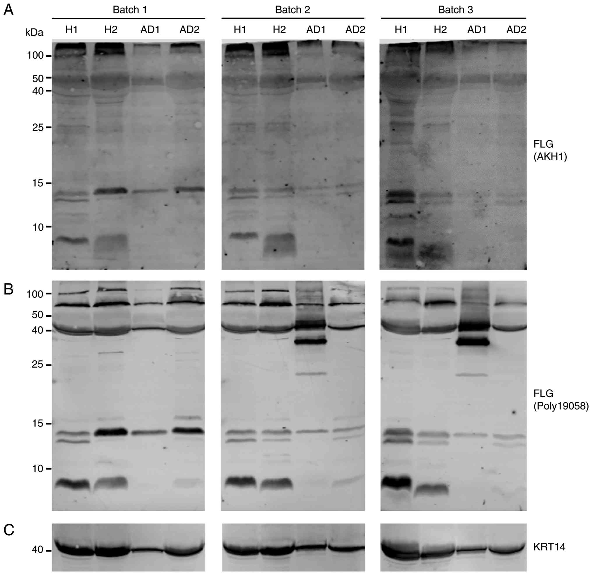 | Figure 2Expression of FLG in buccal cells. (A)
Immunoblotting with mouse anti-FLG monoclonal antibody AKH1
demonstrates stable expression of FLG protein in buccal cells, with
major bands observed at positions ~100, ~50, ~40, ~25, ~15 and ~8
kDa. (B) Immunoblotting with rabbit anti-FLG polyclonal antibody
Poly19058 also reveals stable expression of FLG protein in buccal
cells, with major bands detected at positions ~100, ~70, ~40, ~15,
and ~8 kDa. (C) KRT14 was used as a protein loading control.
Representative immunoblots of four participants (H1, H2, AD1 and
AD2) are shown; expanded cohort data are presented in Fig. S4. H, healthy control; AD, atopic
dermatitis; FLG, filaggrin; KRT14, keratin 14. |
Results
Phenotypic manifestations of palmar
hyperlinearity and buccal cell pellet viscosity in participants
with AD
In the present study, 36 patients with AD and 36
healthy controls, aged 20-40 years, were recruited. Initial
exploratory analyses, including phenotype observation, gene
expression analysis and protein profiling, were performed in 2
healthy controls (H1 and H2) and 2 patients with AD (AD1 and AD2),
with AD2 reporting a family history of AD.
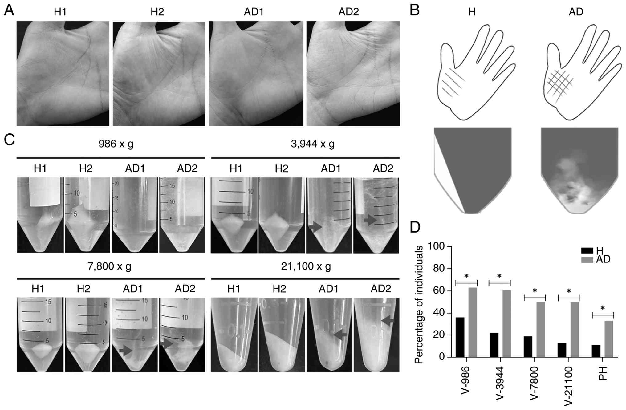
PH, characterized by prominent palmar creases, is
frequently associated with FLG deficiency (7,8). Among
the representative participants, H2 and AD1 showed no evident PH,
H1 exhibited mild PH, and AD2 displayed a pronounced PH phenotype
(Fig. 1A and B). Notably, during buccal cell collection,
a marked difference in pellet properties was observed between
groups. Samples from the patients with AD consistently exhibited
increased viscosity and poorly defined pellet boundaries, whereas
control samples formed compact pellets with sharp edges (Fig. 1B and C). Even at high centrifugal force,
AD-derived pellets remained diffuse and difficult to compact.
Across the full cohort, mild PH was observed in 11% of healthy
controls compared with 33% of patients with AD (Fig. 1D). Similarly, increased pellet
viscosity was more frequently observed in AD samples at all tested
centrifugation conditions.
To investigate potential contributors to this
phenotype, mucin expression was assessed; however, no marked or
consistent differences were observed between the AD and control
groups (Fig. S3), suggesting that
mucins are unlikely to account for the increased viscosity.
Limited consistency of FLG gene
expression in buccal cells
FLG gene expression was evaluated in a subset
of participants (H1, H2, AD1, and AD2) across four independent
collections over different months (Fig. S2). Expression levels were
detectable in all samples but exhibited substantial intra- and
inter-individual variability over time. Given the limited sample
size and exploratory nature of this analysis, these data are
presented descriptively to illustrate variability rather than for
statistical inference. Subsequent analyses therefore focused on
protein-level characterization.
Stable FLG protein expression with
altered degradation profiles in AD samples
To investigate FLG protein expression, two widely
cited antibodies were utilized for FLG protein detection: AKH1
(mouse monoclonal) and Poly19058 (rabbit polyclonal, raised against
residues 24-39 of filaggrin) (6,22-25).
Across all participants, signals corresponding to putative
FLG-derived fragments were consistently detected. By contrast,
distinct differences in FLG degradation profiles were observed
between patients with AD and healthy controls (Fig. 2). Notably, low-molecular-weight
fragments, particularly around ~8 kDa, showed clear differences
between groups and were consistently detected by both antibodies.
Additional variability was observed in intermediate fragments
(20-40 kDa), suggesting altered processing or degradation of FLG in
AD samples.
Analysis of an expanded cohort (Fig. S4; Table SI) confirmed that FLG degradation
profiles differed between patients with AD and healthy controls.
Notably, the putative terminal ≤10 kDa fragment was less frequently
detected in AD samples (19% with AKH1 and 4% with Poly19058) than
in the healthy control samples (50% with both antibodies), while
greater diversity in the 20-40 kDa region was observed in a subset
of AD samples. Together, these findings indicate that while overall
FLG protein expression remains stable in buccal cells, its
degradation profile is altered in AD.
Discussion
FLG stands as the most robustly identified
genetic predisposition for AD (1,5,19,26),
yet investigations into FLG protein expression among patients with
AD remain scarce due to challenges in biopsy acquisition. Despite
nearly two decades of intensive research, most FLG studies have
focused on genetics, transcriptomics, immunohistochemistry, animal
models or cell culture systems (1,2,6,19).
By contrast, direct FLG protein data obtained from human tissues
remain limited, and only a small number of studies have
successfully performed western blot analysis of FLG proteins in
human samples (6,19). This long-standing technical
challenge has hindered efforts to characterize FLG processing and
degradation in vivo. The present study demonstrates that
buccal cells provide a feasible and non-invasive system for
assessing FLG protein expression and degradation profiles in
humans.
Following extensive optimization of protein
extraction methods, a broad spectrum of FLG-immunoreactive species
was reproducibly detected in human buccal cells using two
independent antibodies recognizing distinct epitopes. While
FLG mRNA levels showed variability across samples and time
points, distinct differences in FLG degradation profiles were
observed between patients with AD and healthy controls,
characterized by reduced detection of the terminal ≤10-kDa fragment
and greater diversity in the intermediate fragment range in AD
samples. This pattern points to potential differences in FLG
processing or catabolism in AD, extending beyond simple changes in
overall expression levels. Given the central role of FLG
degradation in generating NMFs, such alterations may have
functional implications for epithelial barrier properties and
warrant further investigation using complementary approaches such
as mass spectrometry and genetic characterization.
In addition to molecular findings, a reproducible
increase in buccal cell pellet viscosity was observed in patients
with AD. This phenotype was not explained by differences in mucin
expression, suggesting that other factors may contribute to this
effect. Several hypotheses may explain the increased viscosity
observed in AD buccal cell pellets. One possibility is that FLG
degradation products, which are abundant in epithelial cells and
show altered hydrolysis profiles in AD, may directly influence the
biochemical and physical properties of the cellular pellet
(27,28). Another is that impaired epithelial
barrier function and altered tissue hydration in AD could affect
cell-cell adhesion or pellet compaction during centrifugation
(29,30). A third possibility is that
differences in the composition of intracellular proteins,
extracellular matrix components or inflammatory exudates may also
contribute to this phenotype (31,32).
Although speculative, these possibilities highlight a previously
underappreciated aspect of epithelial biology in AD that warrants
further investigation.
Several limitations should be acknowledged. First,
due to the non-invasive study design, direct comparisons between
buccal and skin tissues were not performed. Therefore, buccal cells
should be considered a complementary system rather than a direct
substitute for epidermal tissue. Second, comprehensive genotyping
was not conducted, as the highly repetitive structure of the
FLG gene presents substantial technical challenges.
Furthermore, loss-of-function mutations in FLG are rare in
East Asian populations, making genotype-phenotype correlation
difficult even with targeted screening in a cohort of this size.
Third, standardized disease-severity assessments such as Scoring
Atopic Dermatitis tool (33) or the
Eczema Area and Severity Index (34) were not available for the
participants in this study. Fourth, the protein analyses were
primarily qualitative, and densitometric semi-quantification was
not performed. In addition, definitive identification of the
observed immunoreactive bands was not established by orthogonal
methods such as mass spectrometry; however, the detection of the
~8-kDa signal by two independent antibodies recognizing distinct
epitopes provides cross-validation of its FLG-derived origin. These
signals are therefore referred to as putative FLG-derived
fragments. Fifth, phenotypic assessments, including PH and pellet
viscosity, were evaluated using categorical visual scoring without
formal blinding or objective rheological measurement. Future
studies incorporating quantitative protein analysis, genetic
characterization and clinical severity scoring will be necessary to
further elucidate FLG regulation and its association with disease
activity.
In conclusion, the present study establishes buccal
cells as a practical and accessible model for investigating FLG
protein biology in humans. The identification of altered
degradation profiles and increased pellet viscosity in AD provides
new insights into epithelial barrier dysfunction and supports the
potential of non-invasive approaches for studying complex skin
diseases.
Supplementary Material
Participant flow diagram.
FLG gene expression analysis in
buccal cells. (A) FLG gene expression levels in
representative healthy controls (H1 and H2) and patients with AD
(AD1 and AD2) across four biological replicates (batch 1-4)
collected over different months. Expression levels are normalized
to H1 (set as 100%). (B) FLG gene expression levels over 4
months, with each time point normalized to H1 (set as 100%). (C)
Intra-individual variability of FLG gene expression across
batches, with values for each participant normalized to their own
Batch 1 (set as 100%). (D) Annealing positions of FLG primers and
validation of primer specificity by sequencing verification of
amplified products. Both FLG and GAPDH primers were
sourced from PrimerBank. Data are presented as the mean ± SD of
three technical replicates and are shown descriptively to
illustrate temporal and inter-individual variability. No
inferential statistical tests were performed. H, healthy control;
AD, atopic dermatitis; FLG, filaggrin; M, marker.
Expression of mucins in buccal cells.
Immunoblot analysis of mucins expressed in the oral cavity,
including MUC1, MUC4, MUC5B, MUC7, MUC16 and MUC19, in buccal cell
samples from healthy controls and patients with AD. The mucin
immunoblots and the KRT14 loading control shown here are derived
from the same sample set and protein lysates as Fig. 2, and are retained to reflect the
original experimental arrangement. Across all examined mucins,
expression levels were comparable between groups, with no
consistent upregulation in AD samples, indicating that mucin
expression is unlikely to explain the increased viscosity of AD
buccal cell pellets. MUC, mucin; H, healthy control; AD, atopic
dermatitis; KRT14, keratin 14.
FLG hydrolysis profiles in expanded
cohorts: Healthy controls (H3-H14) and patients with AD (AD3-AD26).
(A) Mouse anti-FLG monoclonal antibody AKH1 immunoblots. (B)
Poly19058 antibody immunoblots. (C) KRT14 was used as the control.
Membranes were first probed with AKH1, then Poly19058, and finally
stripped and reprobed with KRT14. Red color indicates samples with
reduced expression levels of major bands or greater diversity
between 20-40 kDa. Additionally, AD3 suffers from both AD and
ichthyosis vulgaris, along with a clear PH phenotype (all
highlighted). Notably, buccal cells failed to pellet at 7,800 x g.
AD4, AD12, AD16 and AD23 have a family history of AD. ‘V-21100’
indicates viscosity at 21,100 x g. H, healthy control; AD, atopic
dermatitis; FLG, filaggrin; PH, palmar hyperlinearity; KRT14,
keratin 14.
Summary of filaggrin protein
degradation patterns detected in buccal cells from healthy controls
(n=14) and patients with AD (n=26).
Acknowledgements
The authors would like to extend their sincere
gratitude to Dr Yongzhen Jin (Wenzhou Central Hospital, Wenzhou,
China) for providing invaluable advice and assistance.
Funding
Funding: This study was funded by the Wenzhou-Kean University
Research Program (grant nos. IRSPK202101 and SSPF2026002).
Availability of data and materials
The data generated in the present study are included
in the figures and/or tables of this article.
Authors' contributions
KL conceived and designed the study, performed data
curation, methodology development and investigation, and drafted
the manuscript. XZhou and XZou performed data curation, methodology
development and investigation. SJ, ZG, XZhu, ZZ and YL contributed
to data curation and investigation. BZ conceived and supervised the
study, contributed to methodology, project administration, funding
acquisition, and manuscript writing and revision. All authors have
read and approved the final manuscript. KL and BZ confirm the
authenticity of all the raw data.
Ethics approval and consent to
participate
The present study protocol was approved by the
Ethics Committee of Wenzhou-Kean University (Wenzhou, China;
approval nos. WKU20210002 and WKUIRB2022017) and all methods were
conducted following the principles of the Declaration of Helsinki.
All participants provided written informed consent.
Patient consent for publication
Not applicable.
Competing interests
The authors declare that they have no competing
interests.
References
|
1
|
Moosbrugger-Martinz V, Leprince C, Méchin
MC, Simon M, Blunder S, Gruber R and Dubrac S: Revisiting the roles
of filaggrin in atopic dermatitis. Int J Mol Sci.
23(5318)2022.PubMed/NCBI View Article : Google Scholar
|
|
2
|
Stefanovic N and Irvine AD: Filaggrin and
beyond: New insights into the skin barrier in atopic dermatitis and
allergic diseases, from genetics to therapeutic perspectives. Ann
Allergy Asthma Immunol. 132:187–195. 2024.PubMed/NCBI View Article : Google Scholar
|
|
3
|
Stamatas GN: Protein degradation in the
stratum corneum. Int J Cosmet Sci. 46:590–597. 2024.PubMed/NCBI View Article : Google Scholar
|
|
4
|
Akiyama M: FLG mutations in ichthyosis
vulgaris and atopic eczema: Spectrum of mutations and population
genetics. Br J Dermatol. 162:472–477. 2010.PubMed/NCBI View Article : Google Scholar
|
|
5
|
Palmer CNA, Irvine AD, Terron-Kwiatkowski
A, Zhao Y, Liao H, Lee SP, Goudie DR, Sandilands A, Campbell LE,
Smith FJ, et al: Common loss-of-function variants of the epidermal
barrier protein filaggrin are a major predisposing factor for
atopic dermatitis. Nat Genet. 38:441–446. 2006.PubMed/NCBI View
Article : Google Scholar
|
|
6
|
Smith FJD, Irvine AD, Terron-Kwiatkowski
A, Sandilands A, Campbell LE, Zhao Y, Liao H, Evans AT, Goudie DR,
Lewis-Jones S, et al: Loss-of-function mutations in the gene
encoding filaggrin cause ichthyosis vulgaris. Nat Genet.
38:337–342. 2006.PubMed/NCBI View
Article : Google Scholar
|
|
7
|
Bradshaw LE, Haines RH, Thomas KS,
Chalmers JR, Irvine AD, Williams HC and Brown SJ: Clinical
examination for hyperlinear palms to determine filaggrin genotype:
A diagnostic test accuracy study. Clin Exp Allergy. 51:1421–1428.
2021.PubMed/NCBI View Article : Google Scholar
|
|
8
|
Brown SJ, Relton CL, Liao H, Zhao Y,
Sandilands A, McLean WH, Cordell HJ and Reynolds NJ: Filaggrin
haploinsufficiency is highly penetrant and is associated with
increased severity of eczema: Further delineation of the skin
phenotype in a prospective epidemiological study of 792 school
children. Br J Dermatol. 161:884–889. 2009.PubMed/NCBI View Article : Google Scholar
|
|
9
|
Kim BE, Goleva E, Kim PS, Norquest K,
Bronchick C, Taylor P and Leung DYM: Side-by-side comparison of
skin biopsies and skin tape stripping highlights abnormal stratum
corneum in atopic dermatitis. J Invest Dermatol. 139:2387–2389.e1.
2019.PubMed/NCBI View Article : Google Scholar
|
|
10
|
Smith SA and Dale BA: Immunologic
localization of filaggrin in human oral epithelia and correlation
with keratinization. J Invest Dermatol. 86:168–172. 1986.PubMed/NCBI View Article : Google Scholar
|
|
11
|
Vigneswaran N, Peters KP, Hornstein OP and
Haneke E: Comparison of cytokeratin, filaggrin and involucrin
profiles in oral leukoplakias and squamous carcinomas. J Oral
Pathol Med. 18:377–390. 1989.PubMed/NCBI View Article : Google Scholar
|
|
12
|
Renò F, Rocchetti V, Migliario M, Rizzi M
and Cannas M: Chronic exposure to cigarette smoke increases matrix
metalloproteinases and Filaggrin mRNA expression in oral
keratinocytes: Role of nicotine stimulation. Oral Oncol.
47:827–830. 2011.PubMed/NCBI View Article : Google Scholar
|
|
13
|
Dale BA, Gown AM, Fleckman P, Kimball JR
and Resing KA: Characterization of two monoclonal antibodies to
human epidermal keratohyalin: Reactivity with filaggrin and related
proteins. J Invest Dermatol. 88:306–313. 1987.PubMed/NCBI View Article : Google Scholar
|
|
14
|
Itoiz ME, Conti CJ, Lanfranchi HE, Mamrack
M and Klein-Szanto AJ: Immunohistochemical detection of filaggrin
in preneoplastic and neoplastic lesions of the human oral mucosa.
Am J Pathol. 119:456–461. 1985.PubMed/NCBI
|
|
15
|
Reibel J, Clausen H, Dale BA and Thacher
SM: Immunohistochemical analysis of stratum corneum components in
oral squamous epithelia. Differentiation. 41:237–244.
1989.PubMed/NCBI View Article : Google Scholar
|
|
16
|
Bierbaumer L, Schwarze UY, Gruber R and
Neuhaus W: Cell culture models of oral mucosal barriers: A review
with a focus on applications, culture conditions and barrier
properties. Tissue Barriers. 6(1479568)2018.PubMed/NCBI View Article : Google Scholar
|
|
17
|
Michalczyk A, Varigos G, Smith L and
Ackland ML: Fresh and cultured buccal cells as a source of mRNA and
protein for molecular analysis. Biotechniques. 37:262–264, 266-269.
2004.PubMed/NCBI View Article : Google Scholar
|
|
18
|
Livak KJ and Schmittgen TD: Analysis of
relative gene expression data using real-time quantitative PCR and
the 2(-Delta Delta C(T)) method. Methods. 25:402–408.
2001.PubMed/NCBI View Article : Google Scholar
|
|
19
|
Sandilands A, Terron-Kwiatkowski A, Hull
PR, O'Regan GM, Clayton TH, Watson RM, Carrick T, Evans AT, Liao H,
Zhao Y, et al: Comprehensive analysis of the gene encoding
filaggrin uncovers prevalent and rare mutations in ichthyosis
vulgaris and atopic eczema. Nat Genet. 39:650–654. 2007.PubMed/NCBI View
Article : Google Scholar
|
|
20
|
Yamamoto-Tanaka M, Makino T, Motoyama A,
Miyai M, Tsuboi R and Hibino T: Multiple pathways are involved in
DNA degradation during keratinocyte terminal differentiation. Cell
Death Dis. 5(e1181)2014.PubMed/NCBI View Article : Google Scholar
|
|
21
|
Deng H, Li F, Li H, Deng Y, Liu J, Wang D,
Han G, Wang XJ and Zhang Q: CtBP1 overexpression in keratinocytes
perturbs skin homeostasis. J Invest Dermatol. 134:1323–1331.
2014.PubMed/NCBI View Article : Google Scholar
|
|
22
|
Kim BE, Kim J, Goleva E, Berdyshev E, Lee
J, Vang KA, Lee UH, Han S, Leung S, Hall CF, et al: Particulate
matter causes skin barrier dysfunction. JCI Insight.
6(e145185)2021.PubMed/NCBI View Article : Google Scholar
|
|
23
|
Miyauchi K, Ki S, Ukai M, Suzuki Y, Inoue
K, Suda W, Matsui T, Ito Y, Honda K, Koseki H, et al: Essential
role of STAT3 signaling in hair follicle homeostasis. Front
Immunol. 12(663177)2021.PubMed/NCBI View Article : Google Scholar
|
|
24
|
Kuechle MK, Presland RB, Lewis SP,
Fleckman P and Dale BA: Inducible expression of filaggrin increases
keratinocyte susceptibility to apoptotic cell death. Cell Death
Differ. 7:566–573. 2000.PubMed/NCBI View Article : Google Scholar
|
|
25
|
Gerritsen MJ, Elbers ME, De Jong EM and
van de Kerkhof PC: Recruitment of cycling epidermal cells and
expression of filaggrin, involucrin and tenascin in the margin of
the active psoriatic plaque, in the uninvolved skin of psoriatic
patients and in the normal healthy skin. J Dermatol Sci.
14:179–188. 1997.PubMed/NCBI View Article : Google Scholar
|
|
26
|
Sandilands A, Sutherland C, Irvine AD and
McLean WHI: Filaggrin in the frontline: Role in skin barrier
function and disease. J Cell Sci. 122:1285–1294. 2009.PubMed/NCBI View Article : Google Scholar
|
|
27
|
Haftek M, McAleer MA, Jakasa I, McLean
WHI, Kezic S and Irvine AD: Changes in nano-mechanical properties
of human epidermal cornified cells in children with atopic
dermatitis. Wellcome Open Res. 5(97)2020.PubMed/NCBI View Article : Google Scholar
|
|
28
|
Thyssen JP, Jakasa I, Riethmüller C, Schön
MP, Braun A, Haftek M, Fallon PG, Wróblewski J, Jakubowski H,
Eckhart L, et al: Filaggrin expression and processing deficiencies
impair corneocyte surface texture and stiffness in mice. J Invest
Dermatol. 140:615–623.e5. 2020.PubMed/NCBI View Article : Google Scholar
|
|
29
|
Hatano Y, Adachi Y, Elias PM, Crumrine D,
Sakai T, Kurahashi R, Katagiri K and Fujiwara S: The Th2 cytokine,
interleukin-4, abrogates the cohesion of normal stratum corneum in
mice: Implications for pathogenesis of atopic dermatitis. Exp
Dermatol. 22:30–35. 2013.PubMed/NCBI View Article : Google Scholar
|
|
30
|
De Benedetto A, Rafaels NM, McGirt LY,
Ivanov AI, Georas SN, Cheadle C, Berger AE, Zhang K, Vidyasagar S,
Yoshida T, et al: Tight junction defects in patients with atopic
dermatitis. J Allergy Clin Immunol. 127:773–786.e1-e7.
2011.PubMed/NCBI View Article : Google Scholar
|
|
31
|
Pfisterer K, Shaw LE, Symmank D and
Weninger W: The extracellular matrix in skin inflammation and
infection. Front Cell Dev Biol. 9(682414)2021.PubMed/NCBI View Article : Google Scholar
|
|
32
|
Szalus K and Trzeciak M: The role of
collagens in atopic dermatitis. Int J Mol Sci.
25(7647)2024.PubMed/NCBI View Article : Google Scholar
|
|
33
|
Stalder JF, Taïeb A, Atherton DJ, Bieber
P, Bonifazi E, Broberg A, Calza A, Coleman R, De Prost Y, Stalder
JF, et al: Severity scoring of atopic dermatitis: The SCORAD index:
Consensus report of the european task force on atopic dermatitis.
Dermatology. 186:23–31. 1993.PubMed/NCBI View Article : Google Scholar
|
|
34
|
Hanifin JM, Thurston M, Omoto M, Cherill
R, Tofte SJ and Graeber M: The eczema area and severity index
(EASI): Assessment of reliability in atopic dermatitis. EASI
evaluator group. Exp Dermatol. 10:11–18. 2001.PubMed/NCBI View Article : Google Scholar
|