Introduction
Nasopharyngeal carcinoma (NPC) is a tumor arising
from the epithelial cells that cover the surface and line the
nasopharynx. The incidence of NPC is higher in Chinese and Tunisian
populations (1). Unfortunately, at
diagnosis, 70% of patients have locally advanced, non-metastatic
stage III or IV disease (2,3).
Despite novel advances in radiotherapy, chemotherapy and
gene-targeting agents, the overall survival rate of patients in the
aggressive phase remains low (4).
Concomitant chemoradiotherapy represents one of the most recent
advances in the treatment of NPC patients. A recent meta-analysis
confirmed the superiority of concurrent chemoradiotherapy to
radiotherapy alone. Cisplatin-based concomitant chemoradiotherapy
is now the standard treatment for locally advanced NPC patients
(2,5,6).
However, intrinsic or acquired resistance to
cisplatin therapy remains a critical problem in the clinical
management of NPC patients. Cisplatin resistance in HNSCC may be
mediated by a number of different mechanisms, including drug
detoxification, up-regulation of DNA repair enzymes or the
overexpression of gene products that provide tumor cells with
survival advantage relative to normal cells (7,8).
Recent studies have shown that Notch may play a role in the
mechanisms of cisplatin resistance (9). Moreover, Notch-1 was found to be
highly expressed in cisplatin-resistant HNSCC patients (8).
The Notch signaling pathway plays an important role
in the proliferation and differentiation of cells. It is an
evolutionarily conserved pathway that regulates critical cell fate
decisions (10). Notch receptor
activation is irreversible as it involves proteolysis-mediated
release of the Notch intracellular domain (NICD) which can be
interrupted by γ-secretase inhibitor, translocation to the nucleus
and transactivation of gene targets associated with the
transcription cofactor CBF1, which in turn affect numerous pathways
involving cell-fate determination (11–14).
Experimental evidence has also revealed that Notch
is involved in anticancer drug resistance, indicating that
targeting Notch may be a novel therapeutic approach for the
treatment of cancer by overcoming drug resistance of cancer cells
(9). Yet, the mechanism of how
Notch functions in intrinsic or acquired cisplatin resistance
remains unclear, particularly in NPC intrinsic resistance to
cisplatin.
In the present study, treatment of poorly
differentiated NPC cells with a γ-secretase inhibitor (DAPT), which
led to the decline in NICD and inhibition of Notch signaling, was
not sufficient to induce pronounced apoptosis of CNE-2 cells, but
did result in the down-regulation of the P-glycoprotein and ERCC1
protein. In contrast, combination of DAPT with cisplatin induced
substantial cell death compared to cisplatin treatment alone.
Materials and methods
Cell cultures and reagents
The human poorly differentiated nasopharyngeal
carcinoma CNE-2 cell line was purchased from the Shanghai Cell
Collection (Shanghai, China) and maintained in RPMI-1640 containing
10% fetal bovine serum (Hyclone, UT, USA), 100 U/ml penicillin and
100 mg/ml streptomycin at 37°C in a humidified atmosphere with 5%
CO2. The γ-secretase inhibitor (DAPT;
N-[N-(3,5-difluorophenacetyl)-L-alanyl]-S-phenylglycinet-butylester,
C23H26F2N2O4)
was purchased from Sigma-Aldrich Co. LLC and cisplatin were
supplied by Qilu Pharmaceutical Co., Ltd.
Cell treatment
Notch signaling pathways were inhibited with the
γ-secretase inhibitor (DAPT) at different concentrations. DAPT was
dissolved in DMSO to a stock concentration of 10 mM, and was
diluted to final concentrations of 25, 50 and 75 μM with
conventional culture medium just prior to use. The control group (0
μM DAPT) was mock treated with conventional medium containing the
mean volume of DMSO carrier only. Cispatin was dissolved in PBS to
a stock concentration of 5 mM and used at a final concentration of
10 μM after a 24-h treatment with various concentrations of DAPT
(15).
Flow cytometric analysis of apoptotic
cells using Annexin V-FITC and propidium iodide (PI)
After 72 h of cell treatment as mentioned above,
cells were gently trypsinized and washed once with serum-containing
media. Each group of cells (1×10⁵ to 2×10⁵)
was collected by centrifugation and then resuspended in 500 μl of
1X Annexin-binding buffer. Annexin V-FITC (5 μl) and 10 μl of PI
were added, and cells were then incubated at room temperature for
10 min in the dark. Annexin V-FITC binding was analyzed by flow
cytometry (Epics Altra, Beckman, USA; Ex=488 nm, Em=530 nm) using
FITC signal detector and PI staining by the phycoerythrin emission
signal detector.
Cell proliferation and cytotoxicity
assay
We dispensed 100 μl of cell suspension (2,000
cells/well) in a 96-well plate. The plate was pre-incubated for 24
h in a humidified incubator. Various concentrations of DAPT (0, 25,
50 and 75 μM) were added to the plate for testing. The plate was
incubated for 24, 48 and 72 h in the incubator, and 10 μl of CCK-8
(Dojindo, Japan) solution was added to each well of the plate. The
plate was incubated for 1 h in the incubator, and the absorbance at
450 nm was measured using a multilable counter (Victor3 1420;
Perkin Elmer).
Flow cytometric analysis of the cell
cycle
The cells were treated similarly as for the analysis
of cell apoptosis. Then, 1×10⁶ cells of each group were
collected and resuspended in 1 ml of 70% ethanol and fixed
overnight at 4°C. Cells were collected by centrifugation again,
washed with PBS twice and then resuspended in 500 μl of 1X binding
buffer containing 0.5% PI. The prepared cells were incubated at 4°C
for 20 min in the dark, and then analyzed by flow cytometry.
Western blot analysis
Cells were harvested from the 60 mm cell culture
plates, and aliquots of cell extracts were separated on an 8–12%
SDS-polyacrylamide gel. Then, proteins were transferred to a
polyvinylidene difluoride membrane and incubated overnight at 4°C
with the rabbit polyclonal antibody anti-NICD and anti-ERCC1 (Cell
Signaling Technology), or mouse monoclonal antibody
anti-P-glycoprotein (Abcam), respectively. Membranes were then
washed and incubated with HRP-conjugated secondary antibodies in
confining liquid for 1 h and the film was developed using an ECL
test kit (Beyotime, China). The density of the bands on the film
were scanned and analyzed with an image analyzer.
Statistical analysis
Statistical comparison of the data was performed
using a Student’s t-test. A P-value of <0.01 was considered to
be significant.
Results
Induction of cell death by the combined
treatment of DAPT and cisplatin in CNE-2 cells
CNE-2 cells were first treated with increasing
concentrations of cisplatin to establish the least effective
concentration which substantially affects cell survival. While 5 μM
cisplatin for 48 h showed little effect, cell death was observed
with 10 μM cisplatin in CNE-2 cells, so this dosage was used for
further drug combination studies. The CNE-2 cells were treated with
increasing concentrations of DAPT, and there was no obvious effect
on cell survival (P>0.01). Cells were initially treated with
various concentrations of DAPT, and then combined with 10 μM
cisplatin after 24 h. DAPT dose-dependent cell apoptosis was
observed after treatment of 10 μM cisplatin for 48 h (Fig. 1).
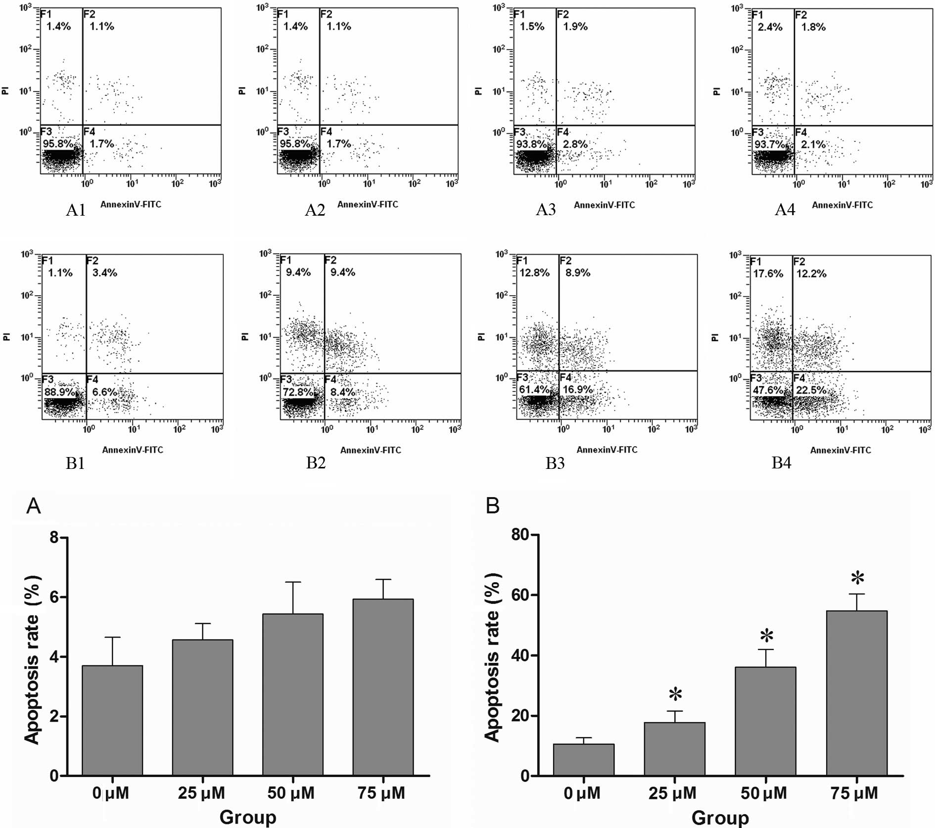 | Figure 1.(A1–4) CNE-2 cells were treated with
different concentrations of DAPT (0, 25, 50 and 75 μM), and the
apoptosis of CNE-2 cells was assessed by FACS after 48 h. (B1–4)
CNE-2 cells were previously treated with various concentrations of
DAPT (0, 25, 50 and 75 μM) before 24 h, and then treated with
cisplatin at the same final concentration of 10 μM. The apoptosis
of CNE-2 cells was detected by FACS after 48 h of treatment with
cisplatin. The control group (0 μM DAPT) was treated with
conventional medium containing the mean volume of DMSO. No
significant apoptosis was noted only with DAPT treatment when
compared to the control group (P>0.01), while pre-treatment with
DAPT enhanced the effect of cisplatin in a dose-dependent manner.
There was a marked difference compared to the control group treated
with cisplatin and DMSO (P<0.01). A1–4 and B1–4 are
respresentative of one of three independent experiments that
yielded similar results. Values in histograms (A) and (B) are the
means ± SD. Proportion of non-apoptotic cells (F3, Annexin
V-FITC−/PI−), early apoptotic cells (F4,
Annexin V-FITC+/PI−), late apoptotic/necrotic
cells (F2, Annexin V-FITC+/PI+) and cell
debris or dead cells (F1). *P<0.01 compared to that
of 0 μM DAPT group by T-test. |
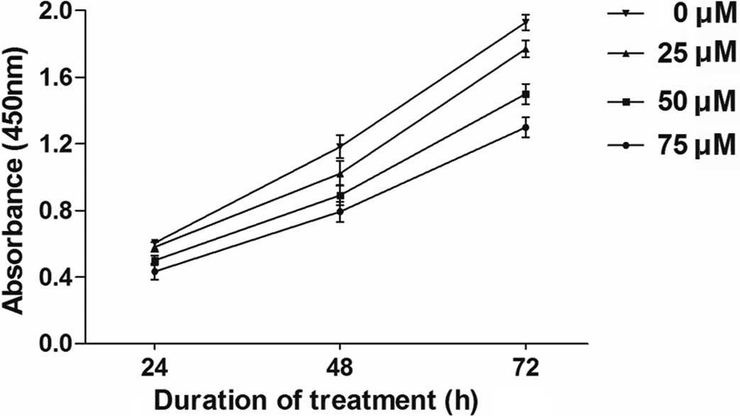
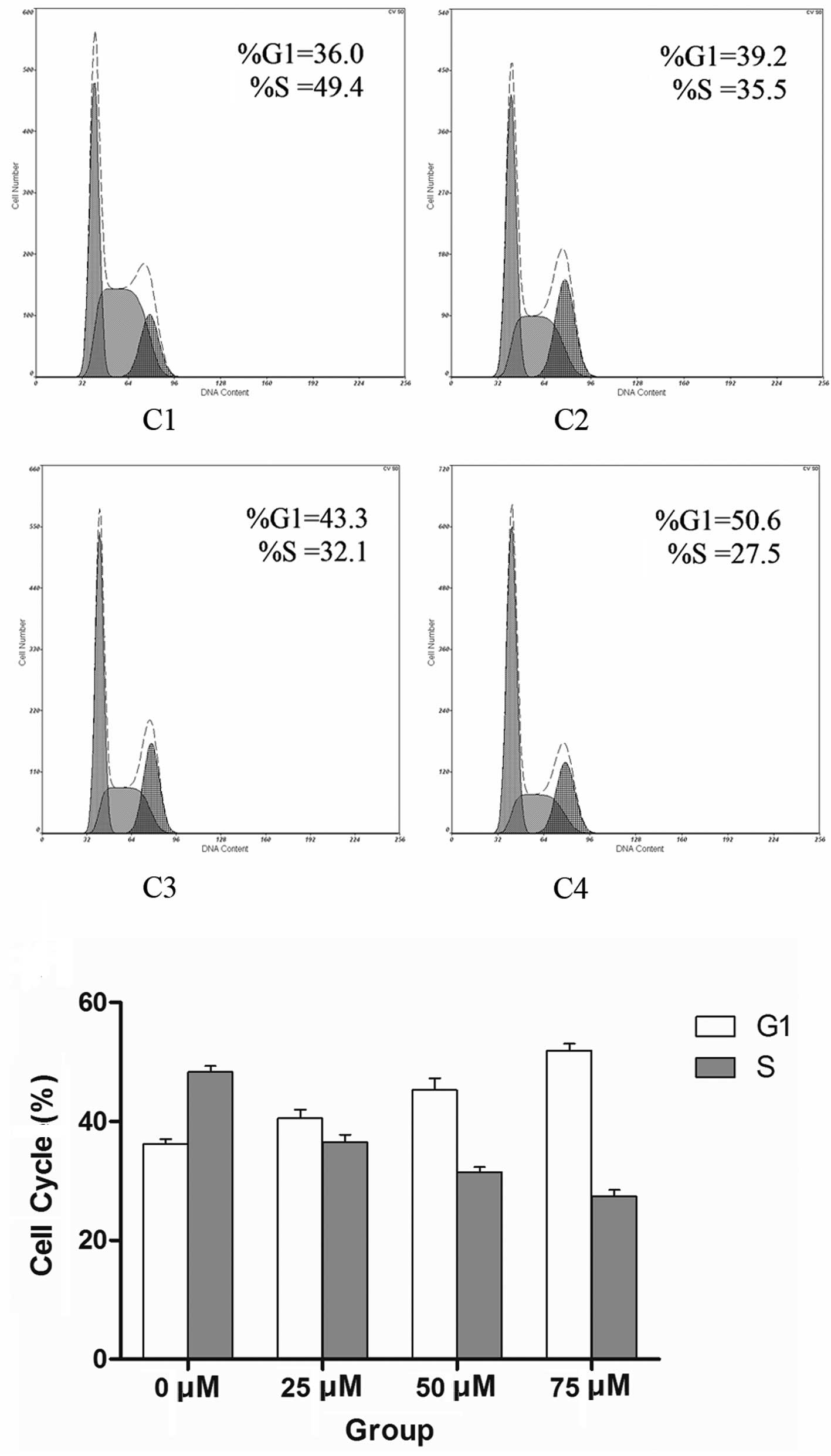
Induction of cell cycle arrest and cell
proliferation is suppressed by DAPT
Although the application of the maximum dose of DAPT
at 75 μM had no obvious effect on cell survival, it substantially
retarded CNE-2 cell proliferation in a dose-dependent manner
(Fig. 2). To assess whether
DAPT-mediated Notch 1 inhibition is associated with changes in the
cell cycle progression of CNE-2 cells, flow cytometry was employed
to detect their allocation at different cell cycle phases in the
presence of various concentrations of DAPT at 48 h. Notch 1
inhibition increased the percentage of cells in the G0/G1 phase at
the expense of S-phase (Fig.
3).
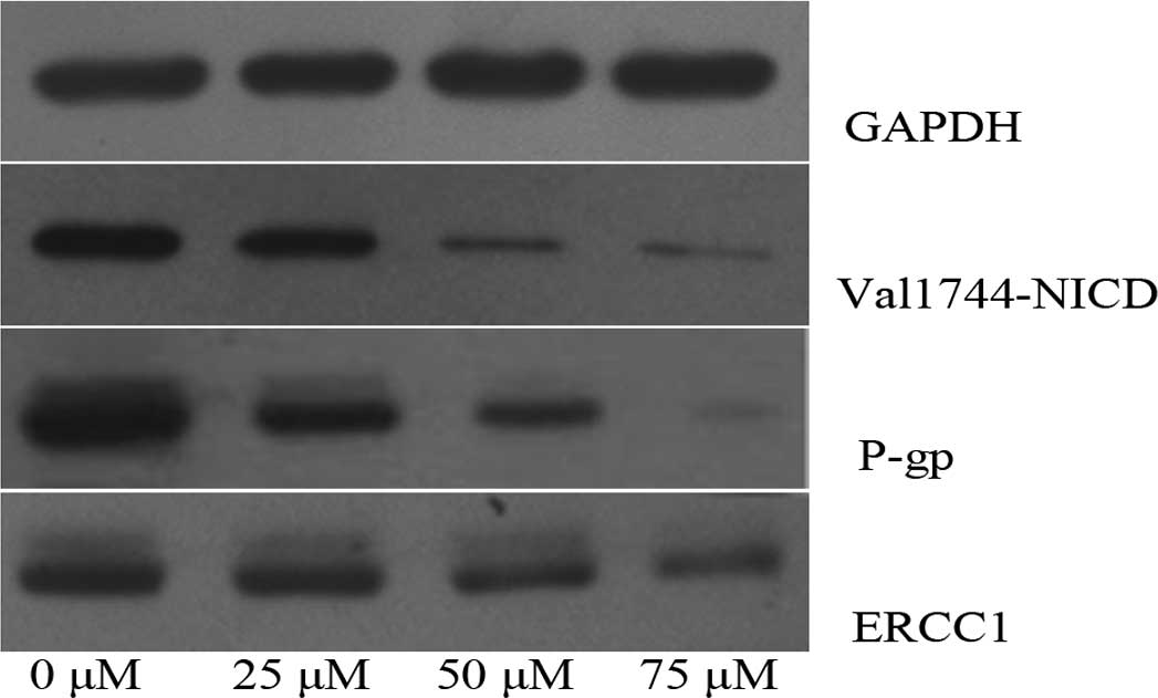
Mechanism of enhanced apoptosis is
induced by the combined treatment of DAPT and cisplatin in CNE-2
cells
CNE-2 cells were treated with increasing
concentrations of DAPT, and the γ-secretase-generated Notch 1
fragment Val1744-NICD was decreased after 48 h in a dose-dependent
manner (P<0.01). The activation of γ-secretase was almost
completely inhibited by DAPT at the concentration of 50 μM.
Meanwhile, the expression of P-gp and ERCC1 protein was detected by
western blotting. There was a significant decrease in the
expression of P-gp and ERCC1 protein in a dose-dependent manner
(P<0.01; Fig. 4).
Discussion
Chemotherapy fails to eliminate all tumor cells due
to intrinsic or acquired drug resistance, which is the most common
cause of tumor recurrence. The mechanisms responsible for drug
resistance are complex and remain poorly understood. Resistance may
be due to either the specific nature and genetic background of the
cancer cell itself or the genetic changes that follow toxic
chemotherapy (9,16,17).
Intrinsic or acquired resistance to cisplatin
therapy remains a critical problem in the clinical management of
NPC patients. Cisplatin resistance in HNSCC may be mediated by a
number of different mechanisms, including drug detoxification,
up-regulation of DNA repair enzymes or overexpression of gene
products that provide tumor cells with survival advantage relative
to normal cells. Studies have demonstrated that Notch regulates the
formation of cancer stem cells and contributes to the acquisition
of the epithelial-mesenchymal transition phenotype, which are also
critically associated with drug resistance (18,19).
There is little evidence indicating that Notch
signaling pathways are involved in cisplatin resistance through
regulating drug detoxification or up-regulating DNA repair enzymes.
In our study, we found that treatment with DAPT (γ-secretase
inhibitor) markedly down-regulated P-gp and ERCC1 protein
expression in CNE-2 cells, and the combination of DAPT and
cisplatin in CNE-2 cells enhanced cisplatin sensitivity, eliminated
the drug resistance and induced greater cell apoptosis compared to
cisplatin treatment alone.
The majority of published data suggest that P-gp
acts as a transmembrane pump which removes drugs from the cell
membrane and cytoplasm (20).
Overexpression of P-gp, the ATP-binding cassette drug transporter,
confers an intrinsic or acquired multidrug resistance (MDR) due to
its capability of transporting a broad range of chemically diverse
anticancer drugs. Occurrence of MDR prevents efficient killing of
cancer cells, leading to chemotherapeutic treatment failure
(21). As a non-specific drug
transporter, the P-gp also removes the γ-secretase inhibitor from
the cytoplasm in theory. In fact, in this study, the concentration
of DAPT applied in CNE-2 cells was higher compared to other studies
(22,23), which indicated more P-gp pumps in
the membrane of drug-resistant cancer cells. The nearly saturated
concentration of DAPT guarantees the inhibition of γ-secretase
activity and the interruption of NICD release, which in turn
affects the transactivation of the MDR1 gene or other target genes
encoding some enzymes or proteins, such as PKC, which activates the
P-gp protein (24).
The expression of ERCC1 was also decreased after
DAPT treatment. ERCC1 activity is involved in resistance to
platinum chemotherapy drugs. Nucleotide excision repair (NER) is
the primary DNA repair mechanism that removes the therapeutic
platinum-DNA adducts from the tumor DNA. ERCC1 activity levels,
being an important part of the NER common final pathway, may serve
as a marker of general NER throughput. One study reported that
ERCC1-positive NSCLC tumors do not benefit from adjuvant platinum
chemotherapy. Thus, down-regulation of ERCC1 enhances the cancer
cell sensitivity to platinum chemotherapy drugs, and ERCC1 may be
involved in the mechanism of Notch-regulated cancer drug resistance
(25–28).
In this study, we also found that application of
γ-secretase inhibitor had no detectable effect on CNE-2 cell
survival, which was not in accord with previous reports (29,30),
but it substantially retarded CNE-2 cell proliferation in a
dose-dependent relationship. Flow cytometry was employed to detect
the allocation to different cell cycle phases in the presence of
various concentrations of DAPT at 48 h. Notch 1 inhibition
increased the percentage of cells in G0/G1 phase at the expense of
S-phase. Borghese et al found similar results which showed
that inhibition of Notch signaling in human embryonic stem
cell-derived neural stem cells delayed G1/S phase transition and
accelerated neuronal differentiation (23). Huang et al (22) also reported that the γ-secretase
inhibitor DAPT induced adipogenesis of adipose-derived stem cells.
Application of γ-secretase inhibitor may alter the differentiation
state of CNE-2 cells and induce CNE-2 cells toward a
well-differentiated state. Whether this change is involved in drug
resistance of CNE-2 cells, requires future research.
In conclusion, inhibition of the Notch signaling
pathway by a γ-secretase inhibitor markedly enhanced the cisplatin
sensitivity of CNE-2 cells, which may be achieved through
down-regulation of P-gp and ERCC1 protein. The combination of GSI
with platinum compounds may provide an option to improve treatment
for a subset of nasopharyngeal carcinoma patients.
Acknowledgements
This study was supported by grants
from the National Natural Science Foundation of China (No. 30872851
and 30901662), and the Science and Technology Department of Hubei
Province (No. 2007AA302B08). This study also received assistance
from Ms. Qing He, the Director of the Central Laboratory of Renmin
Hospital.
References
|
1.
|
B BrennanNasopharyngeal carcinomaOrphanet
J Rare Dis123200610.1186/1750-1172-1-23
|
|
2.
|
S AfqirN IsmailiH ErrihaniConcurrent
chemoradiotherapy in the management of advanced nasopharyngeal
carcinoma: current statusJ Can Res
Ther537200910.4103/0973-1482.4876319293481
|
|
3.
|
RA AlbiruniRazakL LillianFF LiuE ItoB
O’SullivanK ChanNasopharyngeal carcinoma: the next challengesEur J
Cancer4619671978201010.1016/j.ejca.2010.04.004
|
|
4.
|
SM ChenJP LiuJX ZhouSuppression of the
notch signaling pathway by γ-secretase inhibitor GSI inhibits human
nasopharyngeal carcinoma cell proliferationCancer
Lett30676842011
|
|
5.
|
J GuigayAdvances in nasopharyngeal
carcinomaCurr Opin
Oncol20264269200810.1097/CCO.0b013e3282fad84618391624
|
|
6.
|
HM LuaLX PangXB YuanConcurrent
chemoradiotherapy in locally advanced nasopharyngeal carcinoma: a
treatment paradigm also applicable to patients in Southeast
AsiaCancer Treat
Rev35345353200910.1016/j.ctrv.2009.01.00219211192
|
|
7.
|
M KartalouJM EssigmannMechanisms of
resistance to CisplatinMutat
Res4782343200110.1016/S0027-5107(01)00141-511406167
|
|
8.
|
F GuY MaZ ZhangJ ZhaoH KobayashiL ZhangL
FuExpression of Stat3 and Notch1 is associated with cisplatin
resistance in head and neck squamous cell carcinomaOncol
Rep23671676201020127005
|
|
9.
|
ZW WangYW LiA AhmadTargeting Notch
signaling pathway to overcome drug resistance for cancer
therapyBiochim Biophys Acta182582672010
|
|
10.
|
S Artavanis-TsakonasMD RandRJ LakeNotch
signaling: cell fate control and signal integration in
developmentScience284770776199910.1126/science.284.5415.77010221902
|
|
11.
|
T KadeschNotch signaling: the demise of
elegant simplicityCurr Opin
Genet14506512200410.1016/j.gde.2004.07.00715380241
|
|
12.
|
R KopanMX IlaganThe canonical Notch
signaling pathway: unfolding the activation
mechanismCell137216233200910.1016/j.cell.2009.03.04519379690
|
|
13.
|
IM ShihTL WangNotch signaling, γ-secretase
inhibitors, and cancer therapyCancer Res67187918822007
|
|
14.
|
J SarahNotch signaling: a simple pathway
becomes complexNat Rev Mol Cell
Biol7678687200610.1038/nrm200916921404
|
|
15.
|
T AleksicM StephanGamma-secretase
inhibition combined with platinum compounds enhances cell death in
a large subset of colorectal cancer cellsCell Commun
Signal6113200810.1186/1478-811X-6-818950493
|
|
16.
|
G SzakacsJK PatersonJA LudwigC
Booth-GentheMM GottesmanTargeting multidrug resistance in cancerNat
Rev Drug Discov5219234200610.1038/nrd198416518375
|
|
17.
|
MM GottesmanMechanisms of cancer drug
resistanceAnn Rev
Med5615627200210.1146/annurev.med.53.082901.103929
|
|
18.
|
Z WangY LiD KongA AhmadS BanerjeeFH
SarkarCross-talk between miRNA and Notch signaling pathways in
tumor development and progressionCancer
Lett292141148201010.1016/j.canlet.2009.11.01220022691
|
|
19.
|
Z WangY LiS BanerjeeFH SarkarEmerging role
of Notch in stem cells and cancerCancer
Lett279812200910.1016/j.canlet.2008.09.03019022563
|
|
20.
|
LL SuC Yan-ChengD DoloresDrug transporter,
P-glycoprotein (MDR1), is an integrated component of the mammalian
blood-testis barrierInt J Biochem Cell
Biol4125782587200910.1016/j.biocel.2009.08.01519720156
|
|
21.
|
XB ChangA molecular understanding of
ATP-dependent solute transport by multidrug resistance-associated
protein MRP1Cancer Metastasis
Rev261537200710.1007/s10555-007-9041-717295059
|
|
22.
|
Y HuangX YangY Wuγ-secretase inhibitor
induces adipogenesis of adipose-derived stem cells by regulation of
Notch and PPAR-γCell Prolif431471562010
|
|
23.
|
L BorgheseD DolezalovaT OpitzInhibition of
Notch signaling in human embryonic stem cell-derived neural stem
cells delays G1/S phase transition and accelerates neuronal
differentiation in vitro and in vivoStem
Cells28955964201010.1002/stem.40820235098
|
|
24.
|
RL FineTC ChambersCW SachsP-glycoprotein,
multidrug resistance and protein kinase CStem
Cells144755199610.1002/stem.1400478820951
|
|
25.
|
K KirschnerDW MeltonMultiple roles of the
ERCC1-XPF endonuclease in DNA repair and resistance to anticancer
drugsAnticancer Res3032233232201020944091
|
|
26.
|
J BellmuntL Paz-AresM CuelloGene
expression of ERCC1 as a novel prognostic marker in advanced
bladder cancer patients receiving cisplatin-based chemotherapyAnn
Oncol18522528200710.1093/annonc/mdl43517229776
|
|
27.
|
KA OlaussenA DunantP FouretDNA repair by
ERCC1 in non-small-cell lung cancer and cisplatin-based adjuvant
chemotherapyN Engl J
Med355983991200610.1056/NEJMoa06057016957145
|
|
28.
|
QE WangK MilumCH HanDifferential
contributory roles of nucleotide excision and homologous
recombination repair for enhancing cisplatin sensitivity in human
ovarian cancer cellsMol Cancer1024201110.1186/1476-4598-10-24
|
|
29.
|
Y NefedovaM DanielCB SophiaSD WilliamIG
DmitryInhibition of Notch signaling induces apoptosis of myeloma
cells and enhances sensitivity to
chemotherapyBlood11122202228200810.1182/blood-2007-07-10263218039953
|
|
30.
|
S KoduruR KumarS SrinivasanNotch-1
inhibition by Withaferin-A: a therapeutic target against colon
carcinogenesisMol Cancer
Ther9202210201010.1158/1535-7163.MCT-09-077120053782
|