Introduction
Targeted delivery of contrast agents is a highly
desirable strategy for enhancing efficacy and reducing unintended
side effects and toxicity (1–3). Low
et al also stated the need for more sensitive and accurate
tumor markers as well as imaging studies (4). The development of new and improved
tumor selective magnetic resonance imaging (MRI) contrast agents is
clinically desirable as a means of: i) detecting and/or confirming
the presence and location of primary and meta-static lesions; ii)
probing biochemical features of neoplastic tissue that have
implications for tumor staging and/or treatment planning; and iii)
monitoring tumor response to treatment (5).
Macromolecular contrast agents (>20,000 Da), due
to their larger size, enable longer imaging times and possess
higher relaxation rates resulting from their longer rotational
correlation times (6). These
agents have been used to enhance tumors via active targeting.
Active targeting utilizes a specific cell surface molecule that is
either unique or overexpressed in the tissue of interest. This type
of targeting is more challenging due to large amounts of magnetic
label in the tissue of interest required in order to achieve a
sufficient diagnostic signal. Unger et al used an
anti-carcinoembryonic antigen (CEA) antibody conjugated to ∼1.5 Gd
atoms/antibody to enhance human colon carcinoma implanted in the
thighs of hamsters (7). The use of
antibodies as target-specific molecules experience problems that
include rapid clearance by the reticuloendothelial system,
decreased affinity of the antibody following conjugation of the
magnetic label, circulating free antigen, insufficient tumor
penetration, binding of antibodies to non-specific Fc receptors,
and possible changes in the antigen over time (7–9).
To avoid these problems, targeting endogenous
transport mechanisms within cells have been suggested, one of which
is the recycling high-affinity folate receptor (hFR) pathway
(10–12). The folate receptor is a
glycosylphosphatidylinositol-anchored, high-affinity,
folate-binding protein overexpressed in various types of human
tumors (13–17), where it functions by capturing
folate to feed rapidly dividing tumor cells (17–19).
Two folate receptor isoforms have been identified in human cell
membrane, α and β. The type α receptor is frequently overexpressed
in epithelial-lineaged tumors and type β in nonepithelial-derived
tumors. Both isoforms show a high affinity for folic acid.
There has been much interest in exploiting the
folate receptor for tumor-specific-targeted delivery of imaging or
therapeutic agents. Earlier efforts in targeting the folate
receptor were focused on the use of conjugates of antibodies
against the folate receptor. An alternative strategy has been
developed for folate receptor targeting in which the ligand, folic
acid, is covalently linked to the molecule to be delivered. Folic
acid (molecular weight 441.4) is a high-affinity ligand for the
folate receptor (kD ∼10−10 mol/l). Folate conjugates are
taken into cultured tumor cells by binding to the folate receptor
on the cellular surface followed by receptor-mediated endocytosis.
This targeting strategy has been exploited in the receptor-mediated
delivery of proteins, liposomes, gene transfer vectors,
chemotherapeutic agents and radioimaging agents into tumor cells.
By contrast, non-proliferating normal cells are severely restricted
in possessing folate receptors (17,20).
Thus, folate receptors provide highly selective sites that
differentiate tumor cells from normal cells. Folate conjugates are
also suitable for targeted delivery because of folate’s lack of
immunotoxicity and its role in receptor-mediated endocytosis.
In this study, a folate receptor-targeted, polymeric
gadolinium complex was utilized, where the ε-amino groups of
poly-L-lysine were conjugated to ∼56 Gd-DTPA molecules per folic
acid to increase the number of gadolinium (21). Using this folate conjugate, we
successfully produced the contrast-enhanced, folate
receptor-targeted tumor image in vivo in pulmonary tumor
xenografts.
Materials and methods
Contrast agent
All reagents used in the synthesis were commercial
products. Diethylenetriaminepentaacetic acid (DTPA) (10 g) was
added to 30 ml acetic anhydride and 15 ml pyridine and stirred for
6 h at 60°C. CaDTPA in white solid form was obtained after cooling
and drying. Polylysine (10 mg) was dissolved in 3 ml sodium
carbonate buffer (200 mM
NaHCO3/Na2CO3, pH 9.6), to which
CaDTPA solution (polylysine:CaDTPA 1:86 in molar ratio) was added.
PL-DTPA resulted after stirring the mixture solution for 16 h in an
icebath. PL-Gd-DTPA was synthesized by adding GdCl3
solution to PL-DTPA solution (Gd3+:DTPA in equal ratio)
and stirring for 24–30 h at room temperature. Folic acid in 100 mM
NaHCO3/Na2CO3 buffer, pH 9.6, was
conjugated to PL-Gd-DTPA through ECD linkage (folic
acid:PL-Gd-DTPA:ECD 5:1:5.3 in molar ratio). The free folic acid
was separated by gel filtration [Sephadex CL-4B column (1x25 cm)].
The complex of folic acid and PL-Gd-DTPA was collected and
ice-concentrated, and used for animal imaging experiment after
aseptic filtration. ICP-AES and HPLC were used for the
Gd3+ concentration quantification (21).
Cell culture and in vitro specificity
studies
The H460 human lung adenocarcinoma cells, expressing
the hFR were obtained from the Shanghai Cancer Institute (Shanghai,
China). Cell lines were cultured continuously as a monolayer at
37°C in a humidified atmosphere containing 5% CO2 in
folate-free RPMI-1640 medium supplemented with 10% fetal bovine
serum, 50 U/ml penicillin and 50 μg/ml streptomycin. The final
folate concentration (with the fetal bovine serum as the only
source of folate) falls in the range of the physiological
concentration in human serum. Because of their high folate content
(2 μmol/l), which leads to the downregulation and presaturation of
the cellular folate receptor, regular culture media were not
used.
The cultured cells were washed with 1 ml, 0.01 M, pH
7.4, phosphate-buffered saline. Following 3 min of centrifugation
at 2,000 rpm, cells were aliquoted into tubes consisting of
1x10⁶ cells/0.5 ml. Folic acid labeled 125I
(∼5x10⁶) was added to each tube. Then, for the competition
inhibition studies, folic acid or folate-PL-Gd-DTPA 100 μl was
added to each tube in different concentrations, stored at 4°C
overnight. Cells were then washed with 2 ml, 0.01 M, pH 7.4,
phosphate-buffered saline, following 15 min of centrifugation at
3,000 rpm. After blotting supernatant, radioactivity was measured
using the γ-ray emissive counter. Log-curves were drawn between
B/B0 and different concentrations of folic acid or
folate-PL-Gd-DTPA.
Animal model
Pulmonary tumors were induced in BALB/c mice 5–6
weeks of age (Shanghai Cancer Institute, Shanghai, China) by
subcutaneously injecting 107–108 human lung
adenocarcinoma H460 cells expressing the hFR in the subscapular
area on the dorsal surface. Mice were housed in polycarbonate
microisolator cages, and tumor sizes were monitored three times per
week. Tumors reaching a diameter of ∼1 cm after 12–14 weeks of
inoculation were used for MR experiments. One week prior to
imaging, all animals were transferred from their normal rodent diet
to one consisting of a folate-free rodent diet.
In vivo MR imaging
All MR experiments were performed on a Siemens
Vision-Plus 1.5T MR scanner. A head radio frequency coil was used
for all studies. Ten pulmonary tumor animal models (hFR-positive)
were divided into two groups: control group (n=5, Gd-DTPA, 0.2
mmol/Gd3+/kg) and experimental group (n=5,
folate-PL-Gd-DTPA, 0.2 mmol/Gd3+/kg), randomly.
Folate-PL-Gd-DTPA or Gd-DTPA was injected into the caudal vein of
the mice in the study using a 27-gauge catheter (Baxter Healthcare
Corporation, Deerfield, IL, USA). A multi-slice spin-echo imaging
sequence was used to obtain coronal (matrix 114x256; slice
thickness 2 mm) or transverse (matrix 114x256; slice thickness 3
mm) slices under T1-weighted (TR/TE 270/14 msec). Images were
acquired before contrast administration, immediately after contrast
administration, and 12, 24, 48 and 72 h after contrast
administration. The field of view was 160 mm for the coronal images
and transverse images.
Image analysis
The coronal images were used to obtain tumor and
thigh muscle signal intensity and transverse images to liver signal
intensity. Of the five slices obtained for each mouse on coronal
and transverse sections, respectively, the top and bottom slices
were not included in the data analysis to prevent confounding
partial volume effects at the edges of the tumor. The signal
intensities within the region of interest (i.e., tumor or
reference) were obtained. The tumor, muscle and liver signal
intensity was normalized to the noise standard deviation of the
propotional slice reference to obtain a signal-to-noise ratio (SNR)
at each of the imaging conditions (pre-injection, or different scan
time-point post-injection). The percentage of contrast enhancement
(PCE) was calculated according to the following: PCE = (SNRpost -
SNRpre)/SNRpre x 100%. SNRpost was replaced with SNRpost at
different scan time-points to obtain the PCE at different scan
time-points.
Statistical analysis
All data groups were analyzed and data are expressed
as the means ± standard deviation (SD). The differences in tumor,
liver and muscle SNR at different times were tested using analysis
of variance (ANOVA, F-test). Differences were considered
significant at P-value of <0.05. Statistical analysis was
performed using the SPSS statistical software (version 10.0; SPSS,
Inc., Chicago, IL, USA).
Results
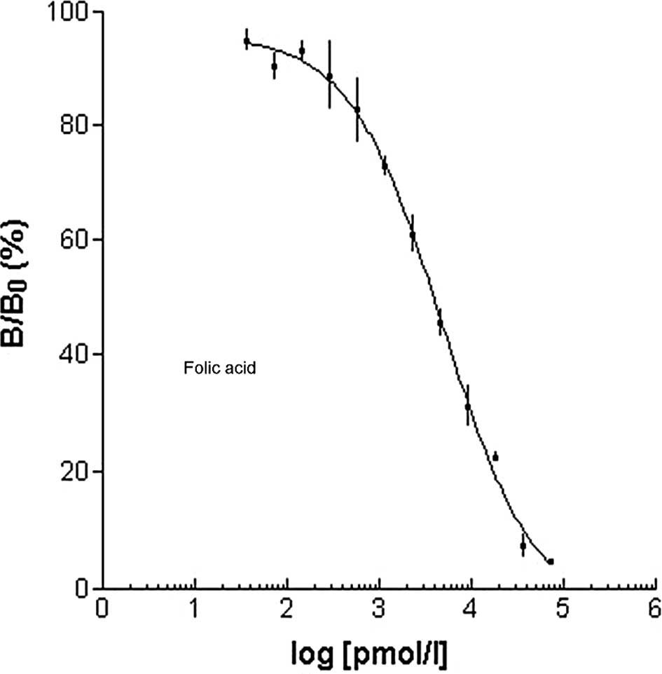
In vitro specificity studies
Prior to showing specificity of the
folate-PL-Gd-DTPA binding in vivo with MRI, radioactive
iodine-125, chelated to folic acid was administered to human
pulmonary adenocarcinoma cells in vitro. We wanted to test
whether folate-PL-Gd-DTPA was able to bind to these cells which
express the folate receptor. In this competition experiment,
folate-PL-Gd-DTPA exhibited a significant effect of competition
inhibition to folic acid binding folate receptor. Results presented
in Fig. 1 show that the folate
receptor-positive cells treated with folate-PL-Gd-DTPA were similar
in binding compared to folic acid.
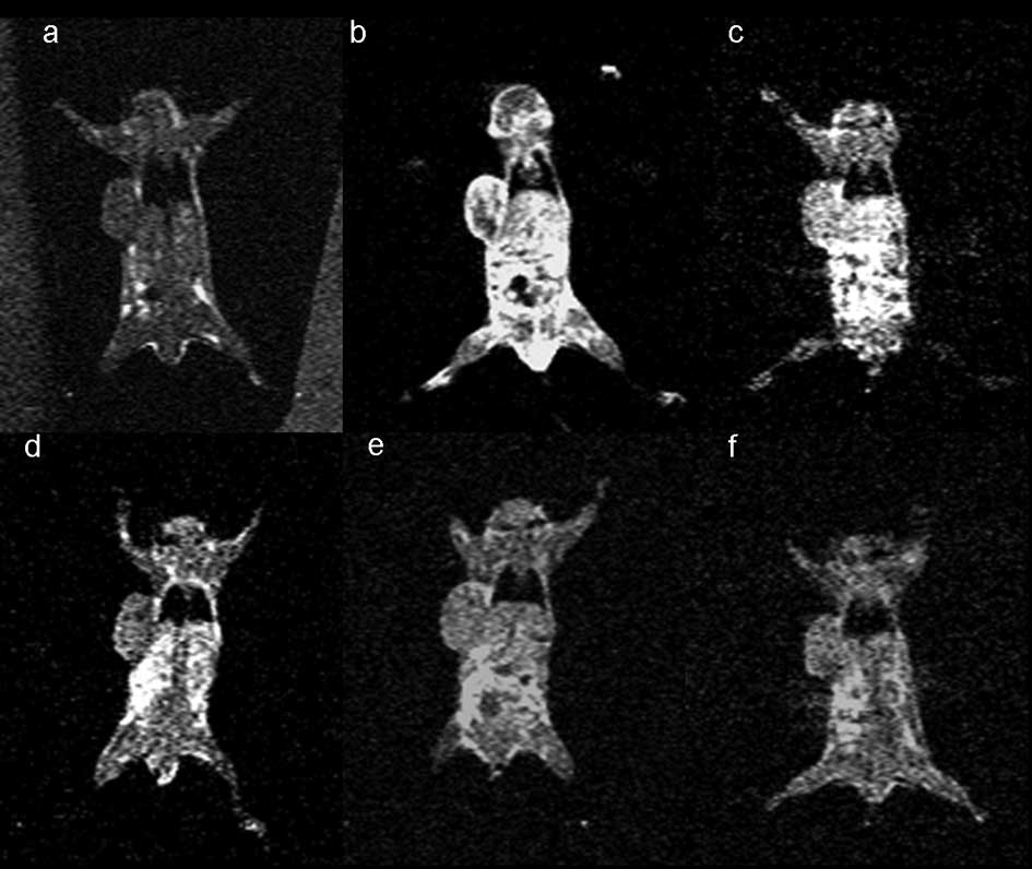
In vivo specificity studies
Following the demonstration of the efficacy of
binding of folate-PL-Gd-DTPA in vitro, we wanted to evaluate
the targeting of folate-PL-Gd-DTPA to pulmonary tumor xenografts
generated in vivo with MRI. Initially, we showed that
folate-PL-Gd-DTPA is able to accumulate in pulmonary tumor
xenografts expressing the hFR resulting in greater enhancement of
the tumors compared to a non-specific agent, Gd-DTPA in the same
tumor type. Forty-eight hours after the injection, a maximum 125.4%
change in PCE was noted in the pulmonary tumors expressing the hFR
(n=5) (Fig. 2 and Table I). However, at the same time-point
in the non-specific agent (Gd-DTPA) group, the change in PCE was
non-significantly different from pre-injection (P>0.05)
(Fig. 3 and Table I).
 | Table I.SNR of tumors pre-injection and
post-injection at different scan time-pointsa. |
Table I.
SNR of tumors pre-injection and
post-injection at different scan time-pointsa.
| Pre-injection | Post-injection | Post-12 h
injection | Post-24 h
injection | Post-48 h
injection | Post-72 h
injection |
|---|
| Folate-PL-Gd-DTPA
(n=5) | 206.2±8.7 | 262.8±37.1b | 305.2±20.6b | 443.8±57.2b | 464.8±31.3b | 424.1±53.4b |
| Gd-DTPA (n=5) | 174.3±6.0 | 394.5±24.5b | 202.5±13.8 | 190.7±16.6 | 188.0±13.9 | 181.4±11.9 |
Next, we tested the specificity of folate-PL-Gd-DTPA
binding to the hFR. To determine whether the PCE observed in the
hFR-positive xenografts following folate-PL-Gd-DTPA administration
was specific, the SNRs of tumor, liver and thigh muscle at
different times were observed as control (n=3). Results of the SNR
of tumor, liver and thigh muscle at pre-contrast and post-injection
different scan time-points are presented in Table II. Early, the tumor and liver had
significant contrast enhancement after injection (P<0.05). The
tumor contrast enhancement achieved a platform at 24–72 h after
injection of folate-PL-Gd-DTPA. Meanwhile, the liver SNR decreased,
and there was no difference between the values before injection and
at 72 h (P>0.05). The muscle had no contrast enhancement
following different scan times after folate-PL-Gd-DTPA injection.
This provided additional evidence that the contrast enhancement
originally observed was most likely due to folate-PL-Gd-DTPA
targeting to the tumor folate receptors.
| Table II.SNR of tumors, liver and thigh muscle
at pre-contrast and post-injection at different scan
time-pointsa. |
Table II.
SNR of tumors, liver and thigh muscle
at pre-contrast and post-injection at different scan
time-pointsa.
| Pre-injection | Post-injection | Post-12 h
injection | Post-24 h
injection | Post-48 h
injection | Post-72 h
injection |
|---|
| Tumor | 209.5±6.3 | 268.6±39.9b | 294.9±26.8b | 453.5±20.4b | 473.4±17.0b | 445.5±51.0b |
| Liver | 240.7±20.9 | 377.6±43.9b | 438.0±18.8b | 388.8±26.9b | 297.5±24.6b | 267.8±12.6 |
| Muscle | 170.3±13.7 | 208.9±37.7 | 213.2±7.7 | 221.7±22.6 | 199.3±10.5 | 232.2±35.1 |
Discussion
Currently, there is a need for contrast agents with
specific localization within targeted tissues of interest in order
to improve lesion to normal tissue contrast, facilitating lesion
diagnosis and disease prognosis. The folate receptor is frequently
overexpressed in human tumors, including pulmonary adeno-carcinoma.
Folic acid conjugates have been shown to bind to the folate
receptor with high affinity and are taken up by tumor cells by
folate receptor-mediated endocytosis. This provides a useful
approach for targeted delivery of imaging or therapeutic agents
into tumor cells. The aim of this study was to show specific
targeting with MRI of the folate-conjugate chelate to pulmonary
tumor xenografts expressing the high-affinity folate receptor.
In this study, folic acid was chosen as the ligand
for the folate receptor. Compared to monoclonal antibody against
the folate receptor, for example Mov18, folate conjugates are
low-molecular-weight agents that have high degrees of tumor
specificity, rapid systemic clearance and are potentially
nonimmunogenic. Additionally, in MRI-targeted diagnosis, large
amounts of the magnetic label in the tissue of interest are
required in order to achieve a sufficient diagnostic signal. Unger
et al (7) used a CEA
antibody conjugated to approximately 1.5 Gd atoms/antibody to
enhance human colon carcinoma implanted in the thighs of hamsters.
In the in vivo MRI study, there was no enhancement of the
tumors due to low amounts of magnetic label in the tissue of
interest. In our study, to increase the number of gadolinium per
folate, polymeric gadolinium complexes were utilized, where the
ε-amino groups of poly-L-lysine were conjugated to approximately 56
Gd-DTPA molecules and then to folic acid (21).
Our competition experiment initially showed that
folate-PL-Gd-DTPA is able to bind to folate receptor-positive cells
similar to folate binding. Next, we tested the specificity of
folate-PL-Gd-DTPA binding to the hFR in vivo. The PCE
increased to 125.4% following 48 h of folate-PL-Gd-DTPA
administration. This increase was not evident in tumors treated
with the non-specific agent (Gd-DTPA) at the same time-point. The
latter result was expected since extracellular, fluid-space agents
are usually not present 24 h following the injection, due to their
short half-life of 20 min in rats and 90 min in humans (22). These results showed that the
folate-conjugated chelates are able to accumulate in pulmonary
tumors expressing the hFR. The mechanisms of enhancement may be as
follows. First, the accumulation could be due to a simple blood
pool effect based on the longer plasma half-life of the
folate-conjugated chelate compared to the low-molecular weight
agent. It may also be due to a passive targeting of the
folate-conjugated chelate into the tumor resulting from the leakage
of the agent into the tumor interstitium via hyperpermeable
capillaries which are often present in tumors (23). We also compared tumor SNR with the
liver and muscle SNRs following different scan times after
folate-PL-Gd-DTPA injection. The results also showed that the
folate-conjugated chelates are able to accumulate in pulmonary
tumors expressing the hFR.
These results, however, do not differentiate between
the binding of the chelate to the hFR on the cell surface, in the
tumor interstitium, or both. It is possible that the enhancement
may result from a combination of a prolonged interstitial residence
time and specific targeting. The hFR is bound to the extracellular
surface of the plasma membrane by a glycosyl-phosphatidylinositol
anchor (24) and is cleaved by
phospholipase C (25). This
results in high concentrations of the soluble form of the receptor
in the tumor interstitium (26,27).
The interstitial residence time of the targeted agent may increase
with its specific binding to the soluble form of the receptor.
Ideally, the results may also be the result of specific binding of
the agent to the cell surface receptor, coupled with intracellular
accumulation caused by receptor recycling (28) without soluble receptor binding.
Finally, a combination of binding to the cell surface receptor and
the soluble receptor may also occur. Regardless, the targeting is
specific to the hFR and detectable by MRI.
In conclusion, we prepared macromolecular contrast
agent folate-PL-Gd-DTPA, which binds to cells which express the
folate receptor such as folic acid in in vitro studies.
Successful MRI of folate receptor-expressing tumors in vivo
demonstrate that they possess specificity targeting pulmonary tumor
xeno-grafts expressing the high-affinity folate receptor. The
folate/poly-l-lysine chelate appears highly versatile and can also
be used as a common platform, by incorporating other diagnostic
agents and chemical/biomolecular substances, including
chemotherapeutics, in the targeted delivery approach for tumor
diagnosis and therapy.
Acknowledgements
This study was supported by the
National Natural Science Foundation of China (No.
30570534/C03031801), and the Science and Technology Commission of
Shanghai Municipality (No. 0552nm026).
References
|
1.
|
Moghimi SM and Rajabi-Siahboomi AR: Recent
advances in cellular, subcellular and molecular targeting. Adv Drug
Deliv Rev. 41:129–133. 2000. View Article : Google Scholar : PubMed/NCBI
|
|
2.
|
Hood JD, Bednarski M, Frausto R, Guccione
S, Reisfeld RA, Xiang R and Cheresh DA: Tumor regression by
targeted gene delivery to the neovasculature. Science.
296:2404–2407. 2002. View Article : Google Scholar : PubMed/NCBI
|
|
3.
|
Hollopeter G, Jantzen HM, Vincent D, Li G,
England L, Ramakrishnan V, Yang RB, Nurden P, Nurden A, Julius D
and Conley PB: Identification of the platelet ADP receptor targeted
by antithrombotic drugs. Nature. 409:202–207. 2001. View Article : Google Scholar : PubMed/NCBI
|
|
4.
|
Low RN, Saleh F, Song SYT, Shiftan TA,
Barone RM, Lacey CG and Goldfarb PM: Treated ovarian cancer:
comparison of MR imaging with serum CA-125 level and physical
examination – a longitudinal study. Radiology. 211:519–528.
1999.PubMed/NCBI
|
|
5.
|
Ke CY, Mathias CJ and Green MA: The folate
receptor as a molecular target for tumor-selective radionuclide
delivery. Nucl Med Biol. 30:811–817. 2003. View Article : Google Scholar : PubMed/NCBI
|
|
6.
|
Brasch RC: Rationale and applications for
macromolecular Gd-based contrast agents. Magn Reson Med.
22:282–287. 1994. View Article : Google Scholar : PubMed/NCBI
|
|
7.
|
Unger EC, Totty WG, Neufeld DM, Otsuka FL,
Murphy WA, Welch MS, Connett JM and Philpott GW: Magnetic resonance
imaging using gadolinium labeled monoclonal antibody. Invest
Radiol. 20:693–700. 1985. View Article : Google Scholar : PubMed/NCBI
|
|
8.
|
Gohr-Rosenthal S, Schmit-Willich H, Ebert
W and Conrad J: The demonstration of human tumors on nude mice
using gadolinium-labeled monoclonal antibodies for magnetic
resonance imaging. Invest Radiol. 28:789–795. 1993. View Article : Google Scholar : PubMed/NCBI
|
|
9.
|
Zuckier LS, Rodriguez LB and Scharff MD:
Immunologic and pharmacologic concepts of monoclonal antibodies.
Semin Nucl Med. 28:166–186. 1989. View Article : Google Scholar : PubMed/NCBI
|
|
10.
|
Leamon CP and Low PS: Delivery of
macromolecules into living cells: a method that exploits folate
receptor endocytosis. Proc Natl Acad Sci USA. 88:5572–5576. 1991.
View Article : Google Scholar : PubMed/NCBI
|
|
11.
|
Leamon CP and Low PS: Cytotoxicity of
mormordin-folate conjugates in cultured human cells. J Biol Chem.
267:24966–24971. 1992.PubMed/NCBI
|
|
12.
|
Leamon CP, Pastan I and Low PS:
Cytotoxicity of folate-pseudo-monas exotoxin conjugates toward
tumor cells. Contribution of translocation domain. J Biol Chem.
268:24847–24854. 1993.PubMed/NCBI
|
|
13.
|
Antony AC: Folate receptors. Annu Rev
Nutr. 16:501–521. 1996. View Article : Google Scholar
|
|
14.
|
Garin-Chesa P, Campbell I, Saigo PE, Lewis
JL Jr, Old LJ and Rettig WJ: Trophoblast and ovarian cancer antigen
LK26. Sensitivity and specificity in immunopathology and molecular
identification as a folate-binding protein. Am J Pathol.
142:557–567. 1993.PubMed/NCBI
|
|
15.
|
Ross JF, Chaudhuri PK and Ratnam M:
Differential regulation of folate receptor isoforms in normal and
malignant tissues in vivo and in established cell lines.
Physiologic and clinical implications Cancer. 73:2432–2443.
1994.PubMed/NCBI
|
|
16.
|
Lu Y and Low PS: Folate-mediated delivery
of macromolecular anticancer therapeutic agents. Adv Drug Deliv
Rev. 54:675–693. 2002. View Article : Google Scholar : PubMed/NCBI
|
|
17.
|
Sudimack J and Lee RJ: Targeted drug
delivery via the folate receptor. Adv Drug Deliv Rev. 41:147–162.
2000. View Article : Google Scholar : PubMed/NCBI
|
|
18.
|
Xu L, Pirollo KF and Chang EH:
Tumor-targeted p53-gene therapy enhances the efficacy of
conventional chemo/radiotherapy. J Control Release. 74:115–128.
2001. View Article : Google Scholar : PubMed/NCBI
|
|
19.
|
Maziarz KM, Monaco HL, Shen F and Ratnam
M: Complete mapping of divergent amino acids responsible for
differential ligand binding of folate receptors alpha and beta. J
Biol Chem. 274:11086–11091. 1999. View Article : Google Scholar : PubMed/NCBI
|
|
20.
|
Shen F, Ross JF, Wang X and Ratnam M:
Identification of a novel folate receptor, a truncated receptor,
and receptor type in hematopoietic cells: cDNA cloning, expression,
immunoreactivity, and tissue specificity. Biochem. 33:1209–1215.
1994. View Article : Google Scholar : PubMed/NCBI
|
|
21.
|
Yuan Z, Liu SY, Xiao XS, Zhong GR and
Jiang QJ: Folate-poly-L-lysine-Gd-DTPA as MR contrast agent for
tumor imaging via folate receptor-targeted delivery. Zhonghua Yi
Xue Za Zhi. 87:673–678. 2007.(In Chinese).
|
|
22.
|
Brasch RC: Rationale and applications for
macromolecular Gd-based contrast agents. Magn Reson Med.
22:282–287. 1991. View Article : Google Scholar : PubMed/NCBI
|
|
23.
|
Jain RK: Transport of molecules in the
tumor interstitium: a review. Cancer Res. 47:3039–3051.
1987.PubMed/NCBI
|
|
24.
|
Rijnboutt S, Jansen G, Posthuma G, Hynes
JB, Schornagel JH and Strous GJ: Endocytosis of GPI-linked membrane
folate-receptor-[alpha]. J Cell Biol. 132:35–47. 1996.
|
|
25.
|
Luhrs CA and Slomiany BL: A human
membrane-associated folate binding protein is anchored by a
glycosyl-phospatidylinositol tail. J Biol Chem. 264:21446–21449.
1989.PubMed/NCBI
|
|
26.
|
Kane MA, Elwood PC, Portilla RM, Antony AC
and Kolhouse JF: The interrelationship of the soluble and
membrane-associated folate-binding proteins in human KB cells. J
Biol Chem. 261:15625–15631. 1986.PubMed/NCBI
|
|
27.
|
Antony AC: The biological chemistry of
folate receptors. Blood. 79:2807–2820. 1992.PubMed/NCBI
|
|
28.
|
Wiener EC, Konda S, Shadron A, Brechbiel M
and Gansow O: Targeting dendrimer-chelates to tumors and tumor
cells expressing the high-affinity folate receptor. Invest Radiol.
32:748–754. 1997. View Article : Google Scholar : PubMed/NCBI
|