Introduction
Cervical carcinoma, the most common malignant tumor
of the female reproductive system, is a serious threat to the
global physical and mental health of women (1). Surgical techniques, radiation
equipment and technology are constantly improving, and chemotherapy
drugs are continually being developed. However, the impact of
surgery on patients particularly on the physical and mental status
of young patients and the series of systemic side effect associated
with radiation and chemotherapy reduce the quality of life of these
patients (2). Therefore, the
search for a safe, effective and targeted treatment that can ensure
the fertility of young patients is an important research
direction.
With the rapid development in molecular biology and
in the understanding of the pathogenesis of tumors, recently, gene
therapy has become a hotspot of research. Gene therapy is a new
techno logy that was developed in the 1980s, aimed at the cause of
disease. It is a new method of treatment for difficult diseases
with a promising future. The p53 gene is a tumor-suppressor gene
which is currently being extensively studied. Numerous experiments
both in vitro and in vivo have confirmed that this
new method can induce cell apoptosis and inhibit the growth of
tumors by introducing the p53 gene (3). It has been reported that abnormal p53
proteins and elevation of cell proliferative activity are related
to the incidence of cervical cancer and the pathological grade of
tumors (4). Therefore, inhibition
of the growth of cancer can be realized if p53 genes can be
transferred into cancer tissues, so as to realize etiological
therapy in the treatment of cervical carcinoma.
At present, the key problem involving gene therapy
is the lack of safe, effective, tissue-specific and tissue-targeted
gene-loaded systems (5). The use
of viral vectors is considered to exhibit high transfection
efficiency yet poor safety. They were found to pose potential
insertional mutagenesis and interference response (6). Conversely, non-viral delivery systems
are safe and easy to apply, but suffer from low transfection
efficiency, biocompatibility and are biodegradable (7). In order to solve this contradiction,
ultrasound-mediated microbubble technique (UMMT), a new type of
atraumatic gene transfer technology, has achieved great
breakthroughs, which ensures that gene transfection is both
efficient and targeted. It not only realizes the location
‘blasting’ of microbubbles (8),
but also increases the permeability of tissues and cells, which
facilitates the transfer of plasmid DNA or drugs into the cell
(9–11). The intake of genes or drugs into
cells is increased significantly so that the effect of treatment is
markedly improved (9,12). However, when gene transfection is
mediated by ultrasound microbubbles, it may damage cells because of
ultrasonic cavitation (13). Thus,
we attempted to ascertain the optimized ultrasonic parameters in
order to minimize the damage and maximize transfection efficiency.
Although researchers in this field have made many achievements,
studies involving the treatment of gynecological tumors are still
lacking.
Toward this purpose, we initially screened the three
main factors affecting transfection efficiency: ultrasound
intensity, exposure time and microbubble concentration.
Subsequently, different experimental groups were treated with the
optimized parameters, respectively. Afterward, the transfection
efficiency of the p53 gene was assessed. The transfection
efficiency was higher in the ultrasound combined with microbubble
group than that of the other groups. We aimed to explore the
feasibility of gene transfer by UMMT and the effects after
transfection. We hoped to discover a reliable vector for gene
therapy that may provide a new concept for the comprehensive
treatment of cervical carcinoma.
Materials and methods
Chemicals and reagents
RPMI-1640 medium and fetal bovine serum (FBS) were
supplied by Gibco (Grand Island, NY, USA).
3-(4,5-Dimethylthiazol-2-thiazyl)-2,5-diphenyltetrazolium bromide
(MTT) and dimethylsulfoxide (DMSO) were obtained from Sigma (St.
Louis, MO, USA). TRIzol was purchased from Sangon Biological
Engineering Technology and Services (Shanghai, China). Polymerase
chain reaction (PCR) mixture was from Takara Biotechnology Co.,
Ltd., (Dalian, China). Lipofectamine™ 2000 reagent was obtained
from Invitrogen (Carlsbad, CA, USA).
Preparation of microbubbles
Microbubbles used in the experiments were made by
the Institute of Ultrasound Imaging, The Second Affiliated Hospital
of Chongqing Medical University (Chongqing, China). Before use, the
microbubbles were washed with phosphate-buffered solution (PBS)
three times and sterilized by 60Co γ-irradiation. The
density of the microbubbles was ∼2.3×10⁹/ml, with a
diameter of 2–5 μm. The ultrasonic gene transfection instrument
(UGT1025) was able to launch ultrasonic frequencies of 300 kHz at
an acoustic intensity of 0.25–2.5 W/cm2 and was
developed by the Institute of Ultrasound Imaging, The Second
Affiliated Hospital of Chongqing Medical University (Chongqing,
China).
Amplification and extraction of
pEGFP-N1-p53 plasmid and preparation of the mixture for
transfection
Recombinant pEGFP-N1-p53 plasmids were purchased
from Promega Beijing Biotech Co., Ltd., (Beijing, China) and grown
in Escherichia coli (obtained from the Key Laboratory of
Infectious Diseases of Chongqing Medical University, Chongqing,
China). After abundant amplification, the plasmid was purified
using a plasmid extraction kit (Axygen Biosciences, Union City, CA,
USA) and was suspended in 2.5 mM Tris-HCl (pH 8.5) at a
concentration of 1.0 μg/μl. For preparation of the mixture of
plasmid and microbubbles, 4.0 μg of plasmid pEGFP-N1-p53 (4.0 μl)
was lightly blended with 200 μl of the microbubble suspension and
the mixture was gently incubated for a few minutes at 4°C to help
adhesion. Additionally, 4.0 μg pEGFPN1-p53 plasmid (4.0 μl) was
lightly blended with 12 μl liposome (Lipofectamine™ 2000,
Invitrogen) and then mixed at room temperature for 20 min. This was
the mixture of plasmid and liposome used for subsequent
experimentation.
Cell line and cell culture
Human cervical carcinoma HeLa cell line (KG-042;
Biotech Co., Ltd., Nanjing, China) was grown as a monolayer in a
50-ml culture flask and passaged every 2–3 days. Cells were
maintained in RPMI-1640 supplemented with 10% FBS and 100 U/ml
penicillin, 100 μg/ml streptomycin at 37°C in a humidified
atmosphere of 5% CO2 and 95% air. When cells reached
subconfluence, they were counted with a hemocytometer (Burker Turk)
and seeded in 6-well plates at a concentration of 4×10⁵
cells/well.
Transfection and transfection rate
assay
Cell transfections were performed under different
ultrasonic conditions using 4 or 8 μg of pEGFP-N1-p53 plasmids
according to the manufacturer’s instructions. Cells were incubated
for 48 h after transfection and then the expression of enhanced
green fluorescent protein (EGFP) was observed under a fluorescence
microscope. Five horizons were selected randomly for each group.
The intensity of EGFP reflected the transfection efficiency of the
pEGFP-N1-p53 plasmids.
In the experiments, HeLa cells were divided into
five groups as follow: i) control (no treatment); ii) plasmid only;
iii) plasmid with ultrasound; iv) plasmid with microbubbles and
ultrasound; and v) plasmid with liposome. The ultrasound treatment
parameters for the HeLa cells were: continuous wave, 300 kHz, 0.5
W/cm2, 30 sec and a 10% concentration of
microbubbles.
Cell viability assay
Cell viability was evaluated by the MTT reduction
assay. In brief, HeLa cells were seeded at a density of 5×10³
cells/well in 96-well microtiter plates containing 150 μl of
RPMI-1640 medium with 10% FBS. After growing to subconfluence, the
cells were exposed to various concentrations of microbubbles (1, 5,
10 and 20%), different intensities of ultrasound (0.5, 0.75 and 1
W/cm2), different radiation times (10, 30 and 60 sec)
and incubated for 24 h. Then incubation with MTT (5 mg/ml) was
carried out in culture medium for 3 h at 37°C. After that, the
medium was discarded and the formazan blue, which formed in the
cells, was dissolved in 100 μl of DMSO. The absorbance was measured
at 490 nm using a Sunrise Remote Microplate Reader (Grodlg,
Austria), and then normalized according to the value of the control
(untreated cells).
Reverse transcription-polymerase chain
reaction (RT-PCR)
Total RNA was extracted from HeLa cells using
TRIzol. The quantity of RNA isolates was determined
spectrophotometrically using a DNA/RNA Gene-Quant Calculator
(Amersham Biosciences, USA). Reverse transcription was performed in
20 μl of reaction mixture containing 2 μg of total RNA, 5.0 units
of AMV reverse transcriptase, 50 pmol of oligo(dT) primer, 40 nmol
of dNTP mixture, 40 units of RNase inhibitor, 4 μl of 5X RT buffer
(Bioer, Hangzhou, China) at 42°C for 1 h and 95°C for 5 min. The
following primers for RT-PCR were used: p53 (568 bp), sense
5′-AGCATCTTATCCGAGTGGAAGGAA-3′; antisense
5′-TTATGGCGGGAGGTAGACTGACC-3′. β-actin (386 bp): sense
5′-GATGGTGGGAATGGGTCAGA-3′; antisense 5′-GGAGAGCATAGCCCTCGTAGAT-3′.
RT-PCR analysis was performed in 20 μl of reaction mixture
containing 1 μl of cDNA reaction mixture, 10 nmol of dNTP mixture,
10 pmol of sense and antisense primers and 2 units of BioReady rTaq
polymerase (Bioer, Hangzhou, China). For PCR product analysis, 6 µl
of each reaction mixture was electrophoresed on 1.5% agarose gel
containing 1% Gold-View™. The band intensity was
analyzed using the Geldoc 2000 system (Bio-Rad, USA) and presented
as a percentage of β-actin expression.
Flow cytometry for cell cycle
analysis
HeLa cells growing in 25-ml culture flasks were
harvested, washed and fixed with ice cold alcohol (75%) for >24
h. After further being washed twice, cells were incubated with PBS
(pH 7.4) containing RNase (5 units) and PI (50 μg/ml) for 15 min at
37°C. Flow cytometry was performed using a FACS Vantage SE flow
cytometer.
Statistical analysis
All data are expressed as the means ± SD.
Statistical analyses were performed using the SPSS 13.0 package
(SPSS Inc., Chicago, IL, USA). Comparisons between groups were
performed by the Student’s t-test and one-way analysis of variance
(ANOVA). Statistical significance was accepted at a p-value
<0.05.
Results
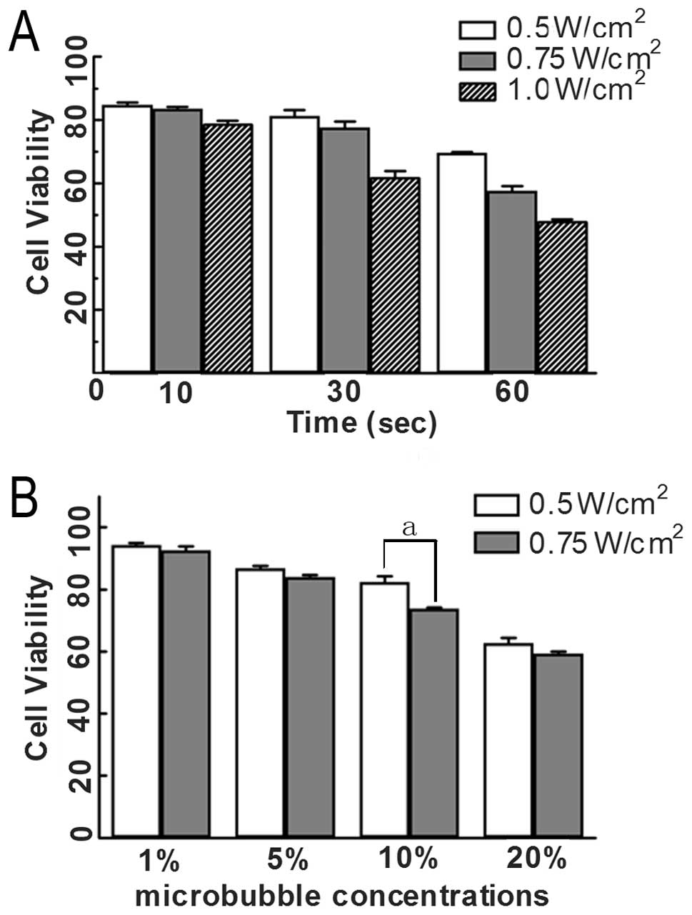
Effect of different ultrasonic conditions
on the viability of HeLa cells
The viability of HeLa cells under different
ultrasonic conditions, as assessed by MTT method, is shown in
Table I and Fig. 1A. When ultrasound was conducted
with 0.5, 0.7, 1.0 W/cm2 and 10 sec, 0.5, 0.75
W/cm2 and 30 sec, cell survival was approximately 80%.
When ultrasound was 0.5 W/cm2 and 60 sec, viability was
approximately 70%. As the intensity and time increased
simultaneously, cell viability was decreased significantly.
 | Table I.Effect of different ultrasonic
conditions on the viability of HeLa cells. |
Table I.
Effect of different ultrasonic
conditions on the viability of HeLa cells.
| Ultrasound intensity
(W/cm2)
|
|---|
| Exposure time
(sec) | 0.5 | 0.75 | 1 |
|---|
| 10 | 84.75±0.75 | 83.43±0.67 | 78.84±0.87 |
| 30 | 81.19±2.00 | 77.61±1.93 | 61.97±1.86 |
| 60 | 69.56±0.32 | 57.67±1.51 | 48.05±0.57 |
Transfection rate of the wtp53 gene in
HeLa cells under different ultrasonic conditions
Based on the results of the MTT assay, we chose
these ultrasound parameters (30 sec, 0.5 and 0.75 W/cm2;
60 sec, 0.5 W/cm2; 10 sec, 1 W/cm2) as the
transfection conditions of the wtp53 gene, for which cell viability
was >70%. Cells were incubated for 48 h after transfection and
then the expression of EGFP was examined using fluorescence
microscopy. As presented in Fig.
2A, when ultrasound was 0.5 W/cm2 and 30 sec,
expression of EGFP in HeLa cells was highter than that in the other
three groups. The result demonstrated that using this acoustic
intensity and exposure time, the transfection efficiency of the
wtp53 gene was highest.
Effect of different microbubble
concentrations on HeLa cell viability
As shown in the result of the MTT assay (Table II and Fig. 1B), when the microbubble
concentration was 20%, and ultrasound parameters were 0.5
W/cm2, 30 sec or 0.75 W/ cm2, 30 sec, cell
survival was <70%. No significant difference was noted between
the two groups (p>0.05). When micro-bubble concentration was 1
or 5% and ultrasound parameters were 0.5 W/cm2, 30 sec
or 0.75 W/cm2, 30 sec, cell survival was >80%. That
is to say, cell viability was not significantly affected. When the
microbubble concentration was 10%, the difference between 0.5
W/cm2, 30 sec and 0.75 W/cm2, 30 sec was
statistically significant (p<0.05). At the same time, we found
that when the ultrasound parameters were 0.5 W/cm2, 30
sec or 0.75 W/cm2, 30 sec, the difference in cell
viability was significant for the four concentrations of
microbubbles.
| Table II.Effect of different microbubble
concentrations on HeLa cell viability. |
Table II.
Effect of different microbubble
concentrations on HeLa cell viability.
| Microbubble
concentrations (%)
|
|---|
| Ultrasound intensity
(W/cm2) | 1 | 5 | 10 | 20 |
|---|
| 0.5 | 94.25±0.76b | 86.78±0.87b |
82.41±1.81a,b | 62.72±1.71b |
| 0.75 | 92.59±1.32c | 84.00±0.69c |
73.74±0.44a,c | 59.35±0.58c |
Due to the slight effect on cell viability and high
transfection efficiency, we chose 0.5 W/cm2, 30 sec,
continuous wave and a microbubble concentration of 10% as the
optimal transfection condition for transfection of the wtp53 gene
in subsequent experiments.
wtp53 transfection rate in each
experimental group
Fig. 2B indicates
that the two groups, plasmid with microbubbles and ultrasound and
plasmid with liposome, showed greater EGFP expression in HeLa cells
than that in the other three groups. The transfection rate in the
former was higher than that in the latter (p<0.05), while it was
higher in the plasmid and liposome group than that in the plasmid
only and plasmid and ultrasound groups (p<0.01).
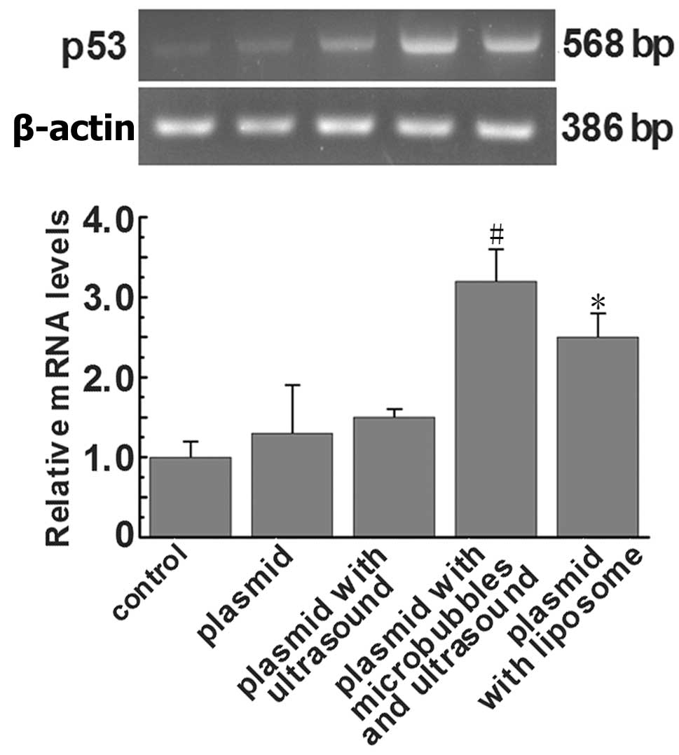
Expression of wtp53 mRNA in each
experimental group
The RT-PCR results (Fig. 3) showed that wtp53 expression was
detected in the plasmid with microbubbles and ultrasound group and
the plasmid with liposome group, while in the other three groups
the expression was not apparent. The wtp53 mRNA level was also
increased in the plasmid with microbubbles and ultrasound group
compared to the plasmid with liposome group. This demonstrated that
the carrier of ultrasound micro-bubbles increased the transfection
rate of the wtp53 gene.
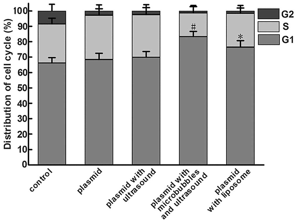
Transfection of wtp53 results in cell
cycle arrest
To test the role of wtp53 in the cell cycle
progression of HeLa cells, the cell cycle distribution was assessed
by flow cytometry. As indicated in Table III and Fig. 4, in the plasmid with microbubbles
and ultrasound group, 83.33±3.43% of the total number of cells was
in the G1 phase of the cell cycle. Compared to the other groups,
the number of G1 phase cells was increased significant, while the
number of cells in the G2 phase was decreased. This indicated that
the wtp53 gene arrested the cell cycle of HeLa cells in the G1
phase.
| Table III.Flow cytometry for cell cycle
analysis. |
Table III.
Flow cytometry for cell cycle
analysis.
| Groups | G1 | S | G2 |
|---|
| Control | 66.17±3.50 | 25.35±3.70 | 8.48±4.32 |
| Plasmid | 68.42±4.12 | 28.81±4.28 | 2.77±3.91 |
| Plasmid with
ultrasound | 69.92±3.71 | 27.69±4.53 | 2.39±4.11 |
| Plasmid with
microbubbles and ultrasound | 83.33±3.43a | 15.34±4.15 | 1.33±3.50 |
| Plasmid with
liposome | 76.51±4.23b | 21.91±3.53 | 1.58±3.64 |
Discussion
Cervical cancer is a common gynecological
malignancy. Its therapy faces the task of reducing recurrence by
the killing of most tumor cells and the removal of the tumor
tissue, but which simultaneously reduces the fertility of the
patient. Therefore, researchers need to explore better methods with
which to optimize the treatment of cervical cancer.
One of the most common tumor-suppressor genes
involved in human malignancies is p53 (14). When there is a mutation in the p53
gene, its monitoring capacity is lost and it becomes an oncogene.
Mutated or inactivated p53 can lead to selective growth advantage
and tumor formation (4). In view
of the high mutation rate of the p53 gene in human malignant
neoplasms, the use of the wtp53 gene in cancer therapy has become a
main focus of research. The wtp53 gene inhibits tumor cell
proliferation by blocking the cell cycle and accelerating cell
apoptosis (15,16). It has been reported that wtp53 also
increases tumor sensitivity to radiotherapy and chemotherapy
(17) and reduces the side effects
of radiotherapy and chemotherapy (18).
Gene therapy is a promising new method. The main
difficulty involves the choice of gene vector. As a novel gene
transfer vector, the microbubble contrast agent can be broken down
by ultrasound exposure in designated areas in order to release the
gene it carries (19,20). At present, researchers working on
gene transfection mediated by UMMT have achieved some important
breakthroughs. It has been demonstrated that genes could be highly
transfected into various types of tissues using this technique
(21). UMMT plays an ever
increasing role in enhancing the delivery of therapeutic agents
into various tissues, such as the myocardium, blood vessels,
skeletal muscle, tumor and even fetal tissues. These deliverable
agents currently include genetic material, proteins and
chemotherapeutic drugs (22–28).
However, a high concentration of microbubbles may
have a negative impact on cell activity, and the use of ultrasound
at a certain range of sound intensity and period of time when used
to break the microbubbles, can harm cells and even cause cell death
(29). Thus, in the present study,
different ultra-sound intensities, ultrasound exposure times and
different microbubble concentrations were used in preliminary
experiments to identify the optimal experimental conditions. MTT
assay showed that ultrasound microbubbles had a certain inhibitory
effect on the proliferation of the HeLa cell line. From the result
of our preliminary experiments, we chose an ultrasound intensity of
0.5 W/cm2, an ultrasound exposure time of 30 sec and a
microbubble concentration of 10% as the optimum experimental
condition for wtp53 plasmid transfection into HeLa cells.
To further investigate the transfection efficiency
of ultra-sound combined with microbubbles, RT-PCR analysis was used
to examine the mRNA level of p53. Under different transfection
conditions, the transfection efficiency of wtp53 varied. It was
clear that the transfection efficiency in the plasmid plus
microbubbles and ultrasound group was significantly higher than
that in the plasmid with liposome group. No expression was
detected, however, in the control group or plasmid with ultrasound
group. This indicates that microbubbles as a novel transfection
medium may meet the need for efficient gene transfer. Flow
cytometric evaluation of the cell cycle showed that as a novel gene
transfer vector, ultrasound microbubble contrast agents can
increase the intracellular levels of the wtp53 gene, affect the
cell cycle, and thereby inhibit tumor cell growth.
In fact, there are many factors relevant to
ultrasound microbubble-mediated gene transfer. Relevant conditions
can be further optimized to improve transfection efficiency and
reduce damage to normal cells. Our study was limited to experiments
in vitro. Experiments in vivo will be the focus of
further research. This technology in gynecological disease is still
in its infancy. Based on the existing technology, more in-depth
study and investigation are warranted.
Acknowledgements
This study was supported by the
National Nature Science Foundation of China (project no.
30770566).
References
|
1.
|
Jemal A, Siegel R, Ward E, Hao Y, Xu J and
Thun MJ: Cancer statistics, 2009. CA Cancer J Clin. 59:225–249.
2009. View Article : Google Scholar
|
|
2.
|
Green J, Kirwan J, Tierney J, Vale C,
Symonds P, Fresco L, Williams C and Collingwood M: Concomitant
chemotherapy and radiation therapy for cancer of the uterine
cervix. Cochrane Database Syst Rev. CD0022252005.
|
|
3.
|
Nishizaki M, Meyn RE, Levy LB, Atkinson
EN, White RA, Roth JA and Ji L: Synergistic inhibition of human
lung cancer cell growth by adenovirus-mediated wild-type p53 gene
transfer in combination with docetaxel and radiation therapeutics
in vitro and in vivo. Clin Cancer Res. 7:2887–2897. 2001.PubMed/NCBI
|
|
4.
|
Oki E, Tokunaga E, Nakamura T, et al:
Genetic mutual relationship between PTEN and p53 in gastric cancer.
Cancer Lett. 227:33–38. 2005. View Article : Google Scholar : PubMed/NCBI
|
|
5.
|
Lu QL, Liang HD, Partridge T and Blomley
MJ: Microbubble ultrasound improves the efficiency of gene
transduction in skeletal muscle in vivo with reduced tissue damage.
Gene Ther. 10:396–405. 2003. View Article : Google Scholar : PubMed/NCBI
|
|
6.
|
Chen S, Ding JH, Bekeredjian R, et al:
Efficient gene delivery to pancreatic islets with ultrasonic
microbubble destruction technology. Proc Natl Acad Sci USA.
103:8469–8474. 2006. View Article : Google Scholar : PubMed/NCBI
|
|
7.
|
Oberle V, de Jong G, Drayer JI and
Hoekstra D: Efficient transfer of chromosome-based DNA constructs
into mammalian cells. Biochim Biophys Acta. 1676:223–230. 2004.
View Article : Google Scholar : PubMed/NCBI
|
|
8.
|
Shohet RV, Chen S, Zhou YT, Wang Z,
Meidell RS, Unger RH and Grayburn PA: Echocardiographic destruction
of albumin microbubbles directs gene delivery to the myocardium.
Circulation. 101:2554–2556. 2000. View Article : Google Scholar : PubMed/NCBI
|
|
9.
|
Xing W, Gang WZ, Yong Z, Yi ZY, Shan XC
and Tao RH: Treatment of xenografted ovarian carcinoma using
paclitaxel-loaded ultrasound microbubbles. Acad Radiol.
15:1574–1579. 2008. View Article : Google Scholar : PubMed/NCBI
|
|
10.
|
Chen Z, Xie M, Wang X, Lv Q and Ding S:
Efficient gene delivery to myocardium with ultrasound targeted
microbubble destruction and polyethylenimine. J Huazhong Univ Sci
Technolog Med Sci. 28:613–617. 2008. View Article : Google Scholar : PubMed/NCBI
|
|
11.
|
Bekeredjian R, Kroll RD, Fein E, et al:
Ultrasound targeted microbubble destruction increases capillary
permeability in hepatomas. Ultrasound Med Biol. 33:1592–1598. 2007.
View Article : Google Scholar : PubMed/NCBI
|
|
12.
|
Anwer K, Kao G, Proctor B, et al:
Ultrasound enhancement of cationic lipid mediated gene transfer to
primary tumors following systemic administration. Gene Ther.
7:1833–1839. 2000. View Article : Google Scholar : PubMed/NCBI
|
|
13.
|
Nie F, Xu HX, Tang Q and Lu MD:
Microbubble-enhanced ultra-sound exposure improves gene transfer in
vascular endothelial cells. World J Gastroenterol. 12:7508–7513.
2006.PubMed/NCBI
|
|
14.
|
Tomkova K, Tomka M and Zajac V:
Contribution of p53, p63, and p73 to the developmental diseases and
cancer. Neoplasma. 55:177–181. 2008.PubMed/NCBI
|
|
15.
|
Nork TM, Poulsen GL, Millecchia LL, Jantz
RG and Nickells RW: p53 regulates apoptosis in human
retinoblastoma. Arch Ophthalmol. 115:213–219. 1997. View Article : Google Scholar : PubMed/NCBI
|
|
16.
|
Ghule P, Kadam PA, Jambhekar N, et al: p53
gene gets altered by various mechanisms: studies in childhood
sarcomas and retinoblastoma. Med Sci Monit. 12:BR385–BR396.
2006.
|
|
17.
|
Xu L, Pirollo KF, Tang WH, Rait A and
Chang EH: Transferrin liposome mediated systemic p53 gene therapy
in combination with radiation results in regression of human head
and neck cancer xenografts. Hum Gene Ther. 10:2941–2952. 1999.
View Article : Google Scholar : PubMed/NCBI
|
|
18.
|
Haupt S and Haupt Y: Importance of p53 for
cancer onset and therapy. Anticancer Drugs. 17:725–732. 2006.
View Article : Google Scholar : PubMed/NCBI
|
|
19.
|
Tiukinhoy SD, Mahowald ME, Shively VP, et
al: Development of echogenic, plasmid-incorporated, tissue-targeted
cationic liposomes that can be used for directed gene delivery.
Invest Radiol. 35:732–738. 2000. View Article : Google Scholar
|
|
20.
|
Li T, Tachibana K and Kuroki M and Kuroki
M: Gene transfer with echo-enhanced contrast agents: comparison
between Albunex, Optison, and Levovist in mice-initial results.
Radiology. 229:423–428. 2003. View Article : Google Scholar : PubMed/NCBI
|
|
21.
|
Shimamura M, Sato N, Taniyama Y, et al:
Development of efficient plasmid DNA transfer into adult rat
central nervous system using microbubble-enhanced ultrasound. Gene
Ther. 11:1532–1539. 2004. View Article : Google Scholar : PubMed/NCBI
|
|
22.
|
Zhigang W, Zhiyu L, Haitao R, et al:
Ultrasound-mediated microbubble destruction enhances VEGF gene
delivery to the infarcted myocardium in rats. Clin Imaging.
28:395–398. 2004. View Article : Google Scholar : PubMed/NCBI
|
|
23.
|
Zhang Q, Wang Z, Ran H, et al: Enhanced
gene delivery into skeletal muscles with ultrasound and microbubble
techniques. Acad Radiol. 13:363–367. 2006. View Article : Google Scholar : PubMed/NCBI
|
|
24.
|
Mitragotri S, Blankschtein D and Langer R:
Transdermal drug delivery using low-frequency sonophoresis. Pharm
Res. 13:411–420. 1996. View Article : Google Scholar : PubMed/NCBI
|
|
25.
|
Mitragotri S and Kost J: Low-frequency
sonophoresis: a noninvasive method of drug delivery and
diagnostics. Biotechnol Prog. 16:488–492. 2000. View Article : Google Scholar : PubMed/NCBI
|
|
26.
|
Tachibana K, Uchida T, Tamura K, Eguchi H,
Yamashita N and Ogawa K: Enhanced cytotoxic effect of Ara-C by low
intensity ultrasound to HL-60 cells. Cancer Lett. 149:189–194.
2000. View Article : Google Scholar : PubMed/NCBI
|
|
27.
|
Munshi N, Rapoport N and Pitt WG:
Ultrasonic activated drug delivery from Pluronic P-105 micelles.
Cancer Lett. 118:13–19. 1997. View Article : Google Scholar : PubMed/NCBI
|
|
28.
|
Yu T, Hu K, Bai J and Wang ZB: Reversal of
adriamycin resistance in ovarian carcinoma cell line by combination
of verapamil and low-level ultrasound. Ultrason Sonochem. 10:37–40.
2003. View Article : Google Scholar : PubMed/NCBI
|
|
29.
|
Dalecki D, Raemen CH, Child SZ, Cox C,
Francis CW, Meltzer RS and Carstensen EL: Hemolysis in vivo from
exposure to pulsed ultrasound. Ultrasound Med Biol. 23:307–313.
1997. View Article : Google Scholar : PubMed/NCBI
|