Evaluation of a partial optic nerve crush model in rats

  • Authors:
    • Hai-Bo Tan
    • Xi Shen
    • Yu Cheng
    • Qin Jiao
    • Zi-Jian Yang
    • Yi-Sheng Zhong
  • View Affiliations

  • Published online on: June 22, 2012     https://doi.org/10.3892/etm.2012.619
  • Pages: 401-404
Metrics: Total Views: 0 (Spandidos Publications: | PMC Statistics: )
Total PDF Downloads: 0 (Spandidos Publications: | PMC Statistics: )


Abstract

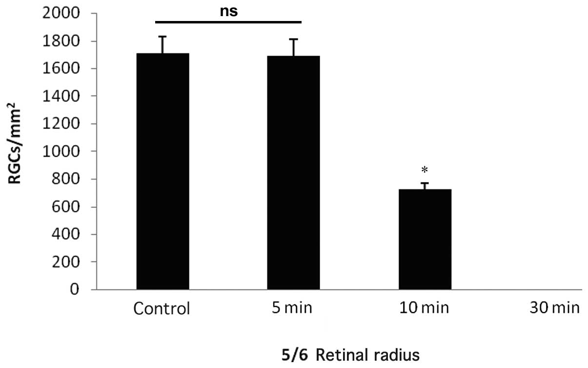
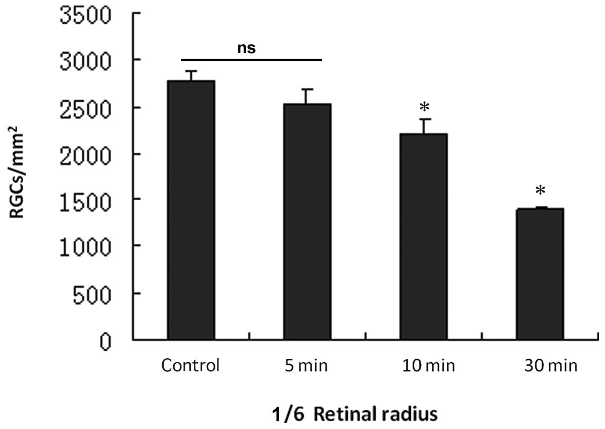
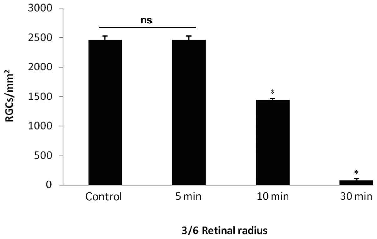
This study was performed to determine whether a partial optic nerve crush (PONC) model in rats is effective and reliable for the study of optic nerve protection and regeneration. Bilateral superior colliculus (SC) retrograde 1,1'-dioctadecyl-3,3,3',3'-tetramethylindocarbocyanine perchlorate (DiI) labeling of retinal ganglion cells (RGCs; n=3) and unilateral SC retrograde labeling of RGCs (n=3) were performed in adult Sprague-Dawley (SD) rats and the results were compared with the bilateral and unilateral SC retrograde-labeled RGCs. Another 40 adult SD rats, three days after bilateral SC retrograde DiI labeling of RGCs underwent crushing with a non‑invasive vascular clip (40 gram power) 1 mm behind the right optic nerve head for 5, 10 and 30 sec (n=10 each), and a sham-operated control group (n=10) was used as a control. The retinas of all 40 rats were flattened by four radial cuts, mounted vitreal side‑up on gelatin-coated slides, and the number of labeled RGCs was counted in four distinct regions per retinal quadrant at three different eccentricities of 1/6, 3/6 and 5/6 of the retinal radius three days later. Bilateral SC retrograde DiI injection labeled the majority of normal RGCs, while unilateral SC injections only labeled a small part of the RGCs; the majority of RGCs were not labeled. In the mild crush (5 sec) injury group, the bilateral SC retrograde DiI injection labeled the majority of RGCs. The RGC densities at 1/6, 3/6 and 5/6 of the retinal radius showed no significant difference compared with the RGC densities at the corresponding region of the retinal radius in the sham-operated control group (P=0.734, 0.461, 0.273, respectively). In the moderate crush injury (10 sec) group, the number of labeled RGCs was significantly lower compared to that of the sham-operated control group, and the RGC densities at 1/6, 3/6, 5/6 of the retinal radius were significantly lower compared to the RGC densities at the corresponding retinal radius in the sham-operated control group (P<0.001). In the severe crush injury (30 sec) group the number of labeled RGCs was significantly decreased, and the labeled RGCs were not observed in the region at 5/6 of the retinal radius. The RGC densities at 1/6 and 3/6 of the retinal radius were significantly lower compared to the RGC densities at the corresponding retinal radius region in the sham-operated control group (P<0.001). Compared with the mild and severe optic nerve crush injury models, the moderate crush injury model is more suitable for the study of optic nerve damage and regeneration.
View Figures
View References

Related Articles

Journal Cover

September 2012
Volume 4 Issue 3

Print ISSN: 1792-0981
Online ISSN:1792-1015

Sign up for eToc alerts

Recommend to Library

Copy and paste a formatted citation
x
Spandidos Publications style
Tan H, Shen X, Cheng Y, Jiao Q, Yang Z and Zhong Y: Evaluation of a partial optic nerve crush model in rats. Exp Ther Med 4: 401-404, 2012
APA
Tan, H., Shen, X., Cheng, Y., Jiao, Q., Yang, Z., & Zhong, Y. (2012). Evaluation of a partial optic nerve crush model in rats. Experimental and Therapeutic Medicine, 4, 401-404. https://doi.org/10.3892/etm.2012.619
MLA
Tan, H., Shen, X., Cheng, Y., Jiao, Q., Yang, Z., Zhong, Y."Evaluation of a partial optic nerve crush model in rats". Experimental and Therapeutic Medicine 4.3 (2012): 401-404.
Chicago
Tan, H., Shen, X., Cheng, Y., Jiao, Q., Yang, Z., Zhong, Y."Evaluation of a partial optic nerve crush model in rats". Experimental and Therapeutic Medicine 4, no. 3 (2012): 401-404. https://doi.org/10.3892/etm.2012.619