Introduction
Breast cancer is considered to be one of the most
commonly diagnosed cancers in women and metastasis remains the
major cause of cancer-related mortality (1,2).
Metastasis is a multi-step process that includes the detachment of
cancer cells from the primary site, migration and invasion of tumor
cells into the blood or lymphatic vessels, as well as motility and
invasion into the new target tissue (3). Cytoskeletal reorganization and cell
movement underlie all the metastatic events, as well as the
disruption of adhesiveness.
Protein 4.1 is a cytoskeletal protein most
extensively studied in red blood cells (4.1R) where it stabilizes
the spectrin-actin network and anchors it to the plasma membrane.
4.1N was originally designated as a neuronal homologue of the
erythrocyte 4.1. The cytoskeletal protein 4.1 family comprising
4.1R (4), 4.1B (5), 4.1G (6) and 4.1N (7) was detected in various cell types and
tissues. However, the functions of protein 4.1 in non-erythroid
cells are not as clear as the functions of 4.1R in the mature red
blood cells. The protein 4.1 family is characterized by the
presence of an N-terminal membrane binding domain (MBD). The MBD of
the 4.1 protein is closely associated, in sequence and in
structure, to the N-terminal domains of ezrin, radixin and moesin
(the ERM proteins), and is, therefore, commonly referred to as the
FERM domain (8–10). A molecule with a FERM domain at the
N-terminus belongs to the protein 4.1 superfamily. Over 40 members
have been identified in this superfamily (11).
Although the membrane-binding domains of the
proteins of the ERM and 4.1 families share a high degree of
sequence homology, and a marked difference in their functions has
been detected. For example, it has been observed that ezrin
promotes cell growth and may be key in tumor metastasis (12,13),
while increasing evidence suggests that members of the 4.1 protein
family act as tumor suppressors (14–17).
Loss of 4.1B was observed in a variety of human tumors, including
meningiomas, non-small cell lung cancers and breast carcinomas
(18–22). Protein 4.1R is also involved in
brain tumors (16). In a screen
for genes involved in breast cancer metastasis, 4.1N expression was
found to be absent in highly metastatic breast cancer MDA-MB-231
cells, whereas poorly metastatic cells, 4.1N was expressed and
predominantly associated with cell-cell junctions. Thus, we
re-introduced protein 4.1N in highly metastatic breast cancer
MDA-MB-231 cells to validate its potential tumor metastatic
suppressive function.
Materials and methods
Cell lines and culture conditions
The human breast cancer cell lines, MCF-7, T-47D and
MDA-MB-231 (all provided by the American Type Culture Collection,
ATCC, Manassas, VA, USA), were used in the present study. MCF-7 is
a low metastatic breast cancer cell line, while T-47D is a middle
and MDA-MB-231 a highly metastatic breast cancer cell line. The
three cell lines were grown in DMEM medium (Gibco, Invitrogen,
Carlsbad, CA, USA) supplemented with 10% fetal bovine serum (FBS)
at 37°C in a humidified atmosphere containing 5% CO2.
The cells were subcultured every 2–3 days to maintain exponential
growth.
Western blot analysis
Western blot analysis was performed for the cell
lines to confirm the presence or absence of the protein 4.1N. Cell
lysates were prepared in RIPA buffer (50 mM Tris, 150 mM NaCl, 0.1%
SDS, 0.5% sodium deoxycholate, 1% NP40) with protease inhibitors.
Total proteins were quantified by the Bradford method using the BCA
protein assay kit according to the manufacturer’s instructions.
Samples were boiled at 100°C in loading buffer for 10 min and 30 μg
protein from each sample were electrophoresed on a 10%
SDS-polyacrylamide gel (SDS-PAGE) and then transferred to PVDF
membranes by semi-dry transfer apparatus (Bio-Rad, Hercules, CA,
USA). The membranes were blocked in TBST (25 mM Tris-HCl, pH 7.5,
137 mM NaCl, 2.7 mM KCl and 0.05% Tween-20) with 5% fat-free milk
for 1 h at 37°C, and then incubated with the primary antibodies
(rabbit anti-4.1N and rabbit anti-GFP, provided by Dr Xiuli An from
the New York Blood Center, Melville, NY, USA) in blocking buffer
overnight at 4°C. After washing three times with TBST, the
membranes were incubated with secondary antibody (goat anti-rabbit
conjugated with HRP; Jackson ImmunoResearch Laboratories, Inc.,
West Grove, PA, USA) for 1 h at room temperature and finally
exposed to Kodak BioMax Film using Super ECL Detection Reagent. The
total protein loading on gel was reconfirmed by blotting with an
antibody against GAPDH (Abcam, Cambridge, MA, USA). Protein
quantification was performed by ImageJ software.
Immunocytochemistry
Immunocytochemistry was used to detect the location
of protein 4.1N. Cells were grown on 12-mm chamber slides for 48 h
and fixed immediately in 1% polyoxymethylene for 15 min, then
permeabilized with 0.1% Triton X-100 in 0.25% PFA/PBS for another
15 min. Cells were blocked using 0.1% Triton X-100 in 0.25% PFA/PBS
with 10% horse serum for 30 min, then incubated with primary
antibody (anti-4.1N) diluted in antibody buffer for 60 min at room
temperature. Goat anti-rabbit IgG secondary fluorescein Alexa
Fluor™ 488-labeled antibody was added and incubated at room
temperature for 40 min. Stained cells were visualized using
appropriate filters on a Nikon Eclipse E800M microscope and imaged
using a Sony Cats Eye Digital Photo Camera and Imaging System.
Cell transfection and screening
MDA-MB-231 cells were seeded into 6-well plates at a
density of 5×10⁵ cells/well and maintained in a 37°C
incubator to obtain 80–90% confluence. Cell transfection was
performed using Lipofectamine® 2000 transfection reagent
according to the manufacturer’s instructions. Briefly, 4 μg of
plasmids pEGFP-4.1N (provided by Dr Xiuli An with sequencing
identification being performed in our laboratory) or pEGFP-3C and 6
μl of Lipofectamine® 2000 (Invitrogen) were gently mixed
with 250 μl of serum-free DMEM without antibiotics for a 10-min
incubation at room temperature. The two mixtures were combined and
kept at room temperature for another 20 min. The complex was then
added to the cells. After incubation at 37°C for 24 h, G418 (800
μg/ml; Invitrogen) was applied to stably screen and isolate the
resistant colonies. MDA-MB-231 cells transfected with pEGFP-4.1N
and pEGFP-C3 were designated as EGFP-4.1N/MDA-MB-231 and
EGFP/MDA-MB-231. Stable transfectant clones with high protein 4.1N
expression were identified by western blot analysis and observed
under fluoro-scope microscopy.
Cell adhesion assay
Cell adhesion was evaluated according to the
modified methods described by Charboneau et al (22). Briefly, the 96-well tissue culture
plates were coated with 5 μg fibronectin (Fn) for each well and
incubated at 4°C overnight. Cells (1×10⁴) suspended in
DMEM containing 0.1% BSA were dispensed into each well of the
96-well plates and incubated with 5% CO2 at 37°C for 60
min, then gently washed twice with PBS to remove the unattached
cells. After fixing with 3.8% PFA for 15 min, the cells were
incubated with 0.2% crystal violet solution to stain the cells for
1 h at room temperature and then washed twice with distilled water.
Dye extraction was performed by adding 100 μl of 10% acetic acid
solution and agitating for 10 min. The absorbance was measured at
570 nm. Each assay was performed in triplicate and repeated at
least twice in independent experiments.
Wound-healing assay
Cells were grown to confluence on culture plates and
a wound was made in the monolayer with a sterile P200 pipette tip
(∼0.5 mm in width). The recovery of these monolayer cells was
dependent on cell proliferation and migration during wound-healing.
After wounding, the medium and debris were removed by washing three
times with PBS, and fresh medium was added to the wells. Images of
the wound were captured at 0 and 20 h after wounding to observe the
changes in migration. A mean wound area was determined using ImageJ
software and the average area of wound closure was calculated.
Cell migration and invasion assay
Cell migration assays were performed using a
Transwell chamber (8.0-μm pore size PET inserts; Becton Dickinson,
Franklin Lakes, NJ, USA). The bottom chamber was coated with 10
μg/ml Fn diluted in PBS and incubated at 37°C overnight. Cells
(5x10⁴/well) suspended in 200 μl of DMEM with 0.1%
bovine serum albumin were seeded into the upper chamber either
uncoated (for migration assay) or coated (for invasion assay) with
Matrigel. Cells were allowed to migrate over 16 h and the cells in
the bottom chamber were fixed with 3.8% PFA, followed by staining
with 500 μl of 0.2% crystal violet solution for 2 h at room
temperature. The migrated cells were counted under bright field
microscopy and photographed. The experiment was performed twice
with each sample in triplicate and cell counting was performed in
five randomly selected fields.
Statistical analysis
Data were analyzed with the software package SPSS
12.0 (SPSS, Chicago, IL, USA). P<0.05 was considered to indicate
a statistically significant result.
Results
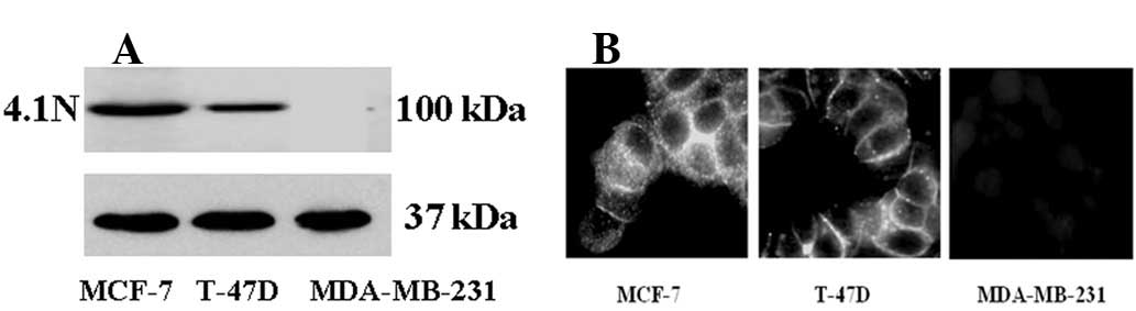
Expression and the cellular location of
the protein 4.1N in breast cancer cells
Immunoblotting analysis was first performed to
examine the expression level of protein 4.1N in three human breast
cancer cell lines with various meta-static abilities. The results
demonstrated that protein 4.1N is expressed in the low and middle
metastatic MCF-7 and T-47D cell lines, respectively, whereas the
protein was not expressed in the highly metastatic MDA-MB-231 cells
(Fig. 1A). Immunocytochemistry was
used to detect the cellular location of protein 4.1N in breast
cancer cell lines with 4.1N antibody. The results showed that 4.1N
was mainly expressed in the cell-cell junctions in the low and
middle metastatic cells. However, 4.1N was not expressed in
MDA-MB-231 cells (Fig. 1B). To
investigate the roles of 4.1N in the metastasis of human breast
cancer cells, the MDA-MB-231 cell line was selected for
transfection and additional study.
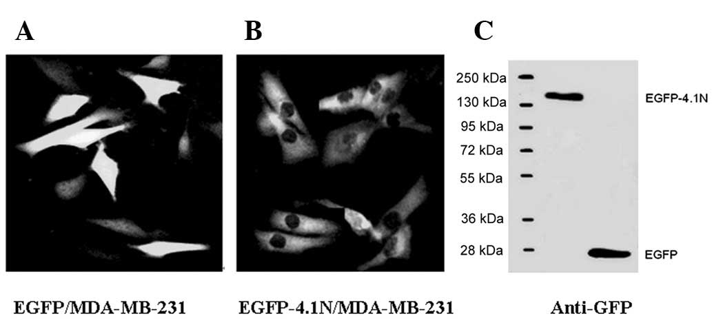
Stable transfection of 4.1N
Based on screening by western blot analysis,
transfection was conducted using MDA-MB-231 cells with pEGFP-4.1N
and an empty pEGFP-C3 vector plasmid was selected for mock
transfectant. After the 14-day selection using 800 μg/ml of G418,
stable cell clones were obtained and pooled populations of clones
were selected to avoid clone variation. Stable transfectant clones
with a high protein 4.1N expression were identified by western blot
analysis and observed under a fluoroscope microscope. Fluorescence
microscopy demonstrated that EGFP is located throughout the cell in
diffuse green fluorescence imaging (Fig. 2A), and the ectogenic EGFP-4.1N
protein was mainly localized in the cytoplasm (Fig. 2B). Western blot analysis confirmed
that EGFP-4.1N fusion protein was highly expressed in MDA-MB-231
cells (Fig. 2C).
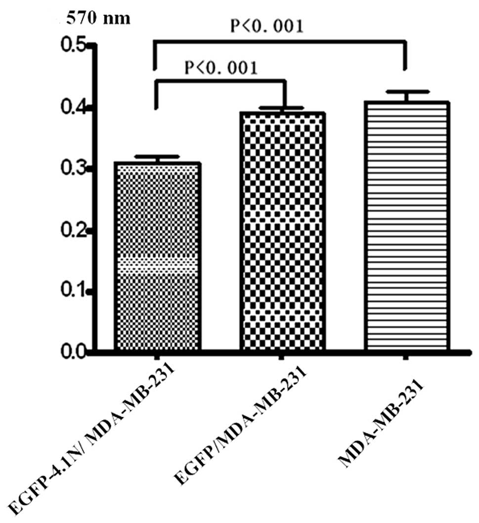
Effect of protein 4.1N on cell
adhesion
Protein 4.1N localizes to the sub-plasma membrane
and, similar to its family members, acts as a linker protein
between the cytoskeleton and the plasma membrane. Thus, this
protein is capable of modulating tumor cell capacity to adhere to
various extracellular matrices, potentially through interaction
with transmembrane proteins and organization of the underlying
cytoskeleton. The effects of the protein 4.1N on cell adhesion were
examined. Results showed that the inhibition of
EGFP-4.1N/MDA-MB-231 cell adhesion ability after incubating cells
for 60 min in 96-well plates coated with Fn (Fig. 3). A significant difference of cell
growth was observed in 4.1N-transfected cells (P<0.001), while
no difference was found between EGFP/MDA-MB-231 and MDA-MB-231
cells, suggesting that protein 4.1N modulated MDA-MB-231 cell
capacity to adhere to Fn.
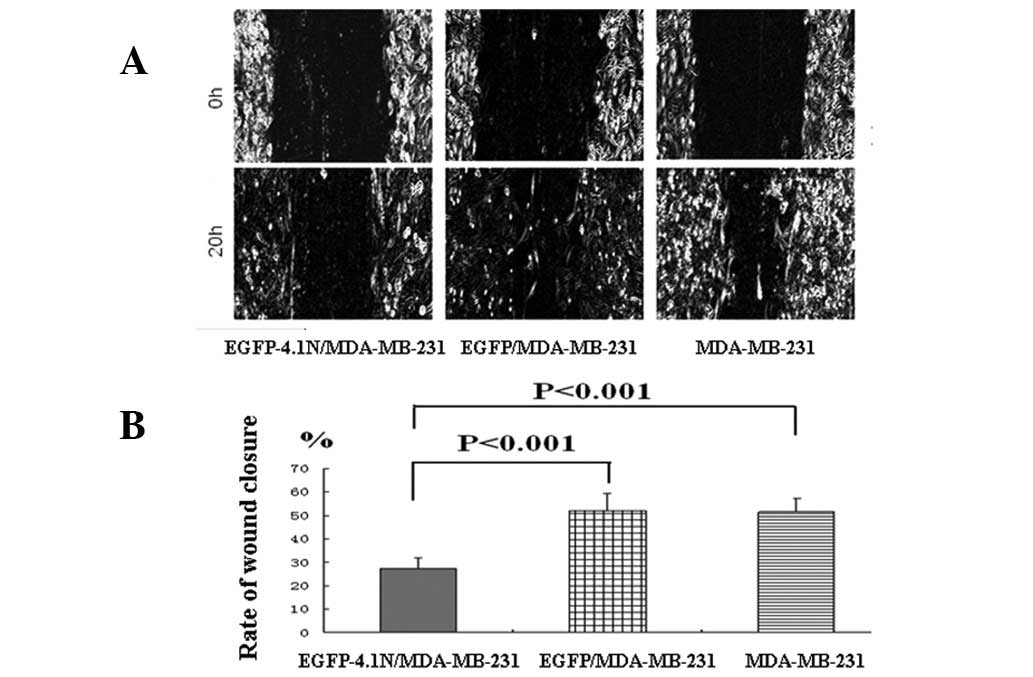
Effect of protein 4.1N on cell
migration
Wound-healing and Transwell assays were used to
investigate whether 4.1N is important in the regulation of cell
migration in MDA-MB-231 cells. In the wound-healing assay, images
were captured at 0 and 20 h to observe the changes in migration
(Fig. 4A). The percentage of wound
area closure at 20 h of wound-healing was then calculated (Fig. 4B). EGFP-4.1N/MDA-MB-231 cells had a
low wound-healing rate when compared to those of EGFP/MDA-MB-231
and MDA-MB-231 cells (P<0.001).
Effect of protein 4.1N on cell
invasion
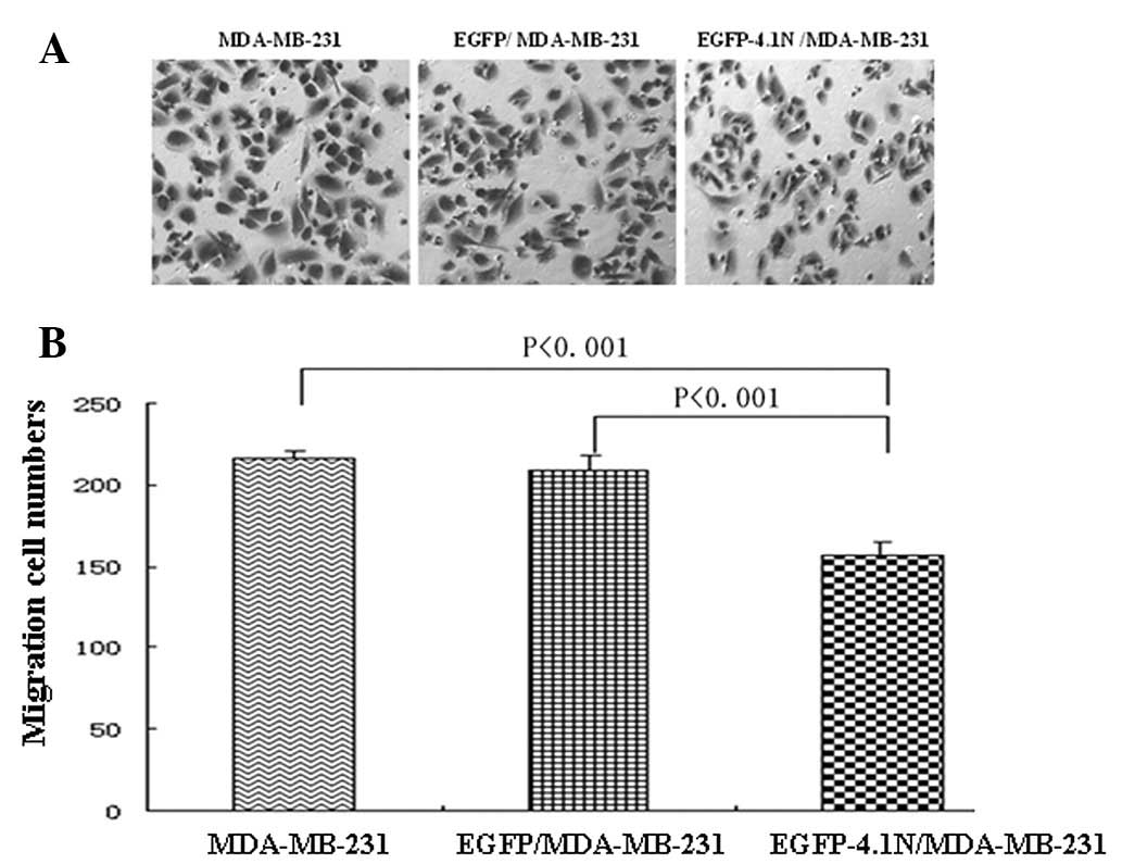
To evaluate the role of protein 4.1N in MDA-MB-231
cell invasion, the ability of cells to permeate through a
reconstituted basement membrane barrier (Matrigel) was tested using
the Transwell assay. EGFP-4.1N/MDA-MB-231 cells (89.3±6.0) showed
significantly reduced invasiveness as compared with EGFP/MDA-MB-231
(163.3±9.1) and MDA-MB-231 cells (166.8±6.0; P<0.001; Fig. 5), indicating that the expression of
4.1N in MDA-MB-231 cells is associated with a reduced invasive
ability.
Discussion
Protein 4.1N was first identified in mouse embryonic
neurons at the earliest stage of differentiation (7). Both 4.1R and 4.1N are known to have
various splice isoforms. The mouse 4.1N predominant isoform in
brain is 135 kDa and a smaller 100-kDa isoform was identified in
peripheral tissues. By analogy with the roles of 4.1R in red blood
cells, 4.1N was considered to confer stability and plasticity to
the neuronal membrane via interactions with multiple binding
partners, including integral membrane receptors and
membrane-associated guanylate kinases. Previously, investigators
identified several proteins that interact with 4.1N which possess
essential roles in cell proliferation, adhesion and signaling
transduction. Typical examples of such proteins include NuMA, PIKE,
NECL1 and AMPA receptor subunit GluR1 (23–27).
Members of the protein 4.1 family that link transmembrane proteins
to the actin cytoskeleton have been demonstrated as tumor
suppressors. 4.1B/DAL-1 was originally identified as a protein
whose expression was reduced in human non-small cell carcinomas
(14). Subsequent studies have
shown that the downregulation of 4.1B/DAL-1 occurs across many
different tumor cell types including brain, breast, prostate,
kidney and sarcoma (28,29). Our previous studies have
demonstrated the complete loss of 4.1N expression in 30% of colon
cancer samples (28/94), particularly in the poorly differentiated
cancers (unpublished data). In the present study, an
anti-4.1N-specific antibody was used to detect 4.1N expression and
subcellular localization in breast cancer cell lines with various
metastatic abilities. Western blot analysis and immunofluorescent
results revealed that the 100-kDa protein 4.1N was expressed and
mainly located at the cell-cell junctions in the poorly metastatic
cell line MCF-7 and middle metastatic cell line T-47D, whereas no
protein 4.1N expression was found in highly metastatic MDA-MB-231
cells. The reintroduction of protein 4.1N by transfection with the
pEGFP-4.1N plasmid attenuated MDA-MB-231 cell adhesion, migration
and invasion, suggesting 4.1N involvement during tumor
progression.
Metastasis is a multi-step process including the
detachment of cancer cells from the primary site, migration and
invasion of tumor cells into the blood or lymphatic vessels,
motility and invasion into the new target tissue. Each step creates
one or more physiological barriers to the spread of malignant
cells. Tumor cells usually have to overcome all of the barriers
including altered adhesiveness, increased motility and invasive
capacity to successfully proceed to metastasis. Since the adhesion
of tumor cells is considered as a key step in the invasive
processes of metastatic tumor cells, the effects of 4.1N on cell
adhesion were examined. One of the observations was that the
reintroduction of 4.1N into MDA-MB-231 cells inhibited cell
adhesion, an observation consistent with the function of NF2, a
member of the protein 4.1 superfamily. In rat schwannoma cells, NF2
expression transiently reduces cell attachment to Fn, with levels
returning to normal after 3 h (30). However, reintroduction of DAL-1
(active segment of 4.1B) into MCF-7 cells increased cell attachment
on all extracellular matrix proteins as measured at 1 h using
similar short-term adhesion assays (22). NF2 and DAL-1 differentially affect
cell adhesion and this is consistent with reports that DAL-1 does
not bind to actin (29). Although
the details of protein 4.1N cell adhesion inhibition require
additional investigation, the data indicate that cells may have an
increased transfection ability in the absence of 4.1N.
Tumor cell migration and invasion through the
basement membranes are key steps in the multi-stage process that
leads to metastatic formation. In the present study,
EGFP-4.1N/MDA-MB-231 cells showed significantly decreased cell
migration and invasion compared to the mock cells. This is the
first evidence suggesting that 4.1N is involved in breast cancer
metastasis. 4.1B reportedly acts as a metastasis suppressor since
its loss supports a reorganization of the F-actin cytoskeleton and
concomitant enhanced cell motility, both of which are likely to be
important in metastasis (17).
4.1N potentially serves as an inhibitor of migration and invasion
by restoring the membrane cytoskeleton.
In conclusion, this is the first study where the
expression of the membrane-cytoskeletal protein 4.1N is associated
with breast cancer metastasis. This study verifies that a 100-kDa
4.1N is crucial in cell adhesion, migration and invasion in breast
cancer cells.
Abbreviations:
|
MBD
|
membrane binding domain
|
|
ERM
|
ezrin, radixin and moesin
|
|
FERM
|
4.1 protein-ezrin-radixin-moesin
homology
|
Acknowledgements
This study was supported by the
National Natural Science Foundation of China (nos. 30972707,
30873002 and 81172784) and the National Science and Technology
Major Projects of New Drugs (2012ZX09103301-0XX).
References
|
1
|
Jemal A, Siegel R, Ward E, Hao Y, Xu J,
Murray T and Thun MJ: Cancer statistics. CA Cancer J Clin.
58:71–96. 2008.
|
|
2
|
Geiger TR and Peeper DS: Metastasis
mechanisms. Biochim Biophys Acta. 2:293–308. 2009.
|
|
3
|
Mahooti S, Porter K, Alpaugh ML, et al:
Breast carcinomatous tumoral emboli can result from encircling
lymphovasculogenesis rather than lymphovascular invasion.
Oncotarget. 2:131–147. 2010.PubMed/NCBI
|
|
4
|
Conboy J, Kan YW, Shohet SB and Mohandas
N: Molecular cloning of protein 4.1, a major structural element of
the human erythrocyte membrane skeleton. Proc Natl Acad Sci USA.
83:9512–9516. 1986. View Article : Google Scholar : PubMed/NCBI
|
|
5
|
Parra M, Gascard P, Walensky LD, et al:
Molecular and functional characterization of protein 4.1B, a novel
member of the protein 4.1 family with high level, focal expression
in brain. J Biol Chem. 275:3247–3255. 2000. View Article : Google Scholar
|
|
6
|
Parra M, Gascard P, Walensky LD, Snyder
SH, Mohandas N and Conboy JG: Cloning and characterization of 4.1G
(EPB41L2), a new member of the skeletal protein 4.1 (EPB41) gene
family. Genomics. 49:298–306. 1998. View Article : Google Scholar : PubMed/NCBI
|
|
7
|
Walensky LD, Blackshaw S, Liao D, et al: A
novel neuron-enriched homolog of the erythrocyte membrane
cytoskeletal protein 4.1. J Neurosci. 19:6457–6467. 1999.PubMed/NCBI
|
|
8
|
Chishti AH, Kim AC, Marfatia SM, et al:
The FERM domain: a unique module involved in the linkage of
cytoplasmic proteins to the membrane. Trends Biochem Sci.
23:281–282. 1998. View Article : Google Scholar : PubMed/NCBI
|
|
9
|
Pearson MA, Reczek D, Bretscher A and
Karplus PA: Structure of the ERM protein moesin reveals the FERM
domain fold masked by an extended actin binding tail domain. Cell.
101:259–270. 2000. View Article : Google Scholar : PubMed/NCBI
|
|
10
|
Han BG, Nunomura W, Takakuwa Y, Mohandas N
and Jap BK: Protein 4.1R core domain structure and insights into
regulation of cytoskeletal organization. Nat Struct Biol.
7:871–875. 2000. View
Article : Google Scholar : PubMed/NCBI
|
|
11
|
Diakowski W, Grzybek M and Sikorski AF:
Protein 4.1, a component of the erythrocyte membrane skeleton and
its related homologue proteins forming the protein 4.1/FERM
superfamily. Folia Histochem Cytobiol. 44:231–248. 2006.PubMed/NCBI
|
|
12
|
Ohtani K, Sakamoto H, Rutherford T, Chen
Z, Satoh K and Naftolin F: Ezrin, a membrane-cytoskeletal linking
protein, is involved in the process of invasion of endometrial
cancer cells. Cancer Lett. 147:31–38. 1999. View Article : Google Scholar
|
|
13
|
Khanna C, Wan X, Bose S, et al: The
membrane-cytoskeleton linker ezrin is necessary for osteosarcoma
metastasis. Nat Med. 10:182–186. 2004. View
Article : Google Scholar : PubMed/NCBI
|
|
14
|
Tran YK, Bögler O, Gorse KM, Wieland I,
Green MR and Newsham IF: A novel member of the NF2/ERM/4.1
superfamily with growth suppressing properties in lung cancer.
Cancer Res. 59:35–43. 1999.PubMed/NCBI
|
|
15
|
Gutmann DH, Donahoe J, Perry A, et al:
Loss of DAL-1, a protein 4.1-related tumor suppressor, is an
important early event in the pathogenesis of meningiomas. Hum Mol
Genet. 10:1495–1500. 2000. View Article : Google Scholar : PubMed/NCBI
|
|
16
|
Robb VA, Li W, Gascard P, Perry A,
Mohandas N and Gutmann DH: Identification of a third Protein 4.1
tumor suppressor, Protein 4.1R, in meningioma pathogenesis.
Neurobiol Dis. 13:191–202. 2003. View Article : Google Scholar
|
|
17
|
Cavanna T, Pokorná E, Veselý P, Gray C and
Zicha D: Evidence for protein 4.1B acting as a metastasis
suppressor. J Cell Sci. 120:606–616. 2007. View Article : Google Scholar
|
|
18
|
Sun CX, Robb VA and Gutmann DH: Protein
4.1 tumor suppressors: getting a FERM grip on growth regulation. J
Cell Sci. 115:3991–4000. 2002. View Article : Google Scholar : PubMed/NCBI
|
|
19
|
Kittiniyom K, Gorse KM, Dalbegue F, Lichy
JH, Taubenberger JK and Newsham IF: Allelic loss on chromosome band
18p11.3 occurs early and reveals heterogeneity in breast cancer
progression. Breast Cancer Res. 3:192–198. 2001. View Article : Google Scholar : PubMed/NCBI
|
|
20
|
Ohno N, Terada N, Murata S, et al:
Immunolocalization of protein 4.1B/DAL-1 during neoplastic
transformation of mouse and human intestinal epithelium. Histochem
Cell Biol. 122:579–586. 2004. View Article : Google Scholar : PubMed/NCBI
|
|
21
|
Terada N, Ohno N, Yamakawa H, et al:
Protein 4.1B in mouse islets of Langerhans and beta-cell
tumorigenesis. Histochem Cell Biol. 120:277–283. 2003. View Article : Google Scholar : PubMed/NCBI
|
|
22
|
Charboneau AL, Singh V, Yu T and Newsham
IF: Suppression of growth and increased cellular attachment after
expression of DAL-1 in MCF-7 breast cancer cells. Int J Cancer.
100:181–188. 2002. View Article : Google Scholar : PubMed/NCBI
|
|
23
|
Ye K, Compton DA, Lai MM, Walensky LD and
Snyder SH: Protein 4.1N binding to nuclear mitotic apparatus
protein in PC12 cells mediates the antiproliferative actions of
nerve growth factor. J Neurosci. 19:10747–10756. 1999.PubMed/NCBI
|
|
24
|
Zhou Y, Du G, Hu X, et al: Nectin-like
molecule 1 is a protein 4.1N associated protein and recruits
protein 4.1N from cytoplasm to the plasma membrane. Biochim Biophys
Acta. 1669:142–154. 2005. View Article : Google Scholar : PubMed/NCBI
|
|
25
|
Shen L, Liang F, Walensky LD and Huganir
RL: Regulation of AMPA receptor GluR1 subunit surface expression by
a 4.1N-linked actin cytoskeletal association. J Neurosci.
20:7932–7940. 2000.PubMed/NCBI
|
|
26
|
Binda AV, Kabbani N, Lin R and Levenson R:
D2 and D3 dopamine receptor cell surface localization mediated by
interaction with protein 4.1N. Mol Pharmacol. 62:507–513. 2002.
View Article : Google Scholar : PubMed/NCBI
|
|
27
|
Maximov A, Tang TS and Bezprozvanny I:
Association of the type 1 inositol (1,4,5)-trisphosphate receptor
with 4.1N protein in neurons. Mol Cell Neurosci. 22:271–283. 2003.
View Article : Google Scholar
|
|
28
|
Heller G, Geradts J, Ziegler B, et al:
Downregulation of TSLC1 and DAL-1 expression occurs frequently in
breast cancer. Breast Cancer Res Treat. 3:283–291. 2007. View Article : Google Scholar : PubMed/NCBI
|
|
29
|
Gutmann DH, Hirbe AC, Huang ZY and Haipek
CA: The protein 4.1 tumor suppressor, DAL-1, impairs cell motility,
but regulates proliferation in a cell-type-specific fashion.
Neurobiol Dis. 8:266–278. 2001. View Article : Google Scholar : PubMed/NCBI
|
|
30
|
Gutmann DH, Sherman L, Seftor L, Haipek C,
Hoang Lu K and Hendrix M: Increased expression of the NF2 tumor
suppressor gene product, merlin, impairs cell motility, adhesion
and spreading. Hum Mol Genet. 8:267–275. 1999. View Article : Google Scholar : PubMed/NCBI
|