Introduction
Increasing evidence has shown that early and
aggressive statin therapy decreases the risk of acute myocardial
infarction (MI) and major adverse cardiovascular events (MACE) in
patients with coronary heart disease (1–3).
Several previous studies have also shown that cardiovascular
morbidity and mortality in patients with hypercholesterolemia are
significantly reduced by statins (4,5). A
meta-analysis of six trials in patients with stable angina revealed
that statin pretreatment resulted in a 59.3% reduction of relative
risk of procedural MI and a 20.5% overall reduction in MACE
(6). Several studies have shown
various loading doses of atorvastatin (ATOR) therapy prior to
percutaneous coronary intervention to be associated with a reduced
risk of MACE (7,8). However, the effects of various
loading doses of statins in patients with stable atherosclerotic
plaques have not yet been evaluated. For this reason, a clinical
follow-up study of various loading doses of ATOR on serum lipids,
inflammation and plaque morphology in patients with stable
atherosclerotic plaques was conducted.
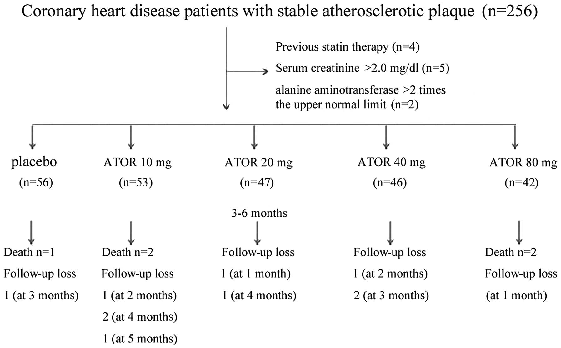
Patients and methods
Study population and design
The patients included in this study were recruited
from Wuxi People’s Hospital, Wuxi City, China, between May 2008 and
December 2010. This study was conducted in accordance with the
declaration of Helsinki and with approval from the Ethics Committee
of Wuxi People’s Hospital, Wuxi City, China. Written informed
consent was obtained from all participants. In total, 256
consecutive patients with stable atherosclerotic plaques who had
undergone diagnostic coronary angiography and intravascular
ultrasound (IVUS) were screened. A total of 11 patients were
excluded due to previous statin therapy, renal insufficiency (serum
creatinine >2.0 mg/dayl) or hepatic disease (history of liver
cirrhosis or alanine aminotransferase greater than twice the upper
limit of normal). Eligible patients were randomly assigned to
receive no statin treatment (placebo group) or to receive ATOR at a
dosage of 10 mg (ATOR 10 mg), 20 mg (ATOR 20 mg), 40 mg (ATOR 40
mg) or 80 mg (ATOR 80 mg). Following six months, five patients had
succumbed to the disease and nine patients were lost to follow-up.
These patients were excluded and the remaining 228 patients were
enrolled in the current study. The study took place after consent
was obtained from all patients. The study design is shown in
Fig. 1.
Aspirin (100 mg/day) was prescribed to all patients
in this study. CK-MB and troponin T levels were measured prior to
coronary angiography and IVUS. Additional cardiac enzyme
measurements were obtained if the patients revealed signs or
symptoms of myocardial ischemia. Low-density lipoprotein
(LDL)-cholesterol, high-density lipoprotein (HDL)-cholesterol and
high-sensitivity C-reactive protein (hs-CRP) levels were assessed
prior to coronary angiography and IVUS. In all patients,
angiotensin-converting enzyme inhibitor (ACEI), angiotensin
receptor blocker (ARB) and beta blockers were administered
according to blood pressure and heart rate.
Patients were followed up for 3–6 months at
one-month intervals, through out-patient contact or by telephone.
Ten patients were lost to follow-up prior to the end of the full
6-month follow-up period, but their data until this point were
included in the statistical analysis. All patients provided written
informed consent.
Coronary angiography and IVUS
analysis
Coronary angiography and IVUS were performed during
inpatient treatment (9). Within
3–6 months of coronary angiography, the site was selected for IVUS
analysis of the coronary artery (10–12).
All IVUS images were acquired using a 20-MHz Volcano Eagle Eye™
IVUS catheter (Volcano Therapeutics Inc,. Rancho Cordova, CA, USA).
Once the coronary lesion had been identified, the IVUS catheter was
inserted distal to the lesion and manually pulled back to assess
the severity and length of the lesion. The IVUS catheter was then
placed distal to a side branch (distal fiduciary landmark site) and
automatic pullback was performed at a rate of 0.5 mm/sec. The
location of the IVUS catheter was determined using continuous
fluoroscopy throughout the time of pullback and by recording
anatomical landmarks observed during IVUS imaging. To create
adequate images, an average of 2 pullbacks per artery were
performed and the best play loop was selected based on imaging
resolution and quality. Continuous EKG monitoring was performed
during the procedure to gate IVUS frames for analysis. IVUS-virtual
histology (VH) data were recorded to the imaging system hard drive
and then extracted and archived for analysis. Analysis was based on
border contour calculation from grayscale. The tissue maps provided
by the software (dark green for fibrous tissue, light green for
fibrofatty tissue, red for necrotic core and white for dense
calcium) were used to analyze each independent frame. Once the
total length of each lesion had been determined, a 20-mm vascular
segment containing the vascular lesion was selected for analysis.
This segment was then divided into equal 2.0-mm subsections,
generating a total of 10 series of cross-sections per vascular
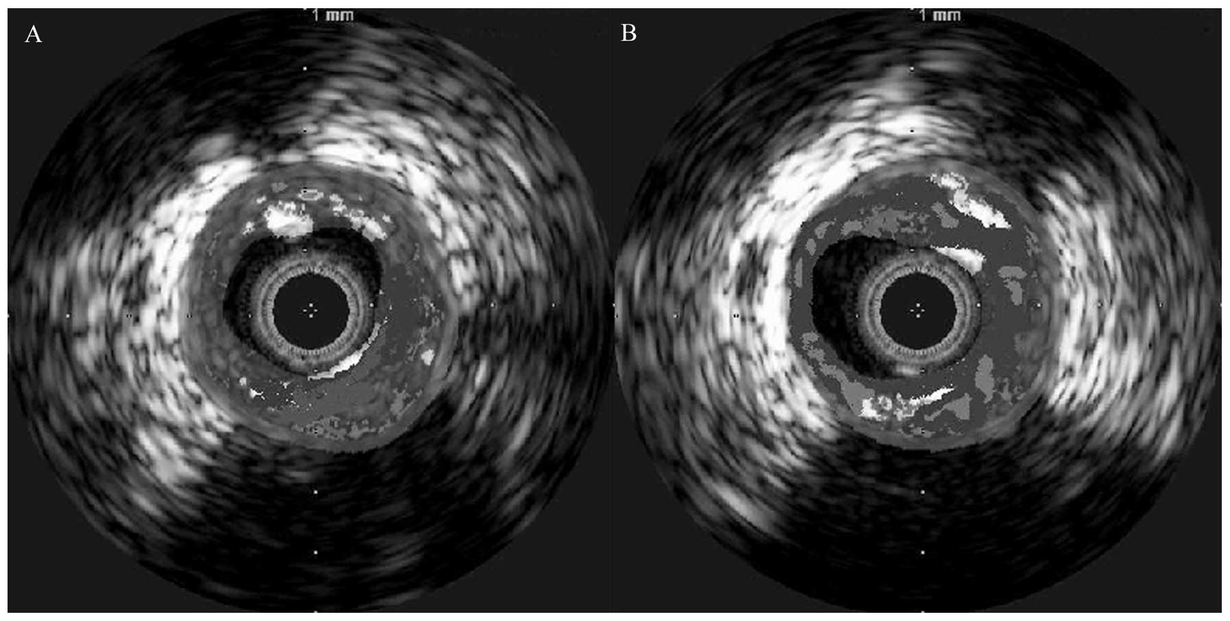
segment. Unstable and stable plaques are shown in Fig. 2.
Endpoints
LDL levels <2.06 mmol/l in patients with coronary
heart disease were defined as normal. HDL levels >1.0 mmol/l and
hs-CRP levels <8 mg/l were defined as normal. Primary endpoints
included changes in LDL, HDL and hs-CRP levels from baseline
following 3–6 months of no treatment or ATOR treatment at the
specified doses. Secondary endpoints included changes in the
percentages of plaque necrosis and in plaque volumes. Stable
plaques were defined as plaques with <10% necrotic tissue.
Plaque volumes = ∑[(external elastic membrane cross-sectional area
- lumen cross-sectional area)/the number of sections] x plaque
length (3).
Statistical analyses
All measurements are shown as mean ± standard
deviation. Continuous variables between two groups were compared by
independent t-tests and Chi-squared tests and multiple groups were
compared by ANOVA. All tests were conducted using SPSS 17.0
software for Windows (Lei An Technology Company, Beijing City,
China). Proportions were compared using Fisher’s exact test when
the expected frequency was <5 and Chi-squared testing in all
other cases. P<0.05 was considered to indicate a statistically
significant result.
Results
Baseline characteristics
There were no significant differences between the
five study groups in baseline and other clinical characteristics as
shown in Table I. ATOR loading was
performed for 3–6 months. VH of IVUS was used with procedural
success achieved in all patients. Medication use was similar among
groups, including the use of ACEI/ARB and beta blockers.
 | Table IBaseline clinical characteristics. |
Table I
Baseline clinical characteristics.
| Characteristics | Placebo (n=54) | ATOR 10 mg group
(n=47) | ATOR 20 mg group
(n=45) | ATOR 40 mg group
(n=43) | ATOR 80 mg group
(n=39) | F(χ2)
P-value |
|---|
| Age (years) | 62.07±8.51 | 62.64±12.00 | 59.18±8.48 | 58.91±12.90 | 58.95±9.68 | 9.085 (0.059) |
| Male/female | 48/6 | 40/7 | 36/9 | 41/2 | 34/5 | 5.00 (0.288) |
| FBG (mmol/l) | 5.26±0.98 | 5.73±1.00 | 5.72±0.82 | 5.00±0.83 | 5.63±0.97 | 0.010 (0.922) |
| Alcohol (yes/no) | 33/21 | 31/16 | 25/20 | 26/17 | 20/19 | 2.246 (0.690) |
| Smoker (yes/no) | 46/8 | 35/8 | 35/10 | 37/6 | 32/7 | 2.962 (0.564) |
| LVEF (%) | 53.65±11.69 | 55.15±13.16 | 58.09±11.10 | 57.14±10.34 | 55.67±10.96 | 1.095 (0.360) |
| Creatinine
(mg/dl) | 85.11±21.63 | 87.33±15.07 | 81.57±16.93 | 83.86±12.90 | 88.11±15.28 | 1.061 (0.377) |
| ACEI (yes/no) | 38/16 | 31/16 | 31/14 | 33/10 | 23/16 | 3.027 (0.524) |
| Beta-blocker
(yes/no) | 28/26 | 29/18 | 21/24 | 21/22 | 15/24 | 4.927 (0.295) |
Primary endpoints
During the follow-up period (3–6 months, mean
4.51±1.23), the endpoints of LDL, HDL and hs-CRP levels in the five
treatment groups revealed significant differences from baseline.
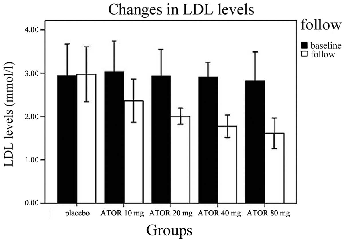
Changes in serum lipids and serum inflammation in groups are shown
in Table II. LDL levels at
follow-up in the placebo group demonstrated no change over baseline
(P=0.813), but LDL levels at follow up in the ATOR 10, 20, 40 and
80 mg groups were lower than their respective baseline levels (all
P<0.01). LDL levels in the ATOR 20 mg group at follow-up were
statistically significantly higher than in the ATOR 40 mg (P=0.048)
and ATOR 80 mg groups at follow-up (P=0.001) and LDL levels in the
ATOR 40 mg group at follow-up were similar to the ATOR 80 mg group
(P=0.168). Changes in LDL at baseline and follow-up are shown in
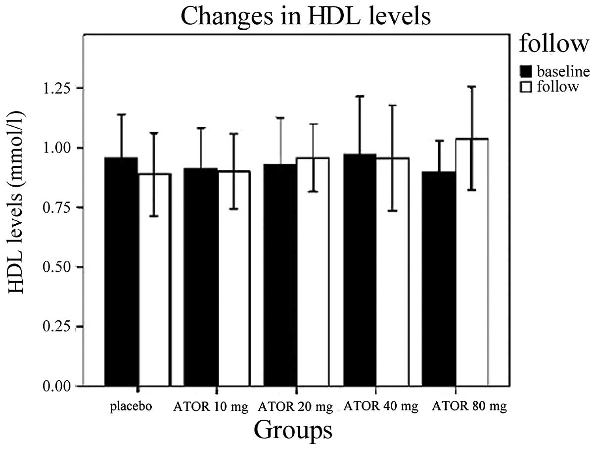
Fig. 3. HDL levels were
significantly higher in the ATOR 80 mg group following treatment
than at baseline (P=0.001). HDL levels were also significantly
higher in the ATOR 80 mg group following treatment than in the
placebo, ATOR 10, 20 or 40 mg groups (P<0.01, P=0.001, P=0.048
and P=0.047, respectively).
| Table IIChanges in serum lipids and serum
inflammation in the study groups. |
Table II
Changes in serum lipids and serum
inflammation in the study groups.
| Variable | Placebo (n=54) | ATOR 10 mg group
(n=47) | ATOR 20 mg group
(n=45) | ATOR 40 mg group
(n=43) | ATOR 80 mg group
(n=39) |
|---|
| HDL (mmol/l) | | | | | |
| Baseline | 0.96±0.18 | 0.90±0.17 | 0.93±0.20 | 0.97±0.24 | 0.90±0.13 |
| Follow-up | 0.89±0.17 | 0.90±0.16 | 0.96±0.14 | 0.96±0.22 | 1.03±0.22 |
| LDL (mmol/l) | | | | | |
| Baseline | 2.94±0.72 | 3.03±0.70 | 2.92±0.62 | 2.90±0.34 | 2.83±0.66 |
| Follow-up | 2.97±0.63 | 2.36±0.50 | 2.01±0.18 | 1.85±0.22 | 1.81±0.32 |
| Hs-CRP (mg/l) | | | | | |
| Baseline | 5.07±1.80 | 6.04±2.52 | 5.09±1.94 | 5.67±2.22 | 6.10±2.12 |
| Follow-up | 6.87±2.62 | 6.74±2.20 | 5.07±1.72 | 4.88±1.52 | 3.59±1.07 |
However, there were no significant differences
between HDL levels amongst the placebo, ATOR 10, 20 or 40 mg
groups. Changes in HDL at baseline and follow-up are shown in
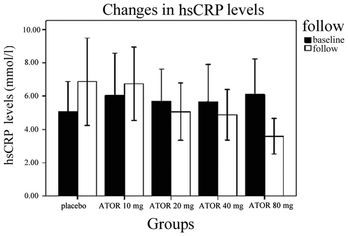
Fig. 4. Hs-CRP levels at follow-up
in the placebo group were higher than baseline (6.87±2.62 vs.
5.07±1.80, P<0.01), while those in the ATOR 80 mg group
following treatment were lower than at baseline (3.59±1.07 vs.
6.10±2.12, P<0.01). There were no statistically significant
differences between hs-CRP following treatment than at baseline for
the ATOR 10 mg, ATOR 20 mg and ATOR 40 mg groups, but higher
dosages of ATOR were generally associated with trends toward
maintaining or decreasing hs-CRP levels over time with treatment.
Changes in hs-CRP at baseline and follow-up are shown in Fig. 5.
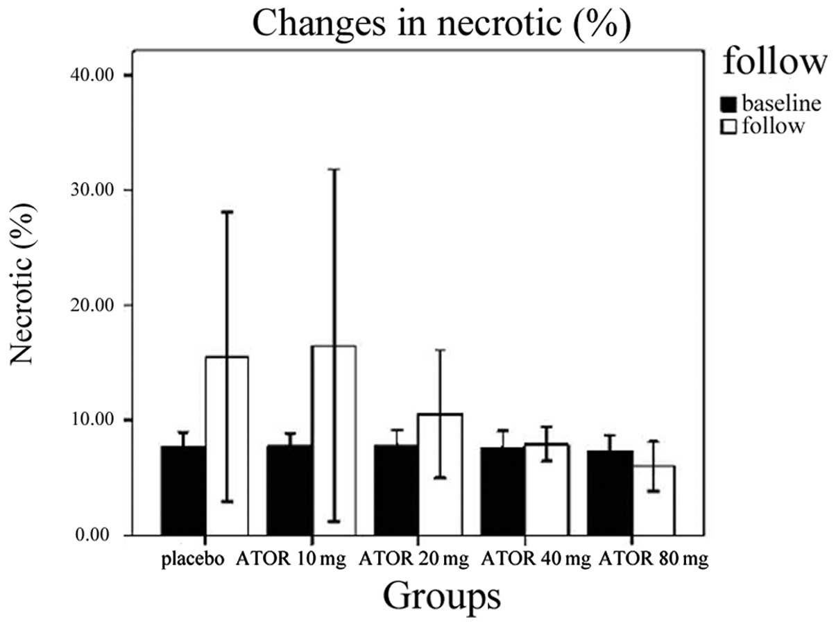
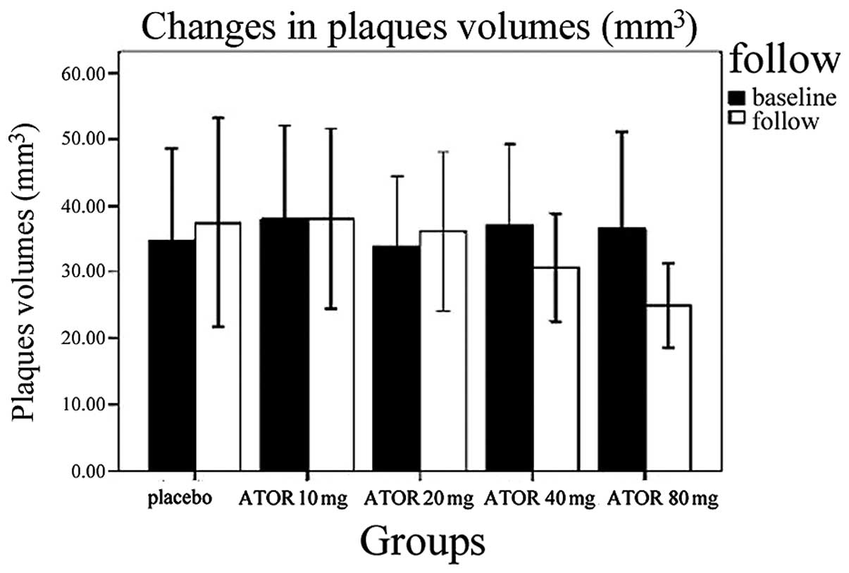
Secondary endpoints
According to the VH of IVUS, the percentages of
plaque necrosis on follow-up increased in the placebo and ATOR 10
mg groups, compared with baseline percentages (15.51±12.56 vs.
7.69±1.31%, 13.54±11.76 vs. 7.83±1.43%, P<0.01), satisfying the
diagnostic criteria for unstable plaques. In the ATOR 20, 40 and 80
mg groups no differences in percentages of plaque necrosis from
baseline were observed (P=0.069, 0.846, 0.643, respectively). In
the placebo, ATOR 10 and 20 mg groups, plaque volumes did not
increase relative to their respective baselines. By contrast, in
the ATOR 40 and 80 mg groups, plaque volumes decreased relative to
their respective baselines (30.69±8.12 vs. 37.09±12.01
mm3, 24.99±1.01 vs. 36.47±14.68 mm3, P=0.019,
P<0.01). Changes in the percentages of plaque necrosis and
plaque volumes in groups are shown in Table III, Figs. 6 and 7
| Table IIIChanges of the percentages of plaque
necrosis and plaque volumes in groups. |
Table III
Changes of the percentages of plaque
necrosis and plaque volumes in groups.
| Variable | Placebo (n=54) | ATOR 10 mg group
(n=47) | ATOR 20 mg group
(n=45) | ATOR 40 mg group
(n=43) | ATOR 80 mg group
(n=39) |
|---|
| Necrotic (%) | | | | | |
| Baseline | 7.69±1.31 | 7.83±1.03 | 7.91±1.27 | 7.64±1.44 | 7.38±1.33 |
| Follow-up | 15.51±12.65 | 16.54±15.76 | 10.55±5.56 | 7.93±1.49 | 6.66±1.92 |
| Plaque volume
(mm3) | | | | | |
| Baseline | 34.83±13.76 | 38.07±13.94 | 33.83±10.56 | 37.06±12.01 | 36.47±14.68 |
| Follow-up | 37.46±15.80 | 38.05±13.56 | 36.12±11.96 | 30.69±8.12 | 24.99±1.01 |
Discussion
In the present dose-ranging study of ATOR in
patients with stable coronary atherosclerotic plaques, the
dose-dependent effects of ATOR on serum lipids, serum inflammation
and plaque morphology were demonstrated. In general, higher dosages
of ATOR up to 80 mg per day for up to 3–6 months of treatment were
associated with a greater potential for beneficial effects by
decreasing LDL, increasing HDL, decreasing hs-CRP, preventing
plaque necrosis and reducing plaque volume in patients with stable
coronary atherosclerotic plaques.
Previous studies have demonstrated that statin
therapy improves the prognosis of patients with coronary heart
disease (13–15). The PROVEIT study reported that a
dose of 80 mg ATOR per day reduced the risk of negative outcomes at
30 days and 2 years (16). The
MIRACL study revealed that 80 mg ATOR per day reduced the risk of
the composite primary endpoints comprising mortality, MI, cardiac
arrest and recurrent ischemia (17). The study indicated that high, early
doses of statin therapy significantly improved the prognosis in
patients with heart disease. A meta-analysis of six trials
(6) in patients with stable angina
revealed that statin pretreatment was associated with a 59.3%
reduction in the relative risk of procedural MI and a 20.5% overall
reduction in MACE. Several studies have been performed to evaluate
the benefits of high doses of statins in patients with heart
disease (18–20). However, to date, no studies have
evaluated the effects of various doses of ATOR on stable
atheroscletotic plaques.
In the current study, the age, male-to-female ratio,
history of diabetes and hypertension, history of alcohol use and
smoking, pre-treatment medication regimen (including ACEI, ARB and
beta blockers), as well as pre-treatment serum lipid levels and
hs-CRP levels, of the patients were not significantly different
between the five groups.
Following treatment, the LDL, HDL and hs-CRP levels
differed among the five groups. For LDL, the use of ATOR at 20
mg/day brought LDL levels down to the standard value, while 40 and
80 mg/day were identified to be similar to each other and more
effective than 20 mg/day in lowering LDL levels. Treatment with 80
mg/day increased HDL levels and no effect was observed with any of
the lower dosages. In patients who were not administered statins,
the hs-CRP levels increased relative to baseline, but 80 mg/day
ATOR lowered hs-CRP levels. Doses of 20 mg/day and higher kept
plaques stable as demonstrated by prevention of progressive plaque
necrosis with treatment, compared with no treatment. In addition,
80 mg/day was revealed to be significantly more effective than 20
or 40 mg/day at decreasing plaque volume. This indicated
dose-dependent effects of ATOR on clinically relevant serum lipid
and inflammatory markers as well as in plaque morphology in
patients with stable atherosclerotic plaques.
The current study has several limitations. Firstly,
the study was not blinded. Secondly, the sample size was relatively
small. Finally, the duration of follow-up was relatively short. Of
note, our original study design called for two years of follow-up,
but the study was stopped early due to statistically significant
results being obtained on interim analysis after 3 and 6 months of
follow-up. Further study is required to confirm our results,
ideally in an independent sample population.
In conclusion, ATOR at a dosage of 80 mg/day for 6
months is associated with improvements in the serum lipid profile,
decrease in serum inflammation and the maintenance of plaque
stability in patients with stable coronary atherosclerotic plaques.
Several of these potentially beneficial effects were also observed
at lower dosages of ATOR, but these effects were more marked and
consistent at 80 mg/day.
References
|
1
|
Schwartz GG, Olsson AG, Ezekowitz MD, et
al Myocardial Ischemia Reduction with Aggressive Cholesterol
Lowering (MIRACL) Study Investigators: Effects of atorvastatin on
early recurrent ischemic events in acute coronary syndromes: the
MIRACL study: a randomized controlled trial. JAMA. 285:1711–1718.
2001. View Article : Google Scholar : PubMed/NCBI
|
|
2
|
Ray KK, Cannon CP, McCabe CH, et al PROVE
IT-TIMI 22 Investigators: Early and late benefits of high-dose
atorvastatin in patients with acute coronary syndromes: results
from the PROVE IT-TIMI 22 trial. J Am Coll Cardiol. 46:1405–1410.
2005. View Article : Google Scholar : PubMed/NCBI
|
|
3
|
Ishizu T, Seo Y, Machino T, et al:
Prognostic impact of plaque echolucency in combination with
inflammatory biomarkers on cardiovascular outcomes of coronary
artery disease patients receiving optimal medical therapy.
Atherosclerosis. 216:120–124. 2011. View Article : Google Scholar
|
|
4
|
Downs JR, Clearfield M, Weis S, et al:
Primary prevention of acute coronary events with lovastatin in men
and women with average cholesterol levels: results of
AFCAPS/TexCAPS. Air Force/Texas Coronary Atherosclerosis Prevention
Study. JAMA. 279:1615–1622. 1998. View Article : Google Scholar
|
|
5
|
Yokoi H, Nobuyoshi M, Mitsudo K, Kawaguchi
A and Yamamoto A; ATHEROMA Study Investigators: Three-year
follow-up results of angiographic intervention trial using an
HMG-CoA reductase inhibitor to evaluate retardation of obstructive
multiple atheroma (ATHEROMA) study. Circ J. 69:875–883. 2005.
|
|
6
|
Ebrahimi R, Saleh J, Toggart E, et al:
Effect of preprocedural statin use on procedural myocardial
infarction and major cardiac adverse events in percutaneous
coronary intervention: a meta-analysis. J Invasive Cardiol.
20:292–295. 2008.
|
|
7
|
Cahoon WD Jr and Crouch MA: Preprocedural
statin therapy in percutaneous coronary intervention. Ann
Pharmacother. 41:1687–1693. 2007. View Article : Google Scholar : PubMed/NCBI
|
|
8
|
Ko DT, Wijerysundera HC, Yun L, Austin PC,
Cantor WJ and Tu JV: Effectiveness of preprocedural statin therapy
on clinical outcomes for patients with stable coronary artery
disease after percutaneous coronary interventions. Circ Cardiovasc
Qual Outcomes. 4:459–466. 2011. View Article : Google Scholar : PubMed/NCBI
|
|
9
|
Wakabayashi K, Mintz G, Delhaye C, et al:
In vivo virtual histology intravascular ultrasound comparison of
neointimal hyperplasia within drug-eluting-versus bare metal
stents. J Invasive Cardiol. 23:262–268. 2011.
|
|
10
|
Mintz GS, Nissen SE, Anderson WD, et al:
American College of Cardiology clinical expert consensus document
on standards for acquisition, measurement and reporting of
intravascular ultrasound studies (IVUS). A report of the American
College of Cardiology Task Force on Clinical Expert Consensus
Documents. J Am Coll Cardiol. 37:1478–1492. 2001.
|
|
11
|
Schoenhagen P, Sapp K, Tuzcu EM, et al:
Variability of area measurements obtained with different
intravascular ultrasound catheter systems: Impact on clinical
trials and a method for accurate calibration. J Am Soc
Echocardiogr. 16:277–284. 2003. View Article : Google Scholar
|
|
12
|
Nasu K, Tsuchikane E, Katoh O, et al:
Effect of fluvastatin on progression of coronary atherosclerotic
plaque evaluated by virtual histology intravascular ultrasound.
JACC Cardiovasc Interv. 2:689–696. 2009. View Article : Google Scholar : PubMed/NCBI
|
|
13
|
Capurso A: The promise of statins. Ital
Heart J. (Suppl 2): 224–229. 2001.(In Italian).
|
|
14
|
Parolari A, Tremoli E, Cavallotti L, et
al: Do statins improve outcomes and delay the progression of
non-rheumatic calcific aortic stenosis? Heart. 97:523–529. 2011.
View Article : Google Scholar : PubMed/NCBI
|
|
15
|
van der Loo B, Spring S and Koppensteiner
R: High-dose ATOR treatment in patients with peripheral arterial
disease: effects on platelet aggregation, blood rheology and plasma
homocysteine. Clin Hemorheol Microcirc. 47:241–251. 2011.PubMed/NCBI
|
|
16
|
Murphy SA, Cannon CP, Wiviott SD, McCabe
CH and Braunwald E: Reduction in recurrent cardiovascular events
with intensive lipid-lowering statin therapy compared with moderate
lipid-lowering statin therapy after acute coronary syndromes from
the PROVE IT-TIMI 22 (Pravastatin or Atorvastatin Evaluation and
Infection Therapy-Thrombolysis in Myocardial Infarction 22) trial.
J Am Coll Cardiol. 54:2358–2362. 2009.
|
|
17
|
Kinlay S, Schwartz GG, Olsson AG, et al
Myocardial Ischemia Reduction with Aggressive Cholesterol Lowering
(MIRACL) Study Investigators: Inflammation, statin therapy, and
risk of stroke after an acute coronary syndrome in the MIRACL
study. Arterioscler Thromb Vasc Biol. 28:142–147. 2008. View Article : Google Scholar : PubMed/NCBI
|
|
18
|
Baigent C, Keech A, Kearney PM, et al
Cholesterol Treatment Trialists’ (CTT) Collaborators: Efficacy and
safety of cholesterol-lowering treatment: prospective meta-analysis
of data from 90,056 participants in 14 randomised trials of
statins. Lancet. 366:1267–1278. 2005. View Article : Google Scholar : PubMed/NCBI
|
|
19
|
Tziakas DN, Chalikias GK, Stakos D, et al:
Statin use is associated with a significant reduction in
cholesterol content oferythrocyte membranes. A novel pleiotropic
effect? Cardiovasc Drugs Ther. 23:471–480. 2009. View Article : Google Scholar
|
|
20
|
Chan DK, O’Rourke F, Shen Q, Mak JC and
Hung WT: Meta-analysis of the cardiovascular benefits of intensive
lipid lowering with statins. Acta Neurol Scand. 124:188–195. 2011.
View Article : Google Scholar : PubMed/NCBI
|