Introduction
Lead acetate [(Pb(CH3COO)2] is
a toxic white crystalline chemical compound made by treating
lead(II) oxide with acetic acid. Lead acetate is used in the
cosmetics industry as a ingredient of hair dyeing products. Lead is
known to damage numerous organs in the human body, including the
brain, liver and kidneys (1–3). One
of the most common sources of exposure to lead is occupational
exposure via inhalation and ingestion (4). Workers may inhale lead dust when
sanding lead-coated surfaces or reclaiming lead from scrap metals.
Certain workers also ingest lead accidentally during work when
basic hygiene measures are not adhered to (4). Contaminated water and food are other
routes of exposure to lead. Lead may be inhaled, ingested or
absorbed through the skin. Lead has been shown to cause damage to
bones, as well as the suppression of haemopoiesis. One study has
shown that lead acetate causes thickening of bone, irregular bony
spicules and mild osteoporotic changes (5). The same study also detected
hypoplasia of bone marrow cells (5). Lead has been shown to cause damage by
inducing oxidative stress. A study demonstrated that lead causes a
high protein carbonyl content (PCC) which is indicative of
oxidative damage and low antioxidant levels (6). Lead inhibits osteoclastic bone
resorption and osteoblastic bone formation and may lead to early
osteoporosis (7). Lead causes
multiple types of damage to the bone and bone marrow and therefore
it is important to identify a method of ameliorating this
toxicity.
Carica papaya, the sole species in the genus
Carica and family Caricaceae, is a fruit tree common
to tropical countries. The fruits are sweet and juicy and the tree
is hollow with large palmate leaves (8). Carica papaya leaves have been
shown to have anti-inflammatory (9), antitumour and immunomodulatory
effects (10) and antioxidant
properties (11). Carica
papaya leaves have been used traditionally to treat dengue as
certain individuals who consume it exhibit healthy increases in
platelet counts. Two studies have supported these findings. The
first identified an increase in the thrombocyte count in mice
following treatment with a papaya leaf suspension (12) and suggested that the extract helped
the bone marrow produce more platelets. Another study on a dengue
patient detected an increase in white blood cell and platelet
counts following treatment with Carica papaya leaf extract
(13). However, no studies have
been performed to assess the stimulatory effect of Carica
papaya in the bone marrow directly or to assess the
haemopoiesis of cells. In the present study, with regard to the
antioxidant properties and stimulatory haemopoietic effects of
Carica papaya, the effects of lead acetate and Carica
papaya in the bone marrow and haemopoiesis were
investigated.
Materials and methods
Lead acetate preparation
Lead acetate was obtained from SYSTERM, ChemAR,
Essen Haus Sdn Bhd (Selangor, Malaysia). A solution was prepared by
dissolving 1 g lead acetate in 10 ml distilled water. The 100 mg/ml
lead acetate solution was administered to rats at 50 mg/kg body
weight. A fresh lead acetate solution was prepared every 2–3
days.
Carica papaya extract preparation
Carica papaya leaves (4 kg) were collected
from nurseries in Sungai Buloh, Malaysia. The leaves were washed
thoroughly and dried in a pre-heated oven at 40°C for 1 day. The
dried leaves were thoroughly blended into powdered form and stored
in a bottle. Powdered dried papaya leaves (50 g) were weighed using
an analytical balance (B204-S, S/N 1119512901; Mettler Toledo,
Schwerzenbach, Switzerland) and transferred to a thimble. By using
a soxhlet extractor and 95% ethanol, the ethanolic extract of the
Carica papaya leaves was obtained. The ethanolic extract was
filtered using filter paper and a conventional glass funnel. The
ethanolic extract was further concentrated using a rotary
evaporator (R-200, S/N 10328564; Buchi Corporation, New Castle, DE,
USA) to remove the ethanol until a dark green viscous solution was
observed. The concentrated green solution was stored overnight at
−80°C. The solution was then freeze-dried using a freeze dryer
(FreeZone 4.5, S/N 040622016S; Labconco, Kansas City, MO, USA) for
2 days. The obtained Carica papaya extract, which was a dark
green powder, was weighed using the same analytical balance. A
total of 50 g of dried papaya leaves yielded ∼10 g of Carica
papaya extract. The Carica papaya extract was then
stored at −80°C until used. To prepare the Carica papaya (50
mg) solution, 1 g Carica papaya extract was dissolved in 10
ml distilled water. The 100 mg/ml Carica papaya solution was
administered to the rats at 50 mg/kg body weight. To prepare the
Carica papaya (200 mg) solution, 4 g Carica papaya
extract was dissolved in 10 ml distilled water. The 400 mg/ml
Carica papaya solution was administered to rats at 200 mg/kg
body weight. The powdered extract did not dissolve fully and
created a dark green suspension. The mixture was agitated
thoroughly before it was force-fed to the rats. The solutions were
freshly prepared and used within 2–3 days.
Experimental animals and experimental
design
A total of 48 male albino rats (Sprague-Dawley
strain) with body weights of 160–180 g and between 6 and 8 weeks
old were obtained from the Animal Housing Facility (AHF) at The
University of Malaysia. The rats were divided into eight groups (6
rats each) and each group (Table
I) was housed in metal cages located in the AHF at the
International Medical University. The rats were kept under normal
laboratory conditions and acclimated for one week. The rats were
given free access to water and fed with standard rat chow. The
weights of the rats were recorded daily. All experimental
procedures were in accordance with ethical guidelines for animal
experimentation and the study protocol was approved by the
Institute Research and Ethics Committee.
 | Table I.Experimental groups. |
Table I.
Experimental groups.
| Group | Substance | Dose (mg/kg body
weight/daya) | Duration (days) |
|---|
| 1 | Distilled water | 1 | 14 |
| 2 | Lead acetate | 50 | 14 |
| 3 | Carica papaya
extract | 50 | 14 |
| 4 | Carica papaya
extract | 200 | 14 |
| 5 | Lead acetate +
Carica papaya | 50+50 | 14+14 |
| 6 | Lead acetate +
Carica papaya | 50+200 | 14+14 |
| 7 | Carica papaya
+ lead acetate | 50+50 | 14+14 |
| 8 | Carica papaya
+ lead acetate | 200+50 | 14+14 |
At the end of the treatment period, the rats were
sacrificed. The femurs were removed and one femur was preserved in
10% neutral buffered formalin. The contents of the other femur were
flushed out using a 5 ml PBS solution. The homogenised contents of
the femur were then centrifuged and the supernatant was stored at
−80°C until the assays were performed.
The assays that were performed on the homogenised
contents of the femur were the glutathione content (GC) and PCC
assays.
For the histopathological analysis, the femur in the
formalin solution was decalcified using a 10% EDTA solution for 2
weeks. The femur was then processed and sectioned into 5-μm
sections using a microtome. The tissue that was made into slides
was stained with haematoxylin and eosin (H&E) and a reticulum
stain and observed under a microscope.
Statistical analysis
All the biochemical findings were expressed as the
mean with standard deviation. The confidence interval was set at
95% with a level of significance, α, set at 0.05. The P-value was
compared with α. P<0.05 was considered to indicate a
statistically significant difference. All groups were compared
using the Kruskal-Wallis non-parametric test. Individual groups
were compared using Mann-Whitney U tests for pair-wise comparisons.
The groups were compared with regard to the antioxidant levels and
the PCC of the bone marrow. This enabled the effects of Carica
papaya and lead acetate on the bone marrow to be compared.
For the histopathological results, the findings were
compared in each group. The findings were confirmed by two
pathologists, CST and SC. Images were obtained as evidence. The
results concerning the observation of haemopoietic cells, fat
cells, extent of fibrosis, sclerosis and reticulum stain fibers
were also summarised in the form of a table. The observations were
compared and given a score of 0, +, ++ or +++, in increasing order
of severity.
Results
PCC assay
Using pair-wise comparison, no significant
differences were observed among the control, Carica papaya
(50 mg) and Carica papaya (200 mg) groups. A significant
increase in PCC was observed in the lead-treated animals
(P<0.05). The rats treated by lead followed by Carica
papaya (post-treatment groups) exhibited PCCs that were
significantly higher than that of the control (P<0.05) but
significantly lower than that of the rats treated with lead alone
(P<0.05). The rats treated with Carica papaya followed by
lead (pre-treatment groups) exhibited PCCs that were significantly
higher than that of the control (P<0.05) but significantly lower
than that of the lead alone group (P<0.05). The decrease in the
PCC was more pronounced in the rats treated with the higher dose of
Carica papaya (200 mg/kg).
GC assay
Using pair-wise comparison, no significant
differences were observed among the control, Carica papaya
(50 mg) and Carica papaya (200 mg) groups. A significant
decrease in GC was observed in the lead-treated animals
(P<0.05). The lead followed by Carica papaya
(post-treatment) groups exhibited GCs that were significantly lower
than that of the control (P<0.05) but significantly higher than
that of the lead alone group (P<0.05). The Carica papaya
followed by lead (pre-treatment) groups exhibited significantly
higher GCs than the lead alone group (P<0.05). The increase in
GC was more pronounced in the groups treated with the higher dose
of Carica papaya (200 mg/kg).
Histopathological analysis
The controls showed healthy cortical bones with
normal and adequate bony spicules. They also exhibited satisfactory
haemopoiesis and no areas of fibrosis were observed. There were
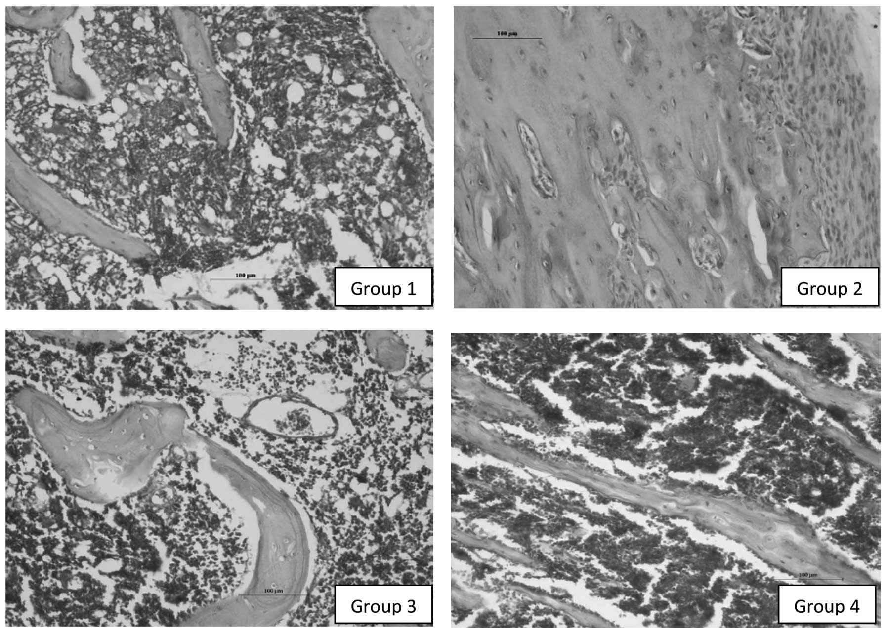
also normal amounts of reticulin fibres. In the lead acetate alone
group, there was strong evidence of fibrosis and focal areas of
sclerosis. Significant marrow hypoplasia was also detected. There
were coarse reticulin fibres with minimal cells (Fig. 1). In the Carica papaya alone
groups, there were healthy cortical bones with normal architecture,
focal areas of mild hyperplasia and normal amounts of reticulin
fibres. One of the distinct features identified was the mild
increase in the levels of eosinophils and their precursors in the
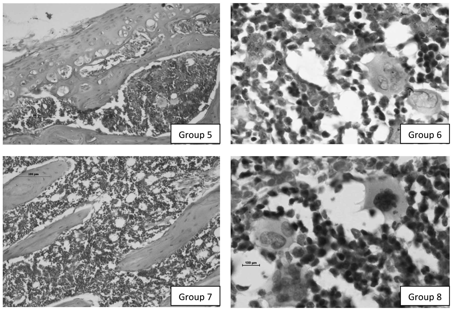
bone marrow. The lead exposure followed by Carica papaya
groups showed abundant osteoid formation, along with hyperplasia of
the marrow, pushing out the cortical bone (Fig. 2). The cortical bone was healthy and
rimmed by active osteoblasts. There were also areas of fibrosis and
sclerosis and an increased amount of reticulin fibres. The
Carica papaya followed by lead exposure groups showed
evidence of bone marrow morphological changes which were suggestive
of lead-induced damage with alternating areas of regenerative
change. There were areas of alternative marrow hypoplasia and focal
islands of hyperplasia of the haemopoietic components. Mild
osteosclerosis and focal areas of fibrosis were observed as well as
an increased amount of reticulin fibres (Table II).
| Table II.Morphological changes in rat femur
sections. |
Table II.
Morphological changes in rat femur
sections.
| Group | BM cells | Fat cells | Fibrosis | Sclerosis | Reticulin fibers |
|---|
| 1 | ++ | + | 0 | 0 | 0 |
| 2 | + | ++ | +++ | +++ | +++ |
| 3 | +++ | + | 0 | 0 | 0 |
| 4 | +++ | + | 0 | 0 | 0 |
| 5 | +++ | + | ++ | + | ++ |
| 6 | +++ | + | + | + | ++ |
| 7 | +++ | + | ++ | + | ++ |
| 8 | +++ | + | + | + | ++ |
Discussion
Lead is known to induce damage via oxidative stress.
This means that lead increases the reactive oxygen species content
of the body and depletes antioxidant enzyme levels (14–16).
PCC is a useful biomarker indicative of oxidative damage (17,18).
PCC are generated by the actions of reactive oxygen species. Hence,
an increase in PCC is directly associated with increased reactive
oxygen species. Glutathione is an important antioxidant in the
body. A low level of glutathione indicates depletion of
antioxidants while a higher level of glutathione signifies higher
antioxidant levels (18).
In the biochemical results, the control and
Carica papaya alone groups exhibited low PCC and high GC,
indicating a low amount of oxidative damage with high antioxidant
levels. High PCC and low GC were observed in the lead acetate alone
group, indicating heavy oxidative damage and low antioxidant
levels. This observation regarding lead acetate is consistent with
previous studies which noted that bone marrow PCC was significantly
higher following treatment with lead acetate and antioxidant levels
were significantly lower (5,6).
In a previous study, Carica papaya leaves
were reported to offer protection against alcohol-induced
oxidatative damage to the gastric mucosa (19). In the present study, the aim was to
observe whether Carica papaya had the ability to ameliorate
oxidative damage induced by lead acetate in the bone marrow.
Carica papaya demonstrated protective effects on lead
acetate-induced oxidative damage in the bone marrow since the
pre-treatment (Carica papaya followed by lead acetate)
groups exhibited significantly lower PCC and significantly higher
GC than the lead acetate alone group (P<0.05).
Carica papaya was also observed to have a
reversal effect on lead acetate-induced oxidative damage in the
bone marrow since the post-treatment (lead acetate followed by
Carica papaya) group showed significantly lower PCC and
significantly higher GC than the lead acetate alone group
(P<0.05). This is consistent with a study that detected the
antioxidant properties of Carica papaya leaves (11). Statistical analysis showing a
significant difference further supported the results.
Histopathological analysis was performed to study
the morphological damage and reversal of any changes in the
experimental groups. It was observed that in the lead alone group
there was extensive damage to the bone and bone marrow, with
fibrosis, sclerosis, hypoplasia and loss of marrow space. This
observation was similar to that of a previous study where the lead
acetate-induced group exhibited thickening of bone, irregular bony
spicules and mild osteoporotic changes (5).
The Carica papaya alone groups were very
similar to the control group and did not show any morphological
changes in terms of damage. The higher dose of Carica papaya
exhibited improved cellularity but this was statistically
insignificant compared with the lower dose (Carica papaya 50
mg); both doses showed adequate cellularity and healthy marrow.
There was also mild hyperplasia in the 2 groups which was more
prominent for the 200 mg dose. This suggests that Carica
papaya alone has a stimulatory effect on the haemopoiesis of
cells in the bone marrow.
The post-treatment (lead acetate followed by
Carica papaya) groups exhibited areas of damage induced by
lead mixed with regenerative areas having good marrow cellularity.
There were zones of fibrosis intermingled with zones of
hyperplastic marrow tissue. This suggests that Carica papaya
has a reversal effect on the lead acetate-induced effects in the
bone marrow, as well as a stimulatory effect on the haemopoiesis of
cells in the bone marrow.
The pre-treatment (Carica papaya followed by
lead acetate) groups also showed good cellularity with focal areas
of damage observed as mild to moderate fibrosis. These groups
showed areas of marrow hypoplasia intermingled with areas of
hyperplasia of the bone marrow. This suggests that Carica
papaya has a protective effect on the lead acetate-induced
effects in the bone marrow and also a stimulatory effect on the
haemopoiesis of cells in the bone marrow. The findings for each
group were supported by reticulum staining.
Of note was the presence of zones of hyperplasia in
the Carica papaya alone, pre-treatment and post-treatment
groups which suggested that Carica papaya had a stimulatory
effect on the haemopoiesis of cells.
Also observed was an increase in the levels of all
blood cell precursors, particularly the megakaryocytes and the
myeloblast series, in all pre treatment and post treatment groups
(groups 5, 6, 7 and 8). Certain areas contained giant
megakaryocytes and there was an increase in eosinophils. Increased
proliferation of blood vessels was also observed which may have
contributed to the hyperplasia of the bone marrow cells. The study
was a double-blind experiment whereby the slides were selected at
random and observed by two researchers.
Studies concerning the effect of Carica
papaya on the lead acetate-induced changes to bone marrow are
lacking. However, a study by Sathasivam et al demonstrated
an increase in thrombocyte count of mice following the
administration of a papaya leaf suspension which may be consistent
with the present observation of an increase in the megakaryocyte
count which may lead to an increased thrombocyte count (12). This observation also agrees with
the finding of Sathasivam et al that platelet counts 72 h
after dosing were significantly higher, indicating an increase in
white blood cells and platelets which normalise clotting and repair
the liver (12). This is
consistent with the present findings of a notable increase in white
blood cells precursors (myeloblast series) and increase in the
megakaryocyte count for platelet production in all groups treated
with Carica papaya.
A study on a dengue patient revealed increased white
blood cell and platelet counts following treatment with Carica
papaya leaf extract (13).
This also agrees with the present findings of increased levels of
white blood cell precursors (myeloblast series) and megakaryocytes
in all groups treated with Carica papaya. These findings
suggest that Carica papaya may protect against and reverse
the lead acetate-induced effects on the bone marrow, such as marrow
hypoplasia, and stimulate haemopoiesis of the cells, particularly
the myeloblasts and megarkaryocytes.
Acknowledgements
The study was performed with a
research grant [no. BMSI01-2011 (07)] provided by the International
Medical University, Kuala Lumpur, Malaysia.
References
|
1.
|
Bellinger DC: Very low lead exposures and
children’s neurodevelopment. Curr Opin Pediatr. 20:172–177.
2008.
|
|
2.
|
El-Nekeety AA, El-Kady AA, Soliman MS,
Hassan NS and Abdel-Wahhab MA: Protective effect of Aquilegia
vulgaris (L.) against lead acetate-induced oxidative stress in
rats. Food Chem Toxicol. 47:2209–2215. 2009.
|
|
3.
|
Sivaprasad R, Nagaraj M and Varalakshmi P:
Combined efficacies of lipoic acid and 2,3-dimercaptosuccinic acid
against lead-induced lipid peroxidation in rat liver. J Nutr
Biochem. 15:18–23. 2004. View Article : Google Scholar : PubMed/NCBI
|
|
4.
|
Bradberry S and Vale A: Lead. Medicine.
31:56–57. 2003. View Article : Google Scholar
|
|
5.
|
Lee KH, Chen YS, Judson JP, Chakravarthi
S, Sim YM and Er HM: The effect of water extracts of Euphorbia
hirta on cartilage degeneration in arthritic rats. Malays J
Pathol. 30:95–102. 2008.
|
|
6.
|
Haleagrahara N, Chakravarthi S, Kulur AB
and Radhakrishnan A: Effects of chronic lead acetate exposure on
bone marrow lipid peroxidation and antioxidant enzyme activities in
rats. AJPP. 5:923–929. 2011.
|
|
7.
|
Haw KY, Chakravarthi S, Haleagrahara N and
Rao M: Effects of Etlingera elatior extracts on lead
acetate-induced testicular damage: A morphological and biochemical
study. Exp Ther Med. 3:99–104. 2012.
|
|
8.
|
Jackie T, Haleagrahara H and Chakravarthi
S: Antioxidant effects of Etlingera elatior flower extract
against lead acetate-induced perturbations in free radical
scavenging enzymes and lipid peroxidation in rats. BMC Res Notes.
4:672011.
|
|
9.
|
Owoyele BV, Adebukola OM, Funmilayo AA and
Soladoye AO: Anti-inflammatory activities of ethanolic extract of
Carica papaya leaves. Inflammopharmacology. 16:168–173.
2008. View Article : Google Scholar : PubMed/NCBI
|
|
10.
|
Otsuki N, Dang NH, Kumagai E, Kondo A,
Iwata S and Morimoto C: Aqueous extract of Carica papaya
leaves exhibits anti-tumor activity and immunomodulatory effects. J
Ethnopharmacol. 127:760–767. 2010.
|
|
11.
|
Seigler DS, Pauli GF, Nahrstedt A and Leen
R: Cyanogenic allosides and glucosides from Passiflora
edulis and Carica papaya. Phytochemistry. 60:873–882.
2002. View Article : Google Scholar : PubMed/NCBI
|
|
12.
|
Sathasivam K, Ramanathan S, Mansor SM,
Haris MR and Wernsdorfer WH: Thrombocyte counts in mice after the
administration of papaya leaf suspension. Wien Klin Wochenschr.
121(Suppl 3): 19–22. 2009. View Article : Google Scholar : PubMed/NCBI
|
|
13.
|
Ahmad N, Fazal H, Ayaz M, Abbasi BH,
Mohammad I and Fazal L: Dengue fever treatment with Carica
papaya leaves extracts. Asian Pac J Trop Biomed. 330–333. 2011.
View Article : Google Scholar
|
|
14.
|
Adonaylo VN and Oteiza PI: Lead
intoxication: antioxidant defences and oxidative damage in rat
brain. Toxicology. 135:77–85. 1999. View Article : Google Scholar : PubMed/NCBI
|
|
15.
|
Upasani CD, Khera A and Balaraman R:
Effect of lead with Vitamins E, C, or Spirulina on malondialdehyde:
conjugated dienes and hydroperoxides in rats. Indian J Exp Biol.
39:70–74. 2001.PubMed/NCBI
|
|
16.
|
Sharma V, Sharma A and Kansal L: The
effect of oral administration of Allium sativum extracts on
lead nitrate induced toxicity in male mice. Food Chem Toxicol.
48:928–936. 2010.
|
|
17.
|
Reznick AZ, Cross CE, Hu ML, et al:
Modification of plasma proteins by cigarette smoke as measured by
protein carbonyl formation. Biochem J. 286:607–611. 1992.PubMed/NCBI
|
|
18.
|
Haleagrahara N, Jackie T, Chakravarthi S,
Rao M and Pasupathi T: Protective effects of Etlingera
elatior extract on lead acetate-induced, changes in oxidative
biomarkers in bone marrow of rats. Food Chem Toxicol. 48:2688–2694.
2010.
|
|
19.
|
Indran M, Mahmood AA and Kuppusamy UR:
Protective effect of Carica papaya L leaf extract against
alcohol induced acute gastric damage and blood oxidative stress in
rats. West Indian Med J. 57:323–326. 2008.
|