Introduction
Henoch-Schönlein purpura nephritis (HSPN) is the
most common secondary renal disease in children and its morbidity
is only less than that of primary nephrotic syndrome and acute
glomerulonephritis. Vasculitis is a clinical manifestation of HSPN
and it has been shown that renal vascular lesions are significant
in HSPN (1). The mechanism may be
endothelial cell damage leading to reduced renal capillary density
and the local occurrence of chronic ischemic changes in the kidney,
thereby increasing renal pathological damage (2). The discovery of circulating
endothelial progenitor cells (EPCs) by Asahara et
al(3,4), and further data suggesting their
participation in postnatal vasculogenesis, provides evidence for
vascular regeneration. EPCs multiply and differentiate into
vascular endothelial cells, which facilitate vascular repair
(5). Studies have revealed that
EPCs differentiate into mature endothelial cells, involved in the
repair of damaged endothelial cells. A decrease in this process and
functional damage may induce vascular lesions and dysfunctional
regeneration (6). A previous study
(7) revealed that levels of EPCs
in the circulation are indicative of risk for vascular disease.
Patients with the highest number of circulating EPCs were least at
risk of coronary artery disease, suggesting that circulating EPC
levels and the maintenance of vascular integrity are associated and
may be of major clinical relevance (8). Recent studies demonstrated the
presence of bone marrow-derived EPCs in the systemic circulation.
They increase in number in response to certain cytokines and/or
tissue ischemia and they target and are incorporated into the site
of neovascularization (9,10). Evidence indicates that the number
and function of EPCs are related to blood vessel damage and repair.
In patients with chronic kidney disease (CKD), a decrease in
circulating EPCs may impair vascular regenerative potential and
thus contribute to a higher cardiovascular risk. The effect of
significantly increased endostatin levels on the endothelial
function and progenitors in patients with CKD requires further
investigation (11–15). In the process of HSPN development,
the correlation between EPCs and renal vascular lesions in HSPN
patients has not been reported, but findings confirming that EPCs
take part in vascular endothelial regeneration suggest that the
progression of HSPN may result from insufficient delivery or
decreased production of EPCs. In the current study, we monitored
changes to EPC number, migration and adhesion function in the
peripheral blood of HSPN patients with different vascular lesions
and the correlation between EPCs and renal vascular lesions to
provide information useful in the clinical diagnosis and treatment
of HSPN.
Materials and methods
Research subjects
Forty-eight children with HSPN, diagnosed by
clinical manifestation and renal biopsy, were observed between June
2004 and August 2009 in the Second Xiangya Hospital of Central
South University, China. All subjects conformed to the standard set
out by the national pediatric renal disease team (16). There were 29 males and 19 females,
aged 2.2–14.2 years. Of the 48 HSPN patients, 19 patients had
hematuria (39.6%), including 9 patients with gross hematuria, 5
patients with microscopic hematuria and 5 patients with isolated
hematuria; 17 patients had isolated proteinuria (35.4%) with serum
albumin lower than normal and 9 patients presented nephrotic
syndrome (18.8%); and 5 patients had typical symptoms of
Henoch-Schönlein purpura, including skin purpura, and an abnormal
renal biopsy, despite the urine routine being normal. The course of
the disease was 2 months to 6 years. According to the degree of
vascular pathology, the 48 HSPN patients were divided into three
groups: mild, moderate and severe. Twenty healthy patients were
simultaneously set as the control group.
Renal biopsy pathological
examination
Tissues from the renal biopsy were examined by light
microscopy and immunofluorescence. For the light microscopic
examination, tissues were embedded in Petroline, sliced to 2
μm thickness and stained by hematoxylin and eosin (H&E),
periodic acid-Schiff (PAS), periodic acid-Schiff metheramine (PASM)
and Masson’s staining. Immunoglobulin (Ig)-A, IgG, IgM, C1q, C3 and
C4 were detected in the frozen sections by direct
immunofluorescence. Glomerular lesions were classified using
International Society for Kidney Disease Community (ISKDC)
guidelines and HSPN was classified into levels I–VI. Vascular
lesion degree was evaluated by the method of Katafuchi et
al(17). Each sample included
>3 renal interstitium vessels (artery) and the condition of
vascular lesions, including the presence or absence of vessel wall
thickening, hardening and hyalinization, was observed. As long as
one vessel demonstrated these changes, it was defined as having
vessel lesions. Vessel wall thickening was defined as vessel inner
diameter/outer diameter <0.5 in the cross-section. The scores
allocated according to the percentage of lesioned vessels were 0, 1
(<10%), 2 (10–25%) and 3 (>25%). The vessel wall
thickening/hardening/hyalinizating was also scored from 0 to 3. The
total score was calculated by adding both scores together; score 0,
no vessel lesions; score 1–2, mild vessel lesions; score 3–4,
moderate vessel lesions and score 5–6, severe vessel lesions. The
48 HSPN patients were divided into mild, moderate and severe groups
according to the degree of vessel pathology.
EPC isolation, culture and
identification
We extracted 5 ml peripheral blood, isolated the
mononuclear cells and inoculated them into a 20-hole culture board
coated with human fibronectin (HFN). The cells were cultivated in
endotheliocyte basic medium (EBM)-2 with 20% fetal bovine serum
(FBS), 50 μg/l vascular endothelial growth factor (VEGF), 50
μg/l stem cell growth factor (SCF), 100 U/ml penicillin and
100 U/ml phytomycin and were placed in a 37°C CO2
incubator. After 4 days, we exchanged the medium and removed the
non-adherent cells. After 7 days, the adherent cells were mixed
with DiI-low density lipoprotein (LDL), with a final concentration
of 24 mg/l. They were incubated for 1 h at 37°C, then fixed with 4%
paraformaldehyde. Fluorescein isothiocyanate-labeled Ulex
europaeus agglutin-I (FITC-UEA-I) was added (final
concentration, 10 mg/l) and placed in an incubator for 1 h at 37°C.
Finally we viewed and counted the cells under an inverted
microscope. Cells stained positive with DiI-LDL and FITC-UEA-I were
differentiating vascular EPCs (18). At the same time, adherent cells
were detected for phycoerythrin (PE)-CD34, PE-CD133 and PE-kinase
insert domain receptor (PE-KDR) expression by flow cytometry,
controlled by the corresponding PE-IgG1.
EPC adhesion and migration
We used trypsin to digest the adherent cells, which
were then collected, added to EBM-2 medium (including 5% FBS),
counted, then inoculated onto a culture board coated with HFN. The
cells were left to culture for 30 min in a 37°C incubator. We then
washed out non-adherent cells with phosphate-buffered saline (PBS)
and counted the number of adherent cells (magnification, ×200)
(19). For the detection of EPC
migration, we collected adherent cells, added them to EBM-2 medium
and counted them. EBM-2 medium and VEGF (50 μg/ml) were
added to the inferior chamber of the modified Boyden chamber and
2×10⁴ EPCs suspended in 50 μl medium were added
to the superior chamber. After cultivating for 24 h, non-moving
cells on the filter membrane were removed. All other cells were
fixed by methanol, stained by Giemsa, then three fields were
selected randomly and cells that had migrated to the underlayer
were counted (magnification, ×200) (20).
Statistical analysis
We used SPSS (SPSS Inc., Chicago, IL, USA) for
Windows 10.0 to analyze data. Numeration data are presented as mean
± standard deviation. Data were compared between the groups and
between several points in the same group. Homogeneity of variance
was analyzed using a one-way analysis of variance (ANOVA),
heterogeneity of variance was analyzed using Kruskal-Wallis and
least significant difference (LSD) and Mann-Whitney U tests were
used to compare data between two groups. P<0.05 was considered
to indicate a statistically significant difference.
Results
Pathological types and vascular lesion
levels of HSPN patients
According to glomerular pathological levels, 11
patients were level II, 21 patients were level III, 12 patients
were level IV and 4 patients were level V. All glomerular
mesenteria in the HSPN patients had deposits of IgA of different
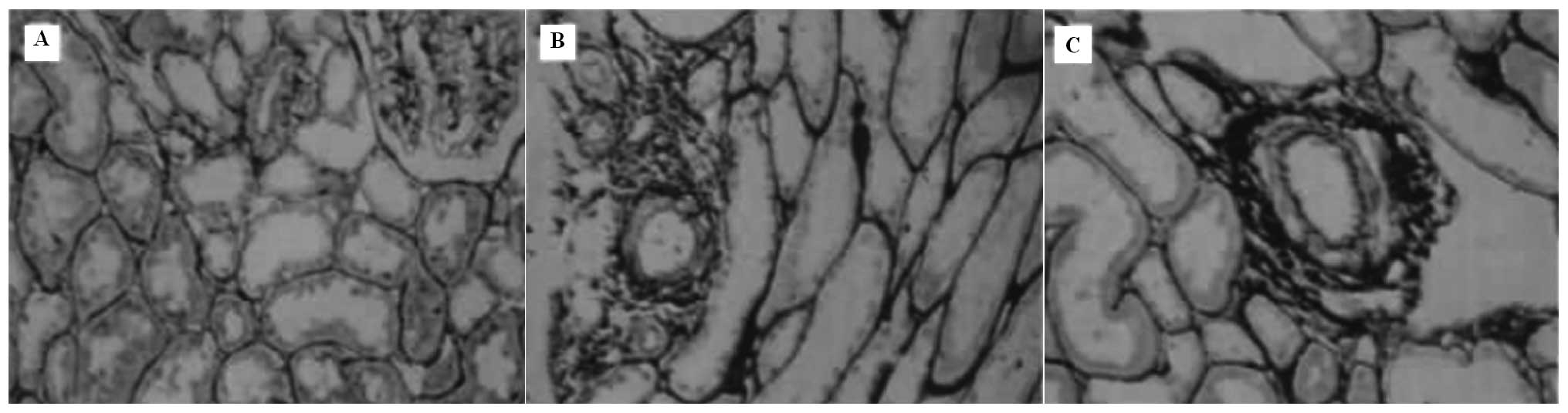
degrees. According to the degree of vascular lesions, 21 patients
belonged to the mild group (1.512±0.306; semi-quantitative score of
vascular damage); 20 patients belonged to the moderate group
(3.517±0.468) and 7 patients belonged to the severe group
(5.416±0.367). Compared with the mild group, pathological
integrations in the moderate and severe groups significantly
increased (both P<0.01) and pathological integration in the
severe group was significantly higher than in the moderate group
(P<0.01). We demonstrated that the more severe the renal lesion,
the more severe the vascular lesion (Table I and Fig. 1).
 | Table I.Renal pathological levels (II–V) of
groups with different degrees of vascular lesions (n). |
Table I.
Renal pathological levels (II–V) of
groups with different degrees of vascular lesions (n).
| Groups | Level II | Level III | Level IV | Level V | Total |
|---|
| Mild group | 9 | 12 | 0 | 0 | 21 |
| Moderate group | 2 | 8 | 9 | 1 | 20 |
| Severe group | 0 | 1 | 3 | 3 | 7 |
| All | 11 | 21 | 12 | 4 | 48 |
Number of peripheral blood EPCs in HSPN
patients with different degrees of renal vascular lesions
After being cultured in vitro for 7 days,
EPCs in peripheral blood changed into endothelioid cells with a
spindle-shape. Under a fluorescent inverted microscope, cells
phagocytizing Dil-LDL evoked a red fluorescence and cells
integrating FITC-UEA-I evoked a green fluorescence. Cells revealing
a positive staining for both expressed a yellow fluorescence, which
confirmed that those adherent cells were differentiating EPCs. The
numbers of CD34+, KDR+ and CD133+
cells were lower in the moderate and severe vascular lesion groups
than that in the control group (all P<0.05). The numbers of
CD34+ and CD133+ cells were higher in the
mild vascular lesion group than in the control group and of
KDR+ cells were lower in the mild group than in the
control group; however, these differences had no statistical
significance (all P>0.05). With the exception of KDR in the
moderate vascular lesion group, the numbers of CD34+,
KDR+ and CD133+ cells were significantly
lower in the moderate and severe vascular lesions groups than in
the mild vascular lesion group (all P<0.05). The numbers of
CD34+, KDR+ and CD133+ cells were
lower in the severe vascular lesion group than in the mild and
moderate vascular lesions groups (all P<0.05; Table II).
| Table II.Comparison of EPC counts in the
peripheral blood of HSPN patients with different degrees of
vascular lesions. |
Table II.
Comparison of EPC counts in the
peripheral blood of HSPN patients with different degrees of
vascular lesions.
| Groups | Number | CD34+
count | CD133+
count | KDR+
count |
|---|
| Mild group | 21 | 57.08±8.25 | 31.14±5.66 | 46.14±8.23 |
| Moderate group | 20 |
45.16±4.38a,c |
21.47±2.79a,c | 39.47±7.82a |
| Severe group | 7 | 38.96±3.74b,d | 11.79±2.01b,d,e | 27.56±5.64b,f |
| Control group | 20 | 53.37±6.41 | 30.21±5.36 | 48.35±9.03 |
| F-value | | 85.79 | 121.47 | 46.38 |
| P-value | | 0.00 | 0.00 | 0.00 |
Adhesion and migration of peripheral
blood EPCs in HSPN patients with different degrees of renal
vascular lesions
The adhesion and migration activity of EPCs was
higher in the control group than in the mild, moderate and severe
groups; however, the difference between the mild and control groups
had no statistical significance (Table
III). The adhesion and migration activities of EPCs were
reduced in turn in the mild, moderate and severe groups. There were
significant differences between the severe group and the mild and
moderate groups (all P<0.05). The adhesion and migration
activities of EPCs in the moderate group were lower than in the
mild group; however, the difference had no statistical
significance.
| Table III.Comparison of adhesion and migration
of peripheral blood EPCs in HSPN children with different degrees of
renal vascular lesions. |
Table III.
Comparison of adhesion and migration
of peripheral blood EPCs in HSPN children with different degrees of
renal vascular lesions.
| Groups | Number | Adhesion
function | Migration
function |
|---|
| Mild group | 21 | 23.25±2.35 | 12.17±2.38 |
| Moderate group | 20 | 18.61±3.09a | 10.19±2.79a |
| Severe group | 7 | 12.47±2.63b,c,e |
8.69±2.20a,d,e |
| Control group | 20 | 25.47±2.79 | 14.56±2.25 |
| F-value | | 76.47 | 63.65 |
| P-value | | 0.00 | 0.00 |
Discussion
Vasculitis is a clinical manifestation of HSPN. The
pathogenesis may be that the impaired vascular endothelial cells
induce a reduction in the renal micrangium density, causing the
kidneys to develop chronic ischemia and renal pathological lesions
to worsen (2). In this study, we
observed that the more severe the renal pathological lesions are,
the worse the vascular lesions are, which demonstrates that renal
vascular lesions play an important role in the occurrence and
development of HSPN. This may be due to the vascular lesions of the
renal interstitium inducing renal interstitial ischemia and
hypoxia, resulting in an infiltration of inflammatory cells.
Inflammatory cytokines and mediators are released and fibrocytes
proliferate, promoting renal tubular epithelial cell apoptosis,
renal tubular atrophy, and an increase in the rate of development
of renal interstitial fibrosis (21,22).
Instantaneously, renal interstitial vascular lesions increase the
resistance of glomerular blood vessels, affecting blood supply to
the glomeruli. This induces further damage to the glomeruli and
renal interstitium, resulting in a cyclic process of damage between
vessels, glomeruli and the renal tubular interstitium. Previous
studies (23) have considered that
immune damage, immune-medium and metabolic abnormalities are the
initiating agents of HSPN vascular lesions. Further research has
shown that vascular epithelial cell damage and the resultant cell
number decrease, induced by various agents, are the most direct
influential factors (24).
EPCs are a group of precursor cells that multiply
and differentiate into mature vascular endothelial cells and are
involved in postnatal vascular growth and the repair of endothelial
damage. Twenty-five percent of epithelial cells in the newborn
vessels are differentiated from EPCs (25). Recent research demonstrates that
there are a number of EPCs in the peripheral blood, and when
cytokine irritation and local ischemia occur, EPCs mobilize to the
impaired site to aid in vascular regrowth (26) and the repair of the vascular
endothelium. Vascular endothelial repair is a complicated regulated
process, including mobilization, adhesion, chemo-taxis, migration,
invasion to the ischemic tissue gaps and then differentiation to
mature vascular endothelium cells and the formation of new vessels
(27). In the current study, we
found that the more severe the renal vascular lesion, the fewer the
number of EPCs in the peripheral blood, suggesting that there is a
correlation between the decrease in EPC number and renal vascular
lesions in HSPN patients. However, in the mild group, the numbers
of CD34+ and CD133+ cells were higher than in
the control group, which may be a reverse feedback mechanism in the
stress repair of autogenous vascular lesions. KDR is a type of
event marker for when EPCs differentiate into endothelial cells.
The KDR count in the peripheral blood was less in the mild group of
HSPN patients than in the normal group. This indicates that EPCs
recruited by stress recovery are not able to supply enough
endothelial cells in the case of vascular damage. We found that EPC
adhesion and migration activity in the peripheral blood of the
control group was higher than in the mild, moderate and severe
groups and EPC adherence and migration were in turn decreased in
the mild, moderate and severe groups. This suggests that the renal
vascular regrowth and endothelial repair are related not only to
the number of EPCs, but also to the decrease of adhesion and
migration activities.
In this study we found that renal vascular lesions
are involved in the occurrence and development of HSPN and the
number of EPCs, as well as the migration and adhesion of EPCs, are
important factors in renal vascular lesions.
Acknowledgements
This study was supported by the
National Natural Science Foundation of China (No. 30500546).
References
|
1.
|
Zhao MF, Yan YC and Qian JQ: Relationship
between decreased renal function and renal vascular changes. Acta
Universitatis Medicinalis Secondae Shanghai. 25:505–507. 2005.(In
Chinese).
|
|
2.
|
Kang DH, Anderson S, Kim YG, et al:
Impaired angiogenesis in the aging kidney: vascular endothelial
growth factor and thrombospondin-1 in renal disease. Am J Kidney
Dis. 37:601–611. 2001. View Article : Google Scholar : PubMed/NCBI
|
|
3.
|
Asahara T, Murohara T, Sullivan A, et al:
Isolation of putative progenitor endothelial cells for
angiogenesis. Science. 275:964–967. 1997. View Article : Google Scholar : PubMed/NCBI
|
|
4.
|
Asahara T, Takahashi T, Masuda H, et al:
VEGF contributes to postnatal neovascularization by mobilizing bone
marrow-derived endothelial progenitor cells. EMBO J. 18:3964–3972.
1999. View Article : Google Scholar : PubMed/NCBI
|
|
5.
|
Chung Y, Abou-Nassar KE, Li Y, et al:
Vascular progenitor recruitment in critically ill patients with
acute kidney injury. Clin Invest Med. 34:3042011.PubMed/NCBI
|
|
6.
|
Waksman R and Baffour R: Bone marrow and
bone marrow derived mononuclear stem cells therapy for the
chronically ischemic myocardium. Cardiovasc Radiat Med. 4:164–168.
2003. View Article : Google Scholar : PubMed/NCBI
|
|
7.
|
Werner N, Kosiol S, Schiegl T, et al:
Circulating endothelial progenitor cells and cardiovascular
outcomes. N Engl J Med. 353:999–1007. 2005. View Article : Google Scholar : PubMed/NCBI
|
|
8.
|
Rosenzweig A: Circulating endothelial
progenitors - cells as biomarkers. N Engl J Med. 353:1055–1057.
2005. View Article : Google Scholar : PubMed/NCBI
|
|
9.
|
Aicher A, Zeiher AM and Dimmeler S:
Mobilizing endothelial progenitor cells. Hypertension. 45:321–325.
2005. View Article : Google Scholar : PubMed/NCBI
|
|
10.
|
Murayama T, Tepper OM, Silver M, et al:
Determination of bone marrow-derived endothelial progenitor cell
significance in angiogenic growth factor-induced neovascularization
in vivo. Exp Hematol. 30:967–972. 2002. View Article : Google Scholar : PubMed/NCBI
|
|
11.
|
Wątorek E, Paprocka M, Duś D, Kopeć W and
Klinger M: Endostatin and vascular endothelial growth factor:
potential regulators of endothelial progenitor cell number in
chronic kidney disease. Pol Arch Med Wewn. 121:296–301.
2011.PubMed/NCBI
|
|
12.
|
Futrakul N, Butthep P, Laohareungpanya N,
Chaisuriya P and Ratanabanangkoon K: A defective angiogenesis in
chronic kidney disease. Ren Fail. 30:215–217. 2008. View Article : Google Scholar : PubMed/NCBI
|
|
13.
|
Rydzewska-Rosolowska A, Borawski J and
Mysliwiec M: High plasma endostatin level unaffected by
low-molecular weight heparin in hemodialysis patients - a
preliminary report. Adv Med Sci. 54:199–202. 2009. View Article : Google Scholar : PubMed/NCBI
|
|
14.
|
O’Riordan E, Mendelev N, Patschan S, et
al: Chronic NOS inhibition actuates endothelial-mesenchymal
transformation. Am J Physiol Heart Circ Physiol. 292:285–294.
2007.PubMed/NCBI
|
|
15.
|
Kohagura K, Ohya Y, Miyagi S, et al:
rHuEPO dose inversely correlated with the number of circulating
CD34+ cells in maintenance hemodialysis patients.
Nephron Clin Pract. 108:c41–c46. 2008. View Article : Google Scholar : PubMed/NCBI
|
|
16.
|
Yi ZW: Henoch-Schönlein purpura nephritis.
Practical Handbook Kidney Disease in Children. People’s Medical
Publishing House; Beijing: pp. 384–390. 2005
|
|
17.
|
Katafuchi R, Kiyoshi Y, Oh Y, et al:
Glomerular score as a prognosticator in IgA nephropathy: its
usefulness and limitation. Clin Nephrol. 149:1–8. 1998.PubMed/NCBI
|
|
18.
|
Kalka C, Masuda H, Takahashi T, et al:
Transplantation of ex vivo expanded endothelial progenitor cells
for therapeutic neovascularization. Proc Natl Acad Sci USA.
97:3422–3427. 2000. View Article : Google Scholar : PubMed/NCBI
|
|
19.
|
Walter DH, Rittig K, Bahlmann FH, et al:
Statin therapy accelerates reendothelialization: a novel effect
involving mobilization and incorporation of bone marrow-derived
endothelial progenitor cells. Circulation. 105:3017–3024. 2002.
View Article : Google Scholar
|
|
20.
|
Vasa M, Fichtlscherer S, Aicher A, et al:
Number and migratory activity of circulating endothelial progenitor
cells inversely correlate with risk factors for coronary artery
disease. Circ Res. 89:E1–E7. 2001. View Article : Google Scholar : PubMed/NCBI
|
|
21.
|
Zhang B, Liang X, Shi W, et al: Role of
impaired peritubular capillary and hypoxia in progressive
interstitial fibrosis after 56 subtotal nephrectomy of rats.
Nephrology (Carlton). 10:351–357. 2005. View Article : Google Scholar : PubMed/NCBI
|
|
22.
|
Hu F and Hu WX: Microvascular disease in
progressive renal disease. J Med Postgraduates. 11:1034–1036.
2004.(In Chinese).
|
|
23.
|
Mrowka C, Heintz B and Sieberth HG:
VCAM-1, ICAM-1, and E-selectin in IgA nephropathy and
Schönlein-Henoch syndrome: differences between tissue expression
and serum concentration. Nephron. 81:256–263. 1999.PubMed/NCBI
|
|
24.
|
Wu H, Chen H and Hu PC: Circulating
endothelial cells and endothelial progenitors as surrogate
biomarkers in vascular dysfunction. Clin Lab. 53:285–295.
2007.PubMed/NCBI
|
|
25.
|
Suzuki T, Nishida M, Futami S, et al:
Neoendothelialization after peripheral blood stem cell
transplantation in humans: a case report of a Tokaimura nuclear
accident victim. Cardiovasc Res. 58:487–492. 2003. View Article : Google Scholar : PubMed/NCBI
|
|
26.
|
Shintani S, Murohara T, Ikeda H, et al:
Mobilization of endothelial progenitor cells in patients with acute
myocardial infarction. Circulation. 103:2776–2779. 2001. View Article : Google Scholar : PubMed/NCBI
|
|
27.
|
Urbich C and Dimmeler S: Endothelial
progenitor cells: characterization and role in vascular biology.
Circ Res. 95:343–353. 2004. View Article : Google Scholar : PubMed/NCBI
|