Introduction
Blunt pancreatic injury is uncommon with a 1–5%
incidence rate in blunt trauma (1). Due to its retroperitoneal location,
early physical signs and symptoms are commonly non-specific. Also,
blunt pancreatic injury may be overlooked in patients with
extensive multi-organ trauma. A delay in diagnosis leads to
complications, including pancreatitis, pancreatic
abscesses/necrosis, pancreatic fistulae and pseudocysts, which
result to a high mortality rate of nearly 20% (1). Therefore the prompt and accurate
diagnosis of pancreatic injury is imperative (2).
Abdominal computed tomography (CT) scans are the
modality of choice for evaluating stable patients with suspicious
abdominal injuries. CT is efficient for the diagnosis of
contusions, pancreatic disruptions and associated complications
(3). The reported sensitivity and
specificity rates are as high as 80% (4), with suspicious findings including
peripancreatic hematomas and fluid in the lesser sac or
retroperitoneum (5). CT grading of
pancreatic trauma has been widely used in the clinic (6). However, CT has its disadvantages; it
is expensive, not performed in real-time, requires exposure to
X-ray irradiation, requires patient transfer away from the
resuscitation area and in the case of contrast-enhanced CT (CECT)
examinations, certain patients may suffer allergic reactions
(7).
Due to being portable, less costly and usable at the
bed-side, ultrasound (US) has been used for the diagnosis and
follow-up of blunt abdominal trauma. However, conventional US is
not able to provide an accurate evaluation of the site and the
severity of pancreatic parenchymal injury and active bleeding. With
the development of a second generation of sonographic contrast
agents and contrast-specific technology, contrast-enhanced US
(CEUS) has been shown to be an effective method in the diagnosis of
hepatic, splenic and nephric trauma (8–10).
Certain studies have shown significant differences in the detection
rate between conventional US and CEUS (P<0.01) (11). CEUS was reported to clearly show
the location, size, extension and active bleeding of trauma, which
improved the detection rate from a range of 45.7–63% for
conventional US to a range of 80–91.4% (9,12).
By contrast, the use of CEUS imaging to define the
features of blunt pancreatic injury has not been well documented
and the value of CEUS in the diagnosis of pancreatic injury has not
been studied. The purpose of the present study was to present CEUS
imaging findings and discuss the utility of CEUS in the evaluation
of blunt pancreatic injury.
Materials and methods
Animal model
The experimental protocols of the present study were
approved by the Institutional Animal Care and Use Committee (IACUC)
of the Chinese People’s Liberation Army General Hospital and
performed in accordance with the regulations for animal experiments
defined by the ethics committee of The Chinese People’s Liberation
Army General Hospital. In total, 40 healthy, male Chinese Guangxi
Bama miniature pigs were housed individually in a windowed room at
26±1°C, with a relative humidity of 40–50%. All the animals were
appropriately acclimatized and observed for a period of 1 week.
Food was withdrawn the evening prior to the experiment. The pigs
were anesthetized by an intra-muscular injection of pentobarbital
sodium (30 mg/kg) and 1,000 ml Ringer’s solution was intravenously
administered via the vena auricularis magna during the experiment.
A midline laparotomy incision was made under aseptic conditions,
the gastrocolic ligament was divided and the lesser sac was
entered. The pancreas was then exposed and injuries were
established randomly using a hemostatic pincette to crush the
pancreas against the spine or posterior abdominal wall. Subsequent
to establishing the injuries the incision was sutured. The present
study was carried out in strict accordance with the recommendations
made in the National Institutes of Health Guide for the Care and
Use of Laboratory Animals (13).
The protocol for animal use was reviewed and approved by the IACUC
of the Chinese People’s Liberation Army General Hospital.
Sonographic equipment and contrast
agent
The baseline US and CEUS were performed with an
Acuson Sequioa 512 Scanner (Siemens Medical Solutions, Mountain
View, CA, USA). A 3–5 MHz transducer (4V1, Acuson) was also used.
The contrast-enhanced studies were performed using the
contrast-specific contrast pulse sequencing (CPS) technique at a
low mechanical index (MI) of 0.15–0.17.
The contrast medium administered was SonoVue
(Bracco, Milan, Italy), a second-generation blood-pool US contrast
agent, which was previously reported to be useful in the detection
of injuries caused by abdominal trauma (8–12).
The contrast agent was injected as a bolus using a 21-gauge
catheter placed in the vena auricularis magna. Immediately
thereafter, a 5-ml saline solution flush (0.9% NaCl) followed
(using a three-way stopcock).
CT equipment and contrast agent
A helical Twin scan (GE Healthcare, Piscataway, NJ,
USA) was used in the present study. The median slice-thickness for
the contrast-enhanced images was 3 mm (range, 2–4 mm). A total of
40–50 ml IV contrast agent (iohexol, Omnipaque 300; GE Healthcare)
was injected at 2.5 ml/sec. Scanning was performed from 20 sec
after the onset of the contrast injection.
Methods of examination
Conventional US (3.5–5.0 MHz, 30–50 mm deep) was
performed within 30 min of the establishment of the pancreatic
trauma. The focus was set at the deeper level of the lesion. The
location, size, extent and characteristics of the laceration were
recorded. Depending on the conventional US findings, CEUS was
performed as a focused examination of the area of interest.
Subsequent to changing to CPS at a low MI (0.15–0.17), a contrast
agent bolus of 1.2 ml was injected into the vena auricularis magna.
The region of interest was then slowly and continuously scanned for
up to 2 min, until the enhancing effect began to disappear. At the
moment of injection, a timer was pushed and video archiving was
started. All images were analyzed by two sonographers, each with a
minimum of 5 years CEUS experience, who were blinded to the
establishment of the traumatic models.
A CECT scan was performed within 30 min subsequent
to the CEUS examination. The sweep range ran from the diaphragmatic
muscle to the inferior margin of the kidney. CT images were
reviewed by two experienced radiologists with 5 and 7 years of
experience in abdominal imaging. They were kept blinded to the
results obtained from the US and CEUS imaging modalities and other
techniques.
Following the examination, a laparotomy was
performed in each pig by a surgeon who was not involved in the
process of injury establishment and examination. The pancreatic
duct injuries were identified by the injection of methylene blue
through the duodenal papilla. All the lesions were then classified
as deep or superficial. Deep lesions were defined as hematomas or
lacerations that were ≥50% of the thickness of the pancreas.
Superficial lesions were described as hematomas or lacerations that
were <50% of the thickness of the pancreas.
Statistical analysis
Measurements are presented as mean ± SD. Differences
between group means were compared by a Student’s t-test. Ratio
proportions were compared using a Chi-square test with continuity
corrections or a Fisher’s exact test when appropriate. The level of
statistical significance was set at P<0.05. Two-sided
significance tests were used throughout.
Results
Injuries
In total, forty male pigs with a mean weight of
22.2±1.7 kg (range, 20–25 kg) were used. A total of forty injury
sites were established, including twenty deep sites with an average
depth of 1.5±0.4 cm (range, 0.9–2.2 cm) and twenty superficial
sites with an average depth of 0.5±0.2 cm (range, 0.2–0.9 cm).
Of the twenty deep lesions, eighteen lesions with
main pancreatic duct injuries (MPD+) and two lesions
without main pancreatic duct injuries (MPD−) were
identified in the laparotomy. Of the twenty superficial lesions,
one MPD+ lesion was detected.
The levels of serum amylase and lipase were observed
to increase during the initial one hour period following pancreatic
trauma (Tables I and II). However, there were no significant
differences between the levels in the superficial and deep lesions
(P>0.05).
 | Table ISerum amylase mean values during the
initial 1 h period following pancreatic trauma. |
Table I
Serum amylase mean values during the
initial 1 h period following pancreatic trauma.
| Lesions | Amylase (IU/l)
|
|---|
| Pre-injury | 1st hour |
|---|
| Total | 1373±349 | 2845±520 |
| Deep | 1295±370 | 2874±480a |
| Superficial | 1451±316 | 2714±608a |
| Table IISerum lipase mean values during the
initial 1 h period following pancreatic trauma. |
Table II
Serum lipase mean values during the
initial 1 h period following pancreatic trauma.
| Lesions | Lipase (IU/l)
|
|---|
| Pre-injury | 1st hour |
|---|
| Total | 1088±246 | 1646±417 |
| Deep | 1080±235 | 1591±281a |
| Superficial | 1095±262 | 1705±512a |
Sonographic findings
In the contrast enhancement analysis, two dynamic
phases were considered: the arterial phase, 10–30 sec subsequent to
injection; and the venous phase, 30–120 sec subsequent to
injection.
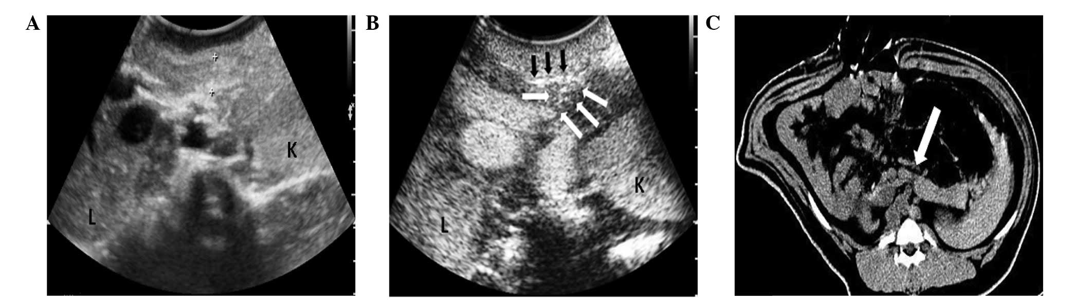
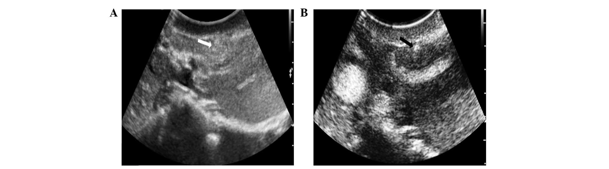
A total of twenty-one lesions were detected using
conventional US (Figs. 1 and
2), while nineteen lesions
remained undetected. The detection rate of conventional US in
pancreatic trauma was 52.5% (Table
III). The pancreatic lacerations appeared as slit- or
stripe-shaped hyperechoic, hypoechoic or anechoic areas on the
baseline US image. The pancreatic contusions appeared as lamellar
hyperechoic or hypoechoic areas with unclear margins in the
parenchyma on conventional US images.
| Table IIIComparison of the detection rate
between US and CEUS in the diagnosis of pancreatic lesions. |
Table III
Comparison of the detection rate
between US and CEUS in the diagnosis of pancreatic lesions.
| Group | Lesions
|
|---|
| Superficial | Deep | Total |
|---|
| US, n (%) | 7 (35) | 14 (70) | 21 (52.5) |
| CEUS, n (%) | 14 (70) | 20 (100) | 34 (85) |
| P-value | 0.0418 | 0.0202 | 0.0038 |
Following the injection of the contrast agent, 34
lesions were detected and 6 lesions remained undetected by CEUS.
The detection rate of CEUS in pancreatic trauma was 85%, which was
significantly higher than that of conventional US, (P<0.05;
Table III). The six lesions that
were not revealed by CEUS were MPD− lesions. The
pancreatic lacerations showed no enhancement in the two phases,
however a high enhancement was observed in the two phases due to
active bleeding (Figs. 1 and
2). The pancreatic contusion
showed low enhancement in the two phases. The margins of the
pancreatic injuries were clearly observed compared with the normal
parenchyma by CEUS.
Peripancreatic fluid collections were identified in
all pigs by US and CEUS.
CECT findings
CECT was performed in all pigs; 33 lesions were
diagnosed as pancreatic injuries and 7 were not. The detection rate
of CECT in pancreatic trauma was 82.5% (Table IV). A total of twenty deep lesions
and thirteen superficial lesions were identified by CECT imaging.
All lesions that were not revealed using CECT were MPD−
lesions. All pigs with hemoperitoneum were successfully identified
by CECT imaging.
| Table IVComparison of the detection rate in
the diagnosis of pancreatic lesions between CEUS and CECT. |
Table IV
Comparison of the detection rate in
the diagnosis of pancreatic lesions between CEUS and CECT.
| Group | Lesions
|
|---|
| Superficial | Deep | Total |
|---|
| CECT, n (%) | 13 (65) | 20 (100) | 33 (82.5) |
| CEUS, n (%) | 14 (70) | 20 (100) | 34 (85) |
| P-value | 1.0000 | - | 1.0000 |
The lacerations were visible as low-attenuation
lines oriented perpendicularly to the long axis of the pancreas
(Fig. 1). The diagnosis of a
pancreatic parenchymal fracture by CECT was based on the
visualization of a clear separation of the pancreas. Certain
fractures or lacerations of the pancreas produced little separation
of the fragmented tissue and showed no enhancement in the two
phases. The contusions appeared as diffuse areas of low attenuation
or focal areas of low attenuation within the normal parenchyma.
There was no significant difference in the detection
rate of pancreatic lesions between CEUS and CECT (P>0.05;
Table IV). The main CEUS and CECT
findings from the forty pigs with blunt pancreatic injuries are
presented in Table V.
| Table VCEUS and CECT findings in the forty
pigs with blunt pancreatic injuries. |
Table V
CEUS and CECT findings in the forty
pigs with blunt pancreatic injuries.
| Main signs | CEUS | CECT |
|---|
| Fracture of the
pancreas | 5 | 5 |
| Pancreatic
laceration | 28 | 29 |
| Contusion | 15 | 13 |
| Focal or diffuse
pancreatic enlargement | 29 | 29 |
| Peripancreatic fluid
collection | 40 | 40 |
Discussion
Injuries to the pancreas are rare. The initial signs
and symptoms of pancreatic injury may be subtle and laboratory
findings of pancreatic injury are nonspecific, which may
consequently lead to delayed or missed diagnoses and substantial
morbidity and mortality rates. Matsuno et al(14) reported that a delayed serum amylase
and lipase measurement may be useful to detect pancreatic injuries.
In the data from the present study, elevated serum amylase and
lipase levels were observed in 36 and 35 pigs, respectively. This
indicated that amylase and lipase measurements are useful to detect
pancreatic injuries, but that the elevated level is not correlated
with the severity of the injury. This result conforms with the
findings of other studies (15,16).
Blunt pancreatic injuries are usually caused by a
direct force applied across the upper abdomen by a seat belt, the
handlebar of a bicycle or motorcycle or by a steering wheel, which
compresses the pancreas against the spine (17). Therefore, a pancreatic parenchymal
compression method was adopted to establish the pancreatic injury
model in the present study and to imitate the mechanism by which
blunt pancreatic injuries usually occur.
In the early phase of the trauma (72 h post-injury),
the identification of the pancreatic trauma was difficult. Although
CT has advantages in the diagnosis of pancreatic trauma (18,19),
it is not available to the first choice for hemodynamically
unstable patients. Due to its characteristics of being noninvasive,
versatile, easily accessible and less costly, US plays a
significant role in assessing pancreatic trauma in the early phase.
US effectively complements CT as an auxiliary examination for
patients who are unstable or allergic to diodone.
Nevertheless, the sensitivity of conventional US in
the diagnosis of pancreatic trauma is reported to be low (45.7–63%)
(9–12), which conforms with the results of
the present study. CEUS is more sensitive than conventional US in
identifying the characteristics of the injury, including the size
and the tear thickness (20). In
the present study, the sites of laceration often appeared with low
enhancement. Occasionally the sites appeared with high enhancement,
due to the presence of a hemorrhage caused by injury to the small
peripancreatic vessels, which was always associated with
extra-pancreatic fluid collections. When the fracture was occupied
by fluid, no enhancement was visible in the CEUS images. These
sonographic depictions were similar to those in the CECT images.
These results suggested that CEUS is almost as sensitive as CECT
for depicting blunt pancreatic injuries.
The detection of pancreatic duct injuries is
critical to the subsequent treatment of the patient. Unfortunately,
the integrity of the pancreatic duct is difficult to determine by
CT and US. Therefore, the depth of the pancreatic injury was used
in the present study, instead of a direct description, to determine
the injury to the pancreatic duct (21). Wong et al(4) suggested that a CT finding of a lesion
of >50% of the thickness of the pancreas indicated a likely
disruption to the pancreatic duct. This theory is in line with the
findings of the present study. In total, 90% of the deep lesions
were confirmed as MPD+ and 95% of the superficial
lesions were confirmed as MPD−. Pancreatic trauma with
MPD+ requires surgical treatment and the results of CEUS
and CECT imaging may help decide what treatment is required. It is
important to separate patients into two groups, those who require
immediate surgery and those who require non-surgical observation.
The American Association for the Surgery of Trauma Organ Injury
Scale for pancreatic injury was not used in the present study, as
only a few surgical or radiological studies remarked upon the
integrity of the pancreatic duct (21,22).
However, false negative results were observed in
CECT and CEUS. This may be associated with the depth or appearance
of the lesion. In total, 7 lesions were not identified by CECT and
6 were not identified by CEUS. Lesions are missed by CECT when the
depth is less than the slice thickness. This problem may be avoided
by using CEUS. The minimum depth shown by CEUS is 2 mm. However,
pancreatic fracture lines are not easily detected by CEUS when the
separation of the fractured pancreatic fragments is minimal or
nonexistent (23). Overestimation
on CEUS may occur as deep lacerations are occasionally not
associated with a disruption of the main duct and transections may
merely disrupt the minor duct, as in the present experiment.
In summary, CEUS is able to successfully diagnose
the majority of acute blunt pancreatic injuries and exactly
evaluate the extent of the trauma. However, a normal appearance in
CEUS is not able to exclude pancreatic injury. Repeated physical
examinations and further imaging studies may aid the identification
of acute pancreatic trauma and its mechanisms.
References
|
1.
|
Kao LS, Bulger EM, Parks DL, Byrd GF and
Jurkovich GJ: Predictors of morbidity after traumatic pancreatic
injury. J Trauma. 55:898–905. 2003. View Article : Google Scholar : PubMed/NCBI
|
|
2.
|
Gupta A, Stuhlfaut JW, Fleming KW, Lucey
BC and Soto JA: Blunt trauma of the pancreas and biliary tract: a
multimodality imaging approach to diagnosis. Radiographics.
24:1381–1395. 2004. View Article : Google Scholar : PubMed/NCBI
|
|
3.
|
Venkatesh SK and Wan JM: CT of blunt
pancreatic trauma: a pictorial essay. Eur J Radiol. 67:311–320.
2008. View Article : Google Scholar : PubMed/NCBI
|
|
4.
|
Wong YC, Wang LJ, Lin BC, Chen CJ, Lim KE
and Chen RJ: CT grading of blunt pancreatic injuries: prediction of
ductal disruption and surgical correlation. J Comput Assist Tomogr.
21:246–250. 1997. View Article : Google Scholar : PubMed/NCBI
|
|
5.
|
Fischer JH, Carpenter KD and O’Keefe GE:
CT diagnosis of an isolated blunt pancreatic injury. AJR Am J
Roentgenol. 167:11521996. View Article : Google Scholar : PubMed/NCBI
|
|
6.
|
Lin BC, Liu NJ, Fang JF and Kao YC:
Long-term results of endoscopic stent in the management of blunt
major pancreatic duct injury. Surg Endosc. 20:1551–1555. 2006.
View Article : Google Scholar : PubMed/NCBI
|
|
7.
|
Tang J, Li W, Lv F, et al: Comparison of
gray-scale contrast-enhanced ultrasonography with contrast-enhanced
computed tomography in different grading of blunt hepatic and
splenic trauma: an animal experiment. Ultrasound Med Biol.
35:566–575. 2009. View Article : Google Scholar
|
|
8.
|
Catalano O, Lobianco R, Sandomenico F and
Siani A: Splenic trauma: evaluation with contrast-specific
sonography and a second-generation contrast medium: second
experience. J Ultrasound Med. 22:467–477. 2003.PubMed/NCBI
|
|
9.
|
Catalano O, Lobianco R, Sandomenico F,
D’Elia G and Siani A: Real-time contrast-enhanced ultrasound of the
spleen: examination technique and preliminary clinical experience.
Radiol Med. 106:338–356. 2003.(In English and Italian).
|
|
10.
|
Catalano O, Lobianco R, Raso MM and Siani
A: Blunt hepatic trauma: evaluation with contrast-enhanced
sonography: sonographic findings and clinical application. J
Ultrasound Med. 24:299–310. 2005.PubMed/NCBI
|
|
11.
|
Poletti PA, Platon A, Becker CD, et al:
Blunt abdominal trauma: does the use of a second generation
sonographic contrast agent help to detect solid organ injuries? AJR
Am J Roentgenol. 183:1293–1301. 2004. View Article : Google Scholar : PubMed/NCBI
|
|
12.
|
McGahan JP, Horton S, Gerscovich EO, et
al: Appearance of solid organ injury with contrast-enhanced
sonography in blunt abdominal trauma: preliminary experience. AJR
Am J Roentgenol. 187:658–666. 2006. View Article : Google Scholar : PubMed/NCBI
|
|
13.
|
Guide for the Care and Use of Laboratory
Animals. National Research Council (US) Committee for the Update of
the Guide for the Care and Use of Laboratory Animals. 8th edition.
Washington (DC): National Academies Press (US); 2011
|
|
14.
|
Matsuno WC, Huang CJ, Garcia NM, Roy LC
and Davis J: Amylase and lipase measurements in paediatric patients
with traumatic pancreatic injuries. Injury. 40:66–71. 2009.
View Article : Google Scholar : PubMed/NCBI
|
|
15.
|
Al-Ahmadi K and Ahmed N: Outcomes after
pancreatic trauma: experience at a single institution. Can J Surg.
51:118–124. 2008.PubMed/NCBI
|
|
16.
|
Subramanian A, Dente CJ and Feliciano DV:
The management of pancreatic trauma in the modern era. Surg Clin
North Am. 87:1515–1532. 2007. View Article : Google Scholar : PubMed/NCBI
|
|
17.
|
Fisher M and Brasel K: Evolving management
of pancreatic injury. Curr Opin Crit Care. 17:613–617. 2011.
View Article : Google Scholar
|
|
18.
|
Malgras B, Douard R, Siauve N and Wind P:
Management of left pancreatic trauma. Am Surg. 77:1–9.
2011.PubMed/NCBI
|
|
19.
|
Teh SH, Sheppard BC, Mullins RJ, Schreiber
MA and Mayberry JC: Diagnosis and management of blunt pancreatic
ductal injury in the era of high-resolution computed axial
tomography. Am J Surg. 193:641–643. 2007. View Article : Google Scholar : PubMed/NCBI
|
|
20.
|
Lv F, Tang J, Luo Y, et al:
Contrast-enhanced ultrasound imaging of active bleeding associated
with hepatic and splenic trauma. Radiol Med. 116:1076–1082. 2011.
View Article : Google Scholar : PubMed/NCBI
|
|
21.
|
Ahmed N and Vernick JJ: Pancreatic injury.
South Med J. 102:1253–1256. 2009. View Article : Google Scholar
|
|
22.
|
Rekhi S, Anderson SW, Rhea JT and Soto JA:
Imaging of blunt pancreatic trauma. Emerg Radiol. 17:13–19. 2010.
View Article : Google Scholar
|
|
23.
|
Soto JA, Alvarez O, Múnera F, Yepes NL,
Sepúlveda ME and Pérez JM: Traumatic disruption of the pancreatic
duct: diagnosis with MR pancreatography. AJR Am J Roentgenol.
176:175–178. 2001. View Article : Google Scholar : PubMed/NCBI
|