Introduction
Foreign-body ingestion is a commonly encountered
problem in the clinic. Although the majority of foreign bodies
ingested pass spontaneously through the gastrointestinal tract,
foreign bodies become lodged in the esophagus in ∼10–20% of cases,
which require immediate intervention, typically by non-surgical
endoscopic management (1). When
esophageal foreign bodies are impacted for longer than 24 h,
complications, including esophageal perforation, esophageal
fistula, aortoesophageal fistula and mediastinal abscesses, may
occur, and mucosal edema or anabrosis around the wounded areas may
complicate or even prevent the endoscopic removal of foreign
bodies.
Foreign-body ingestion is a commonly encountered
clinical problem in China. However, only a few studies concerning
the endoscopic removal of esophageal foreign bodies from Chinese
patients are available in the literature (2). In addition, in the majority of these
studies the standard flexible endoscope, which has limitations in
removing sharp or larger foreign bodies, was generally used for
management in these patients. The dual-channel endoscope has two
advantages over the single-channel endoscope. The dual-channel
endoscope is slender and flexible, and has a larger inner diameter
that expands the esophageal wall thereby protecting the mucosa,
while the single channel endoscope is stiff. The dual-channel
endoscope also allows the insertion of a balloon catheter with a
pincer, which may increase the success rate of foreign-body removal
while minimizing complications.
In the present study, we report our experience and
the outcomes achieved by using a more powerful dual-channel
endoscope to remove impacted esophageal foreign bodies from 19
Chinese patients admitted to the Digestive Endoscope Center
(Lishui, China) between September 2008 and July 2011. Our study
suggests that dual-channel endoscopic management may be the optimal
choice for removing impacted esophageal foreign bodies, including
those that are large and/or sharp, from patients over a broad range
of ages.
Patients and methods
Patients
Among 19 patients with impacted foreign bodies
admitted to the People’s Hospital of Lishui City (Lishui, China),
13 were male and 6 were female, with ages ranging from 7 to 78
years (Table I). The impacted
foreign bodies were identified at different locations with 11 cases
in the upper esophagus, 5 cases in the middle and 3 cases in the
lower esophagus. The delay time between ingestion and initial
examination varied from 2 to 18 h and the endoscopic interventions
were performed within 24 h. The types of foreign bodies found in
these cases included mammalian bones (6 cases), peach kernels (2
cases), a jujube kernel (1 case), a denture (1 case), fish bones (3
cases), encapsulated pills (1 case), toothpicks (2 cases), a key (1
case), a toy (1 case) and a beer cap (1 case).
 | Table I.Patient information and management
methods. |
Table I.
Patient information and management
methods.
| Age | Gender | Management |
|---|
|
|
|
|---|
| Child | Adult | Elderly | Male | Female | Endoscopy | Surgery |
|---|
| No. of patients | 3 | 7 | 9 | 13 | 6 | 18 | 1 |
Endoscopic management
A dual-channel endoscope (Fujinon EG-250D5; Fujifilm
(China) Investment Co., Ltd. Pudong New Area, Shanghai, China) was
used to remove the impacted foreign bodies. Based on the shapes and
properties of the foreign bodies, suitable accessories were chosen,
including rat-toothed forceps, an alligator clamp, snare, trielcon,
basket and 2.8-mm biopsy forceps. A Boston CRE™ balloon dilatation
catheter (Model Number: M00558380; Boston Scientific, Raffles city,
Shanghai, China) with a diameter of 6–15 mm was used for the
majority of cases.
Results
Initial management
The patients were closely monitored for symptoms,
including subcutaneous emphysema, cough and neck tenderness prior
to endoscopic management. X-ray and CT scanning methods were used
to identify foreign-body shapes, and monitor the impacted areas and
adjacent tissues and organs for possible complications such as
esophageal inflammation, perforation and fistula. Cardiothoracic
surgeons were consulted for potential risks and written consent
forms were obtained from the patients or their guardians after they
had been informed of the planned treatment.
Endoscopic management
Each patient received continuous low-flow oxygen
inhalation through nasal catheters and intravenous injection of
propofol (1.5–2.0 mg/kg). When the patient were under anesthesia
with no eyelash reflex, the endoscope was used to intubate the
esophagus to locate the foreign body. The impacted area and damaged
esophageal mucosa were closely examined prior to a balloon-tipped
catheter being gently inserted and moved to a position 1.0–1.5 cm
away from the impacted foreign body. The inner diameter of the
right side clamp channel of the dual-channel endoscope was larger
than the channel of a single-channel endoscope. The balloon was
inserted via the channel of the right side clamp while the left
side was used to insert other accessories, namely rat-toothed
forceps, to aid in the foreign-body removal. In general, one side
of the foreign body detached when the esophagus was expanded
following balloon inflation. The appropriate accessory was then
used to clamp the detached part of the foreign body, which was then
removed gently. The endoscope, the foreign body and the inserted
device were slowly removed simultaneously. During the procedure,
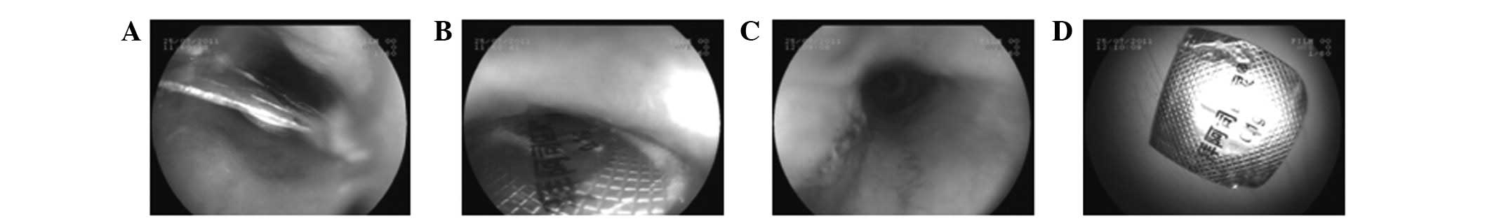
efforts were made to minimize esophageal mucosal damage. An example
of the removal of an encapsulated pill is shown in Fig. 1.
Following successful foreign-body removal, the
affected areas were routinely checked with the endoscope for any
mucosal damage, bleeding, residual foreign bodies, perforations and
complicating diseases, including pharyngalgia and subcutaneous
emphysema. In addition, antibiotics and other necessary medications
were administered to the patients following endoscopy.
Outcomes of endoscopic management
Foreign bodies were successfully removed in 18 cases
using the dual-channel endoscope. Among these 18 cases, two foreign
bodies had sizes (length or diameter) >12 cm and one foreign
body had perforated the esophagus, and so were considered to be
difficult to remove using a standard flexible endoscope. In
addition, no complications were observed in our dual-channel
endoscopic management. Dual-channel endoscopic removal failed in
only one patient in whom the two ends of a denture (∼30×25 mm size)
had perforated into the middle esophagus ∼25 cm away from the
incisors. The foreign body did not separate from the mucosa
following balloon expansion at the esophagus. Endoscopic management
was finally abandoned due to the risk of heavy bleeding from the
aortic arch to which the foreign body was adjacent, and the patient
was then subjected to surgical removal.
Discussion
Esophageal perforation resulting from impacted
foreign bodies is a serious hazard encountered in the clinic
(3). However, there are no
reported epidemiological data on esophageal foreign bodies in
China, and only few studies have been published on esophageal
foreign body impactions in Chinese patients (4,5). In
the present study, we report our experience and the outcomes of the
removal of foreign bodies using a dual-channel endoscope with the
aid of a balloon catheter. The balloon catheter is useful for
dual-channel endoscopic foreign-body management as the inflated
balloon causes the esophageal wall to expand, following which one
side of the foreign body often detaches from the esophagus. This
then facilitates clamping the foreign body for removal without
further damage to the mucosa. In addition, the balloon catheter
method also greatly reduces the risk of heavy bleeding or
esophageal perforation following foreign-body removal as the
inflated balloon suppresses esophageal bleeding while emergency
surgery is in progress. We observed that the optimal location of
the inserted balloon in the esophagus is ∼1.0–1.5-cm away from the
far end of the endoscope. If the balloon is too close to the
foreign body, it may worsen the condition by pressing the foreign
body into the esophageal walls or the balloon may be perforated by
a sharp foreign body.
The dual-channel endoscope has certain advantages
over the standard flexible endoscope for foreign-body removal. A
flexible endoscope that is generally 9–10 mm in diameter is not
capable of removing a sharp or hooked foreign body or a larger
foreign body (6–8). By contrast, the dual-channel
endoscope has a greater diameter which greatly facilitates the
removal of sharp or larger foreign bodies. In addition, a
dual-channel endoscope may be used with a balloon catheter to
minimize mucosal damage by expanding the esophagus. In contrast to
a complication rate of ∼5% for standard flexible endoscopic
management (2), our dual-channel
endoscopic management for foreign-body removal in 19 Chinese
patients aged between 7 and 78 years did not cause any
complications in 18 patients. One patient presented with an
impacted foreign body close to the main aortic arch and underwent
surgery due to the risk of heavy bleeding. These results indicate
that the dual-channel endoscope may prove to be a useful tool for
removing ingested foreign bodies, particularly sharp or large
foreign bodies, from the esophagus.
References
|
1.
|
Llompart A, Reyes J, Ginard D, et al:
Endoscopic management of foreign bodies in the esophagus. Results
of a retrospective series of 501 cases. Gastroenterol Hepatol.
25:448–451. 2002.(In Spanish).
|
|
2.
|
Li ZS, Sun ZX, Zou DW, et al: Endoscopic
management of foreign bodies in the upper-GI tract: experience with
1088 cases in China. Gastrointest Endosc. 64:485–492. 2006.
View Article : Google Scholar : PubMed/NCBI
|
|
3.
|
Tang Y, Jiang F, Shi R, et al: Threading
method to remove ingested foreign bodies at the esophagus. Chinese
Journal of Digestive Endoscopy. 24:151–152. 2007.(In Chinese).
|
|
4.
|
Port JL, Kent MS, Korst RJ, et al:
Thoracic esophageal perforations: a decade of experience. Ann
Thorac Surg. 75:1071–1074. 2003. View Article : Google Scholar : PubMed/NCBI
|
|
5.
|
Wang C, Zeng L, Yang G, et al: Application
of painless gastroscope for removing foreign bodies at the upper
gastrointestinal. Chinese Journal of Digestive Endoscopy.
24:455–457. 2007.(In Chinese).
|
|
6.
|
Athanassiadi K, Gerazounis M, Metaxas E
and Kalantzi N: Management of esophageal foreign bodies: a
retrospective review of 400 cases. Eur J Cardiothorac Surg.
21:653–656. 2002. View Article : Google Scholar : PubMed/NCBI
|
|
7.
|
Sittitrai P, Pattarasakulchai T and
Tapatiwong H: Esophageal foreign bodies. J Med Assoc Thai.
83:1514–1518. 2000.
|
|
8.
|
Ren X, Xu Z, Zhang X, et al: 112 Cases of
clinical analysis of risky foreign bodies at the esophagus. Chinese
Journal of Otorhinolaryngology-Skull Base Surgery. 10:307–309.
2004.(In Chinese).
|