Introduction
Cavernous hemangiomas are benign vascular tumors
that may develop in any part of the body. They are composed of
large vessels with dilated lumina and thin walls. The vessels may
have abnormal walls and cannot be identified as arterial or venous.
Thrombosis and calcification are commonly observed in cavernous
hemangiomas.
Testicular cavernous hemangiomas are a rare type of
benign testicular tumor and distinguishing them from other common
testicular tumors prior to surgery is challenging. The chief
presenting symptom of testicular cavernous hemangioma is testicular
enlargement. In the current study, we report a case of cavernous
hemangioma of the left testis, which mimicked a testicular teratoma
and was treated with radical orchiectomy, and provide a review of
the literature.
Case report
A 42-year-old male presented with a sudden episode
of left testicular fullness for three months. The patient denied
any history of hematuria, fever, scrotal trauma or urinary tract
infection. The patient’s past medical history and family history
were non-contributory.
Physical examination revealed a palpable,
non-tender, left testicular mass ∼3×2.5 cm in size. The left testis
was swollen and stiff. The epididymis and spermatic cord were
normal. The patient had a normal blood cell count and urinalysis.
Laboratory examinations, including relevant tumor markers,
particularly α-fetoprotein and β-human chorionic gonadotrophin,
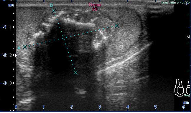
were normal. Scrotal ultrasound revealed a roundish,
well-demarcated, hypoechoic mass in the left testicle (Fig. 1) and several calcifications were
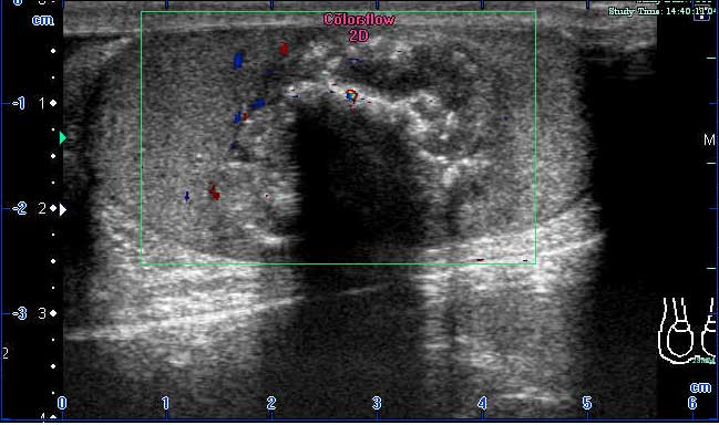
visible within the mass. The mass demonstrated blood flow in color
Doppler sonography (Fig. 2). The
patient was diagnosed with a testicular teratoma and a left radical
orchidectomy, using an inguinal approach, was performed. However,
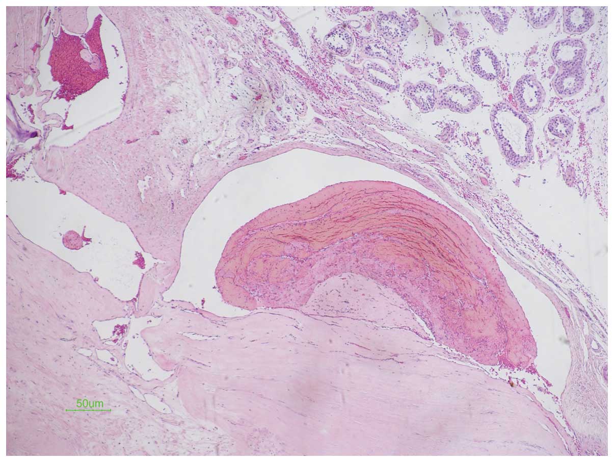
pathological evaluation of the mass revealed that is was a
testicular cavernous hemangioma with thrombus organization and
calcifications (Fig. 3). The study
was approved by the ethics committee of the First Affiliated
Hospital, College of Medicine, Zhejiang University, Zhejiang,
China. Written informed patient consent was obtained from the
patient.
Discussion
Cavernous hemangiomas are benign vascular tumors,
which may develop in any part of the body. The occurrence of
cavernous hemangioma in the testis is rare. The first case of
testicular cavernous hemangioma was reported in 1944 and, to date,
23 cases have been reported (1–13).
The majority of subtypes of vascular tumors of the testis have been
described as cavernous, capillary, histiocytoid and juvenile, with
the most common being cavernous hemangioma (2). Hemangiomas most likely arise from the
inner layer of the tunica albuginea, which contains blood vessels
and lymphatics and sends septa into the testicular parenchyma
(9). A hemangioma may extend into
the testicular parenchyma by way of these septa. Cavernous
hemangiomas are composed of large vessels with dilated lumina and
thin walls. They may be composed of vessels whose walls are
abnormal and cannot be identified as arterial or venous. Thrombosis
and calcification are common in cavernous hemangiomas (9).
The age of onset for testicular cavernous
hemangiomas reported in the literature varies from 17 weeks to 77
years (1–13). Testicular enlargement, with or
without tenderness, is the chief presenting symptom, which is
similar to that of malignant testicular tumors on clinical
presentation. However, there are reports of testicular hemangiomas
presenting as testicular torsion or associated with testicular
infarction (7,11). Distinguishing cavernous hemangioma
of the testis from other common testicular tumors prior to surgery
is not feasible. Doppler ultrasonography is useful for diagnosing
testicular hemangiomas. It demonstrates the nature of the mass and
differentiates it from other testicular neoplasms (14). Hemangiomas in sonographs vary from
hypoechoic to hyperechoic, or they may be heterogeneous (15). Various sizes of calcification are
common, and in this case report we misdiagnosed cavernous
hemangioma of the testis as a testicular teratoma. To date, all
reported vascular testicular tumors have demonstrated benign
behavior, without local recurrence or metastasis (16). Testis-sparing surgery may be
performed if intraoperative examination of frozen sections of
representative tissue is possible (16).
In conclusion, testicular cavernous hemangioma is
rare. When a patient presents with a testicular mass where the
ultrasound reveals a mass with calcifications of various sizes and
negative tumor marker findings, a diagnosis of testicular cavernous
hemangioma should be considered.
Acknowledgements
This study was supported by a grant
from the Zhejiang Provincial Educational Science Foundation of
China (Grant no. Y201226273) and the National Key Clinical
Specialty Construction Project of China.
References
|
1.
|
Kleiman AH: Hemangioma of the testis. J
Urol. 51:548–550. 1944.
|
|
2.
|
Suriawinata A, Talerman A, Vapnek JM and
Unger P: Hemangioma of the testis: report of unusual occurrences of
cavernous hemangioma in a fetus and capillary hemangioma in an
older man. Ann Diagn Pathol. 5:80–83. 2001. View Article : Google Scholar : PubMed/NCBI
|
|
3.
|
Fossum BD, Woods JC and Blight EM Jr:
Cavernous hemangioma of testis causing acute testicular infarction.
Urology. 18:277–278. 1981. View Article : Google Scholar : PubMed/NCBI
|
|
4.
|
Gharpure KJ, Ahmed YB and Bhargava MK:
Cavernous haemangioma of testis with acute testicular infarction -
a case report. Indian J Cancer. 22:73–75. 1985.PubMed/NCBI
|
|
5.
|
Ogawa O, Yoshimura N, Nishimura K, et al:
A case of cavernous hemangioma of the testis. Hinyokika Kiyo.
31:2060–2064. 1985.(In Japanese).
|
|
6.
|
Tada M, Takemura S, Takimoto Y and
Kishimoto T: A case of cavernous hemangioma of the testis.
Hinyokika Kiyo. 35:1969–1971. 1989.(In Japanese).
|
|
7.
|
Lozano V, Alonso P and Marcos-Robles J:
Case report: sonographic appearance of cavernous haemangioma of the
testis. Clin Radiol. 49:284–285. 1994. View Article : Google Scholar : PubMed/NCBI
|
|
8.
|
Frank RG, Lowry P and Ongcapin EH: Images
in clinical urology. Venous cavernous hemangioma of the testis.
Urology. 52:709–710. 1998. View Article : Google Scholar : PubMed/NCBI
|
|
9.
|
Erdag G, Kwon EO, Lizza EF and Shevchuk M:
Cavernous hemangioma of tunica albuginea testis manifesting as
testicular pain. Urology. 68:673.e13–673.e15. 2006. View Article : Google Scholar : PubMed/NCBI
|
|
10.
|
Takaoka E, Yamaguchi K and Tominaga T:
Cavernous hemangioma of the testis: a case report and review of the
literature. Hinyokika Kiyo. 53:405–407. 2007.PubMed/NCBI
|
|
11.
|
Minagawa T and Murata Y: Testicular
cavernous hemangioma associated with intrascrotal testicular
torsion: a case report. Hinyokika Kiyo. 55:161–163. 2009.(In
Japanese).
|
|
12.
|
Venkatanarasimha N, McCormick F and
Freeman SJ: Cavernous hemangioma of the testis. J Ultrasound Med.
29:859–860. 2010.PubMed/NCBI
|
|
13.
|
Hadzi-Djokić J, Pejcić T, Aćimović M and
Andrejević V: The case of cavernous testicular hemangioma. Acta
Chir Iugosl. 57:107–109. 2010.PubMed/NCBI
|
|
14.
|
Carmignani L, Gadda F, Gazzano G, et al:
High incidence of benign testicular neoplasms diagnosed by
ultrasound. J Urol. 170:1783–1786. 2003. View Article : Google Scholar : PubMed/NCBI
|
|
15.
|
Ricci Z, Koenigsberg M and Whitney K:
Sonography of an arteriovenous-type hemangioma of the testis.
174:1581–1582. 2000.PubMed/NCBI
|
|
16.
|
Mazal PR, Kratzik C, Kain R and Susani M:
Capillary haemangioma of the testis. J Clin Pathol. 53:641–642.
2000. View Article : Google Scholar : PubMed/NCBI
|